JAN-FEB, 2013, Vol. – I, Issue-IV
www.srjis.com
Page 1079EEG Brain Mapping of Developmental Dyslexia- A Case Study Report
Nithiya Amirtham S.
Meston College of Education,
(Affiliated to Tamilnadu Teachers Education University)
Royapettah, Chennai. Tamilnadu, India.
K. Saraladevi
Meston College of Education,
(Affiliated to Tamilnadu Teachers Education University)
Royapettah, Chennai. Tamilnadu, India.
Received: 22 February 2013
Reviewed & Received: 28 February 2013
Accepted: 28February 2013
Developmental dyslexia is a specific and significant impairment in reading abilities which is
unexplainable by any kind of deficit in general intelligence, learning opportunity and general
motivation or sensory acuity. The main objective of the study was to measure the absolute
powers of beta, alpha, theta and delta of the dyslexic case with the help of the intervention
programme on relaxation techniques. The experimental sample for the present study was a single
dyslexic case and control group for comparison with quasi experimental approach .Brain waves
of the control samples including the experimental case were recorded for pre test using Electro
Encephalogram (EEG) and relaxation therapy was given for the experimental case alone and
post test was recorded. Statistical analyses were done on the different brain waves. It was found
that increase in absolute powers of Alpha and decrease in Beta brain waves in dyslexic case was
due to relaxation techniques because of the internal mental operations and thereby relaxed and
JAN-FEB, 2013, Vol. – I, Issue-IV
www.srjis.com
Page 1080alert condition of the case was increased. Then decreasing absolute powers of theta and delta
brain waves were mainly due to the changes in cognitive processing during relaxation.
Keywords:Brain Waves, Developmental Dyslexia, EEG, Relaxation Techniques
Introduction:
Developmental dyslexia is defined as a specific and significant impairment in reading abilities,
unexplainable by any kind of deficit in general intelligence, learning opportunity, general motivation or
sensory acuity (Critchley, 1970; World Health Organization, 1993). Children with this condition often
have associated deficits in related domains such as oral language acquisition (dysphasia), writing abilities
(dysgraphia and misspelling), mathematical abilities (dyscalculia), motor coordination (dyspraxia),
postural stability and dexterity, temporal orientation (dyschronia), visuo-spatial abilities (developmental
right-hemisphere syndrome), and attentional abilities (hyperactivity and attention deficit disorder)
(Weintraub & Mesulam, 1983; Rapin & Allen, 1988; Dewey, 1995; Gross-Tsur et al., 1995, 1996;
Fawcett et al., 1996). EEG measures voltage fluctuations resulting from ionic current flows within the
neurons of the brain (Niedermeyer E. & Da Silva, 2004). EEG is a useful source of information on the
background state of the brain, indexing the substrate of cognition and behaviour. EEG studies under
resting state provided important information about basic differences between normal and impaired
readers, a significant advance in the research on electrophysiological correlates of Developmental
Dyslexia has been reached only recently, by using experimental paradigms aimed at stressing (through
specific stimuli or tasks) the functional cognitive processes assumed to be potentially impaired in
dyslexics (i.e., linguistic, perceptual, attentional) (Ackerman, McPherson, Oglesby, & Dykman, 1998;
Rippon & Brunswick, 1998, 2000). Measurement of Absolute power from EEG spectrum is the amount
of EEG in one band without relationship to other bands. The differences between pre and post tests
absolute power of the Developmental Dyslexic case on Beta, Alpha, Theta and Delta are observed with
Relaxation Therapy as an intervention programme, which can be employed as one element of a wider
stress management programme and can decrease muscle tension, lower the blood pressure and slow heart
and breath rates, among other health benefits (Daniel Goleman, 2006).
Colon et al., 1979, aimed to compare EEG power-density spectra between dyslexic and normal
children. Children with age groups 8, 9, 10 of specific reading and writing tasks were selected excluding
organic diseases and mental disorders. In the 8-year-old group the power in the alpha band is higher in
normals, in the 9-year-old group there was a higher power of the mu rhythm in normals and in the
JAN-FEB, 2013, Vol. – I, Issue-IV
www.srjis.com
Page 1081 absolute and relative power and amplitude of EEG spectra (T6-02) of 24 patients with "probable"Alzheimer Disease (AD) at the early stage of the disease and 1 year later and also compared the values to
those of normal elderly controls. The AD patients had significantly higher absolute theta power and lower
beta power values compared to controls but absolute delta and alpha values did not differ. Fein et al.,
1986, studied the resting eyes open and eyes closed EEG in carefully screened samples of 9-13-year-old
dyslexic and control boys within a 2-cohort cross-validation design with repeat testing 1-3 years later. It
was concluded that dyslexia per se is not associated with increased absolute power in the delta and theta
bands; lower power in the high beta band is reliably found in these samples of dyslexics without other
disorders; and alpha power levels are not consistently lower in the dyslexic group.
Objectives of the Study:
To find out the impact of relaxation therapy on absolute powers of beta, alpha, theta and
delta of dyslexic case.
To improve the quality of academic life of dyslexic case.
Research Question:
Does relaxation therapy change the absolute powers of various wave patterns of the
brain?
Design of the Study:
Out of 150 slow learners (age range 14 - 17 years) 18 Dyslexic students were chosen, from which
eight dyslexic students were randomly selected from special school for learning disabled and they are
called as control group. Initially Simple Random Sampling Technique was adopted for control samples’
selection and Selective Sampling Method was used to select the Experimental Sample. Subjects were free
from medical and sleep disorders as determined by history, physical examination, biochemical screening
tests, electrocardiograms, and psychological screening questionnaires. Quasi-Experimental design is used
for the study. Under that Single Case pre test and post test having control group design is framed for the
present study. In order to measure their brain waves permission had been requested from the school
authorities and parents of the selected students. After receiving the permission, Electro Encephalogram
was recorded for all the students to measure their brain activation waves and these values were considered
as pre test values. After that, one student had been taken randomly from the control group as a case for
present research. Relaxation therapy was given to the single case for about a month. Post-test was
recorded on brain waves while doing relaxation therapy for the experimental case at the end of the
JAN-FEB, 2013, Vol. – I, Issue-IV
www.srjis.com
Page 1082 sample statistics as sample statistics (Crawford & Garthwaite, 2002) is used to compute the significanceof difference between pre test and post test of the dyslexic case.
[image:4.612.80.538.159.450.2]Statistical Analyses and Results:
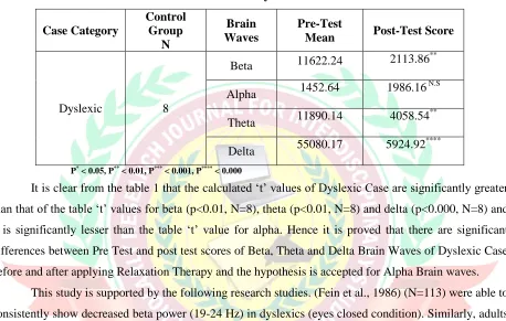
Table 1: Differentiation of pre and post test absolute power of Beta, Alpha, Theta
and Delta from Dyslexic Case
Case Category
Control Group
N
Brain Waves
Pre-Test
Mean Post-Test Score
Dyslexic 8
Beta 11622.24 2113.86
**
Alpha 1452.64 1986.16
N.S
Theta 11890.14 4058.54
**
Delta 55080.17 5924.92
****
P* < 0.05, P** < 0.01, P*** < 0.001, P**** < 0.000
It is clear from the table 1 that the calculated ‘t’ values of Dyslexic Case are significantly greater
than that of the table ‘t’ values for beta (p<0.01, N=8), theta (p<0.01, N=8) and delta (p<0.000, N=8) and it is significantly lesser than the table ‘t’ value for alpha. Hence it is proved that there are significant
differences between Pre Test and post test scores of Beta, Theta and Delta Brain Waves of Dyslexic Case
before and after applying Relaxation Therapy and the hypothesis is accepted for Alpha Brain waves.
This study is supported by the following research studies. (Fein et al., 1986) (N=113) were able to
consistently show decreased beta power (19-24 Hz) in dyslexics (eyes closed condition). Similarly, adults
receiving 15 min of massage therapy showed a pattern of decreased beta power (Field et al., 1996),
suggesting increased relaxation and alertness (Niedermeyer, 1982). (Ikemi, 1988) studied with
Self-Regulation Method (SRM) before vs. during, Self-Self-Regulation Method (SRM) vs. during drowsiness,
having novices (N=12) samples resulted decreased beta power in meditation state.
The EEG signal generated by alpha (8–12 Hz) activity was first described by Hans Berger in
1929, when he demonstrated that closing the eyes decreased sensory input and increased alpha power
over the occipital scalp (Berger, 1929). EEG studies have used these methods to limn the
neurophysiological changes that occur in meditation (Rael Cahn & John Polich, 2006). (Lagopoulos et al.,
2009) observed alpha power increase over the posterior regions. Alpha power increase is one of the more
consistent findings about meditation state effects: alpha is generally associated with relaxation (Aftanas &
JAN-FEB, 2013, Vol. – I, Issue-IV
www.srjis.com
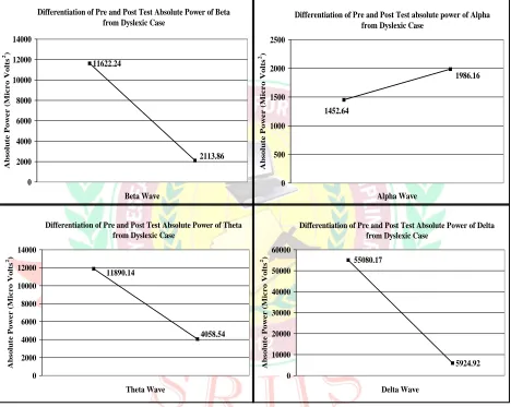
Page 1083 Differentiation of Pre and Post Test Absolute Power of Betafrom Dyslexic Case
11622.24 2113.86 0 2000 4000 6000 8000 10000 12000 14000 Beta Wave A b s ol u te Po w er ( Mi cr o V ol ts 2)
Differentiation of Pre and Post Test absolute power of Alpha from Dyslexic Case
1986.16 1452.64 0 500 1000 1500 2000 2500 Alpha Wave A b s ol u te Po w er ( Mi cr o V ol ts 2)
Differentiation of Pre and Post Test Absolute Power of Theta from Dyslexic Case
4058.54 11890.14 0 2000 4000 6000 8000 10000 12000 14000 Theta Wave A b sol u te Po w er ( Mi cr o V ol ts 2)
Differentiation of Pre and Post Test Absolute Power of Delta from Dyslexic Case
5924.92 55080.17 0 10000 20000 30000 40000 50000 60000 Delta Wave A b sol u te Po w er ( Mi cr o V ol ts 2)
The most dominant effect standing out in the majority of studies on meditation is a state-related
slowing of the alpha rhythm (8–12 Hz) in combination with an increase in the alpha power (Hirai, 1974).
These findings were relatively robust, because they did not depend on either a certain meditation tradition
[image:5.612.74.542.160.533.2]or the experience of the mediators.
Figure 1: Shows Differentiation of Pre and Post Test Absolute Power of Beta, Alpha,
Delta and Theta from Dyslexic Case (from Clockwise Direction)
(Anand et al., 1961) worked with Raj yoga meditation type having the experimental design of
((N=6) and Rest vs. meditation) showed that increased alpha power during Samadhi in meditation state.
(Travis, 1991) studied with Transcendental meditation (TM) type having LTM and STM design (N=20),
showed that increased alpha power in their meditation traits. (Lee et al., 1997) analyzed with Qigong
JAN-FEB, 2013, Vol. – I, Issue-IV
www.srjis.com
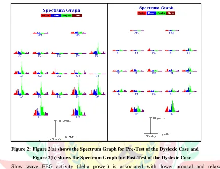
Page 1084 Figure 2: Figure 2(a) shows the Spectrum Graph for Pre-Test of the Dyslexic Case andFigure 2(b) shows the Spectrum Graph for Post-Test of the Dyslexic Case
Slow wave EEG activity (delta power) is associated with lower arousal and relaxation
(Niedermeyer & Rodriguez, 1982), as were decreased in heart rate (Mok & Wong, 2003). (Dunn et al.,
1999) (N=10) used Breath-focused Concentrative vs. Mindfulness Relaxation techniques for the
experimental design of (N=10) (Relaxation and 2 meditation conditions counterbalanced, each practiced
for 15 min) and found that meditation vs. relaxation resulted in decreased delta and theta power in
meditation state. Post-qEEG data from ADHD sample showed that the neurofeedback-trained group but
not the control group showed reduced EEG theta power (Gevensleben et al., 2009 a).
Bootstrap analysis with False Discovery Rate (FDR) corrections for multiple comparisons
analysis of the sensor data indicated decreased delta power during meditation relative to control period at
bilateral frontal electrodes (F3, F4, F7, F8, C3, and C4, all P < 0.05). Frontal delta power at electrodes F3
and F4 was analyzed separately as delta activity at these electrodes was shown to be most significantly
decreased in the bootstrap analysis of the scalp data (Rael Cahn, Arnaud Delorme & John Polich, 2010).
A significant interaction among state, order of experimental sessions (meditation → control vs.
control → meditation), and midline electrode location was found, F (2, 28) = 4.70, P = 0.017). This
JAN-FEB, 2013, Vol. – I, Issue-IV
www.srjis.com
Page 1085 participants doing the control period prior to the meditation period, but not those meditating first.Breakdown of this interaction with Tukey post hoc testing indicated that when the control period occurred
first, midline delta power was decreased in the subsequent meditation session at Fz (P = 0.0004) and Cz
(P = 0.027) but not Pz (P = 0.30) (Rael Cahn, Arnaud Delorme & John Polich, 2010).
A second covariate interaction was found for the state × reported drowsiness × midline electrode
location during meditation, F(2,28) = 3.39, P = 0.06), indicating that only those subjects not reporting
drowsiness during meditation showed a tendency for decreased midline delta power in meditation (Rael
Cahn, Arnaud Delorme & John Polich, 2010).
Discussion:
As a result of the present research findings decrease in beta power and increase in alpha power by
relaxation therapy in dyslexic case is because of the internal mental operations thereby relaxed and alert
condition of the case is increased. Considerable previous research findings indicate that, participants
receiving facial massage therapy exhibited decreased beta power, an EEG pattern that may reflect
attention and alertness (Klimesch et al., 1998; Shagass, 1972; Nunez, 2000). It was shown that a pattern
of decreased beta power using EEG (Field et al., 1996) in massage therapy, suggesting increased
relaxation and alertness (Niedermeyer, 1982). This finding was found to be true in the present
investigation. In Alzheimer’s disease (AD), the decrease in beta power was interpreted as a sign of
compromised function of the affected brain areas (Gianotti, 2007). The structural cause of decrease in
beta power might be a loss of cholinergic and glutamatergic neurons in the course of the disease (Mann,
Oliver & Snowden, 1993). Alpha oscillations are known to arise from an increase of internal attention
(Ray & Cole, 1985) which of course does not only occur due to meditation. Various studies showed an
increase of alpha power related to internally driven mental operations, like the imagery of tones (Ray &
Cole, 1985; Cooper et al, 2003; Cooper et al, 2006) or working memory retention and scanning (Jensen,
2002; Klimesch, 1999). Increase in alpha power was often observed when meditators are evaluated during
meditating compared with control conditions (Aftanas & Golocheikine, 2001; Anand, Chhina, & Singh,
1961; Arambula, Peper, Kawakami, & Gibney, 2001). Several EEG meditation studies reported sleeplike
stages during meditation with increased alpha power (Pagano, Rose, Stivers, & Warrenburg, 1976;
Younger, Adriance, & Berger, 1975). The association between alpha changes and cortical activation had
been assessed with combined EEG and fMRI–PET studies, with increased alpha power related to
decreased blood flow in inferior frontal, cingulate, superior temporal and occipital cortices (Goldman,
Stern, Engel, & Cohen, 2002; Sadato et al., 1998).
In the present research, decreasing theta and delta power are recorded on the experimental case
JAN-FEB, 2013, Vol. – I, Issue-IV
www.srjis.com
Page 1086 due to the changes in cognitive processing during relaxation. Previous research literature also shows thatcortical EEG analyses by Low Resolution Electromagnetic Tomography method (LORETA) revealed
significantly decreased theta activity in the hippocampus, para- hippocampal regions, and the cingulate
cortex areas are known to play a role in cholinergic-associated cognitive functions (Browne et al., 2006).
The pattern of meditation-induced increase in parieto-occipital gamma activity, concomitant decrease in
frontal delta power, and a shift to a more frontal distribution of theta activity suggested that sensory
processing and cognitive processing were altered during meditation relative to the control state (Cahn &
Polich, 2006). Correlation analyses revealed that a decrease in prefrontal delta and, theta power correlated
with an improvement in cognitive performance. Moreover, drowsiness was positively correlated with
theta power in parietal and medial prefrontal regions and beta-1 and beta-2 power in occipital regions
(Saletu, 2007). Neuro feedback aimed at decreasing theta activity might lead to the normalization of
dysfunctional neural network and thus improve clinical symptoms (Koprivova et al., 2007). Power
decreases in the delta and beta-1 bands were found predominantly over the temporo-parieto-occipital
junction, whereas theta power was reduced in the temporo-medial cortex and in fronto-medial regions.
(Riba et al., 2004) suggested the involvement of uni-modal and hetero-modal association cortex and
limbic structures in the psychological effects elicited by ayahuasca (Amazonian beverage ayahuasca).
Acknowledgements:
We mention our deep sense of gratitude to University Grants Commission for Junior Research
Fellowship and also we would like to express our heartfelt thanks to all the children those who
participated in our research, their parents, school authorities, neuro-physician and technicians.
References:
Ackerman, P.T., Mc Pherson., W. B., Oglesby, D. M., & Dykman, R. A. (1998). EEG Power
spectra of adolescent poor readers. Journal of Learning Disabilities, 31, 83-90.
Aftanas, L. I., & Goloecheikine, S. A. (2001). Human anterior and frontal midline theta and
lower alpha reflect emotionally positive state and internalized attention: high-resolution EEG
investigation of meditation. Neuroscience Letters. 310, 57-60.
Anand, B., Chhina, G. S., & Singh, B. (1961). Some aspects of electroencephalographic studies
in yogis. Electroencephalography and Clinical Neurophysiology, 13, 452–456.
Arambula, P., Peper, E., Kawakami, M., & Gibney, K. H. (2001). The physiological correlates of
Kundalini yoga meditation: A study of a yoga master. Applied Psychophysiology and Biofeedback, 26, 147–153.
JAN-FEB, 2013, Vol. – I, Issue-IV
www.srjis.com
Page 1087 Nervenkrankheiten, 87, 527–570.Browne, R.O., Moyal-Segal, L.B., Zumsteg, D., David, Y., Kofman, O., Berger, A., Soreq, H., &
Friedman, A. (2006) Coding region paraoxonase polymorphisms dictate accentuated neuronal
reactions in chronic, sub-threshold pesticide exposure. FASEB Journal, 20(10).
Colon, E.J., Notermans, S.L., de Weerd, J.P., Kap, J. (1979). The discriminating role of EEG
power spectra in dyslexic children. J Neurol, 221 (4), 257-62.
Cooper, N. R., Burgess, A. P., Croft, R. J., & Gruzelier, J. H. (2006). Investigating evoked and
induced electroencephalogram activity in task-related alpha power increases during an internally
directed attention task. Neuroreport, 17, 205–208.
Cooper, N. R., Croft, R. J., Dominey, S. J. J., Burgess, A. P., & Gruzelier, J. H. (2003). Paradox
lost? Exploring the role of alpha oscillations during externally vs. internally directed attention and
the implications for idling and inhibition hypotheses. Int Job satisfaction Psychophysiol, 47, 65–
74.
Crawford, J. R., & Garthwaite, P. H. (2002). Investigation of the single case in neuropsychology:
Confidence limits on the abnormality of test scores and test score differences. Neuropsychologia,
40, 1196-1208.
Crawford, J. R., & Howell, D. C. (1998). Comparing an individual’s test score against norms
derived from small samples. The Clinical Neuropsychologist, 12, 482-486.
Critchley, M. (1970). The dyslexic child (2nd ed.). London: Heinemann Medical.
Daniel Goleman. (1986, May 13). Relaxation: Surprising Benefits Detected. The New York
Times. Retrieved from http://www.nytimes.com.
Dewey, D. (1995). What is developmental dyspraxia? (Review). Brain Cogn, 29, 254–74.
Dunn, B. R., Hartigan, J. A., & Mikulas, W. L. (1999). Concentration and mindfulness
meditations: Unique forms of consciousness? Applied Psychophysiology and Biofeedback, 24, 147–165.
Fawcett, A. J., Nicolson, R. I., & Dean, P. (1996). Impaired performance of children with
dyslexia on a range of cerebellar tasks. Ann Dyslexia, 46, 259–283.