http://dx.doi.org/10.4236/ojrm.2014.33007
How to cite this paper: Karsten, E., Sung, J., Morgan, C., Herbert, B. and Vesey, G. (2014) Evaluation of TransFix® Mediated Stabilisation of Adipose-Derived Stromal Vascular Fraction for Delayed Flow Cytometry Analysis. Open Journal of Regenera- tive Medicine, 3, 54-63. http://dx.doi.org/10.4236/ojrm.2014.33007
Evaluation of TransFix
®
Mediated
Stabilisation of Adipose-Derived Stromal
Vascular Fraction for Delayed Flow
Cytometry Analysis
Elisabeth Karsten*, Judy Sung, Charlotte Morgan, Benjamin Herbert, Graham Vesey
Regeneus Ltd., Sydney, Australia
Email: *[email protected]
Received 28 July 2014; revised 25 August 2014; accepted 2 September 2014
Copyright © 2014 by authors and Scientific Research Publishing Inc.
This work is licensed under the Creative Commons Attribution International License (CC BY). http://creativecommons.org/licenses/by/4.0/
Abstract
The increasing implementation of multicentre studies has led to a need for the optimization of a method that allows for accurate post-hoc analysis of patient biological samples. Assessment of to-tal cell number, viability and immunophenotype can present logistical challenges which can be aided by batch processing. The increased sample storage time that this requires necessitates the use of reagents to preserve cellular integrity, viability and immunophenotype. TransFix® is a
sta-bilising reagent that has been developed for the preservation of cell numbers and cell marker ex-pression in peripheral whole blood for up to ten days. This study investigated the use of TransFix®
reagent for the preservation of the stromal vascular fraction (SVF) of collagenase digested adipose tissue. It was demonstrated that TransFix® was suitable for accurately measuring nucleated SVF
cell numbers for up to seven days as well as back calculating original cell viability. It also stabi-lised three CD markers commonly used to identify populations within SVF (CD90, CD31 and CD45) for up to seven days. There was no significant difference between the number of CD90, CD31 and CD45 positive cells after stabilisation at Day 7 compared to Day 0 unstabilised samples. The re-sults suggest that TransFix® can be used to preserve a biological mixed cell population from
hu-man adipose-derived SVF for up to seven days for accurate post-hoc analysis.
Keywords
TransFix®, Adipose Tissue, Stromal Vascular Fraction, Immunophenotyping
1. Introduction
Mesenchymal stem cells (MSCs) have been investigated for the treatment of a wide range of medical conditions including graft vs host disease [1] and corneal ulceration [2]. MSCs are present in high numbers in the stromal vascular fraction (SVF) of digested adipose tissue which is comprised of a mixed population of cells including erythrocytes, leukocytes, endothelial cells and MSCs [3] [4]. The SVF could be administered immediately for the treatment of a range of conditions [5]-[7] (as opposed to after MSC expansion in culture), allowing for the treatment to be delivered on the same day that the adipose tissue was collected and prepared. However, for use in multicentre studies where analysis is conducted at a core facility, accurate post-hoc analysis of SVF samples can be impeded. For example, shipping and storage can lead to time-dependant alteration of cell makers and cell viability [8]. For this reason, a method of cellular stabilisation would be highly advantageous to allow for de-layed evaluation of total cell number, cell viability and population immunophenotyping by flow cytometry.
TransFix® reagent is a stabilising solution that was designed as a fixative to maintain cellular integrity of whole blood for 7 - 10 days at 2˚C - 22˚C [9]-[11]. Barnett et al.[11] reported that there was no loss in antige-nicity for approximately 20 cell makers including CD45, CD13, CD34 and CD79b over a 10-day period. Ng et al.[12] compared the efficacy of three fixatives and reported that TransFix® was the most effective at preserving extracellular markers in whole blood. On this basis, it was hypothesised that the immunophenotype and perhaps the viability of adipose-derived cells could also be maintained for up to 10 days with the use of TransFix® rea-gent.
This study was performed to evaluate the use of TransFix® reagent for the stabilisation of the SVF of digested adipose tissue for up to 9 days. This was achieved through the comparison of the total nucleated cell count and viability and the analysis of the immunophenotype of CD31, CD45 and CD90 positive cells in the SVF of un-stabilised and un-stabilised patient samples.
2. Materials and Methods
2.1. ReagentsTransFix® reagent (Cytomark, Buckingham UK) was used as a stabilising agent. The monoclonal antibodies used for immunophenotyping are outlined inTable 1. The cellular stains propidium iodide (PI) and Syto 11 were purchased from Sigma Aldrich (NSW, Australia) and Life Technologies (Australia) respectively. Tru-COUNT™ tubes and FACS lysing solution were purchased from Becton Dickinson (Australia) and flow cyto-meter staining buffer was purchased from eBioscience (SA, Australia).
2.2. Patient Samples
Aliquots of SVF were collected from patients receiving a commercial adipose-derived stem cell procedure prior to administration of the therapy. The patients receiving the commercial therapy provided informed consent to provide an aliquot of their SVF for analysis. This study presents the quality control findings from 77 patients. Patient information was kept restricted and confidential at all times. All samples were deidentified and were la-belled in a coded format.
2.3. Adipose-Tissue Processing
The SVF was isolated after collagenase digestion and centrifugation as previously reported [13]. Briefly, the li-poaspirate was digested with 0.5 mg∙mL−1
[image:2.595.91.540.625.722.2]collagenase (human grade, NB6 GMP, SERVA Electrophoresis
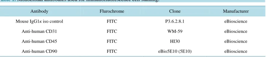
Table 1.Monoclonal antibodies used for immunofluorescence cell staining.
Antibody Flurochrome Clone Manufacturer
Mouse IgG1κ iso control FITC P3.6.2.8.1 eBioscience
Anti-human CD31 FITC WM-59 eBioscience
Anti-human CD45 FITC HI30 eBioscience
GmbH, Heidelberg, Germany) for 20 minutes. The adipose tissue was subsequently washed and the SVF was isolated by centrifugation (1500 g, 5 min, 23˚C) and resuspended in saline. The quantity of adipose tissue col-lected from each patient was dependent upon the individual and varied between 100 - 300 g.
2.4. Sample Preparation
Directly after SVF isolation and resuspension in saline, two or more aliquots (one or more unstablised samples and one or more TransFix® samples) were taken. The unstabilised samples consisted of the prepared SVF re-suspended in saline for a total of 1 mL. For the TransFix®-treated samples, 500 μL of the SVF patient sample was combined with 600 μL of fetal bovine serum (FBS) and 100 μL of TransFix®
reagent. The chosen concen-tration of TransFix® in the stabilised samples were optimised for adipose-derived cells (data not shown). All samples were stored at 4˚C for zero to ten days until required for analysis.
2.5. Flow Cytometry
[image:3.595.148.478.387.708.2]Immunophenotyping, cell count and cell viability of nucleated cells were performed on the samples (Table 2) by flow cytometry on a FACScan flow cytometer (Becton Dickinson) with a 488 nm argon laser. The analysis was carried out using CELLQuest software (ver. 3.3). Before analysis, the control and TransFix®-treated samples were passed through a 35 μm cell strainer (Becton Dickinson) to attain a single cellular suspension. The Day 0 control samples were stored at 4˚C and were assessed within 24 hrs of the adipose digestion. The other unstabi-lised samples and the TransFix® samples were stored at 4˚C for analysis at various time points up to ten days af-ter initial isolation. Daily QC was performed on the flow cytomeaf-ter to standardise instrument settings and flui-dics using flow-check fluorospheres (Beckman Coulter, Australia). The fluorospheres were used according to manufacturer’s instructions.
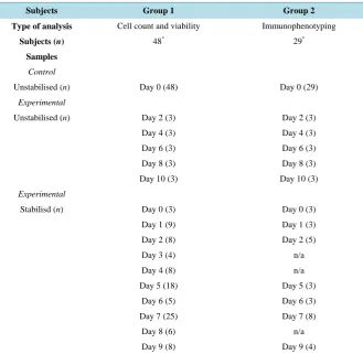
Table 2. Subject table and experimental plan.
Subjects Group 1 Group 2 Type of analysis Cell count and viability Immunophenotyping
Subjects (n) 48* 29*
Samples
Control
Unstabilised (n) Day 0 (48) Day 0 (29)
Experimental
Unstabilised (n) Day 2 (3) Day 2 (3)
Day 4 (3) Day 4 (3)
Day 6 (3) Day 6 (3)
Day 8 (3) Day 8 (3)
Day 10 (3) Day 10 (3)
Experimental
Stabilisd (n) Day 0 (3) Day 0 (3)
Day 1 (9) Day 1 (3)
Day 2 (8) Day 2 (5)
Day 3 (4) n/a
Day 4 (8) n/a
Day 5 (18) Day 5 (3)
Day 6 (5) Day 6 (3)
Day 7 (25) Day 7 (8)
Day 8 (6) n/a
Day 9 (8) Day 9 (4)
*
Cell Count and Viability
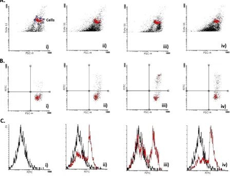
Briefly, 100 μL of the samples were added to individual TruCOUNT™ tubes containing 600 μL of Isoflow, 100 μL of Syto 11 (0.005 mM) and 200 μL of PI (0.015 mM) directly prior to flow cytometry analysis. These prepared tubes were then run on the flow cytometer which was set-up as previously reported [14]. Cells were gated according to FSC characteristics. Live/dead counts were measured by PI exclusion, live cells were denoted by no or little PI staining, whilst dead cells were denoted by high levels of PI staining (Figure 1). Cell counts were calculated to the total nucleated cell count in the original patient sample.
Monoclonal Antibody Labelling (Immunophenotyping)
Samples were divided into four 50 μL aliquots. These were then pelleted (2000 rcf, 5 min) and resuspended in 50 μL of staining buffer. The cellular samples were incubated for 30 min at 4˚C with 5 μL FITC-coupled mo-noclonal antibodies for CD31, CD45, and CD90 or with 5 μL of an isotype control (Mouse IgG1κ IsoControl) to assess the level of background staining. All samples were washed in a staining buffer, and were fixed in 180 μL of a 10% formaldehyde solution (FACS lysing buffer). The cells were then stained with PI to achieve a final stain concentration of 0.0015 mM. The proportion of cells in each sample that were CD31, CD45 and CD90 positive was determined according to quadrant analysis using flow cytometry (Figure 2) [15].
2.6. Statistical Analysis
[image:4.595.148.482.360.656.2]Stabilised and/or unstabilised samples from one patient was compared only to the unstabilised Day 0 controls from that same patient. Data from each pair-wise (control vs experimental sample) comparison was collated, av-eraged (±standard deviation) and assessed for consistency between patients. Two-tailed, paired t-tests were per-formed at each time point (control vs stabilised and/or unstabilised samples) to assess statistical significance.
Figure 2. Primary data and gating strategies for immunophenotyping analysis. (A) Dot plots, (B) dot plots with quadrant analysis gated off the cell population in (A), and (C) overlay histograms prepared to analyse the immunophenotype of unsta-bilised Day 0 samples (subject 105). Samples were stained with FITC conjugated, IgGκ1 antibodies: (i) a mouse IgGκ1 Iso-control (negative Iso-control), (ii) CD31, (iii) CD45, and (iv) CD90. A comparison of the isoIso-control (black) and CD31, CD45 or CD90 (red) was performed using overlay histograms (B).
A p < 0.05 was considered statistically significant. Statistical analysis and graphing of data was performed using GraphPad Prism 6 (vers. 6.01, 2010, California, United States) and Flowing Software 2 (vers. 2.5.1, Fin-land).
3. Results
3.1. Cell Count and Viability
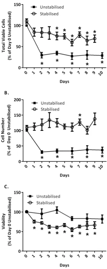
The cell counts and cell viability values attained from the Day 0 unstablised controls were normalised to 100% to present as the baseline. A significant increase in total nucleated cell count after TransFix® stabilisation was observed (Figure 3). After seven days of storage in TransFix® reagent, the mean nucleated cell count for each sample was 127% ± 50.2% of the corresponding Day 0 unstabilised sample.
Total cell viability was 66% ± 11.7% at Day 0, which then decreased and plateaued to 43% ± 13.4% after sta-bilisation for nine days. Normalisation of this data (Day 0 samples normalised to 100%) demonstrated a declin-ing trend of approximately 40% of the initial viability after stabilisation for seven days at 4˚C (Figure 3). A sig-nificantly lower viability was observed for each of the TransFix® stabilised samples at each storage time point when compared to the Day 0 controls.
Figure 3. Cell number and viability of stabilised and unstabilised samples after extended storage. (A) Viable cell numbers, (B) Total cell numbers and, (C) Cell viability of TransFix® stabilised and unstabilised stromal vascular fraction samples af-ter extended storage. Data was normalised to the experimental control (unstabilised, Day 0 sam-ples) which were represented as 100%. Values are mean ± SEM, signifcant difference (*) to Day 0 unstabilised samples if p < 0.05.
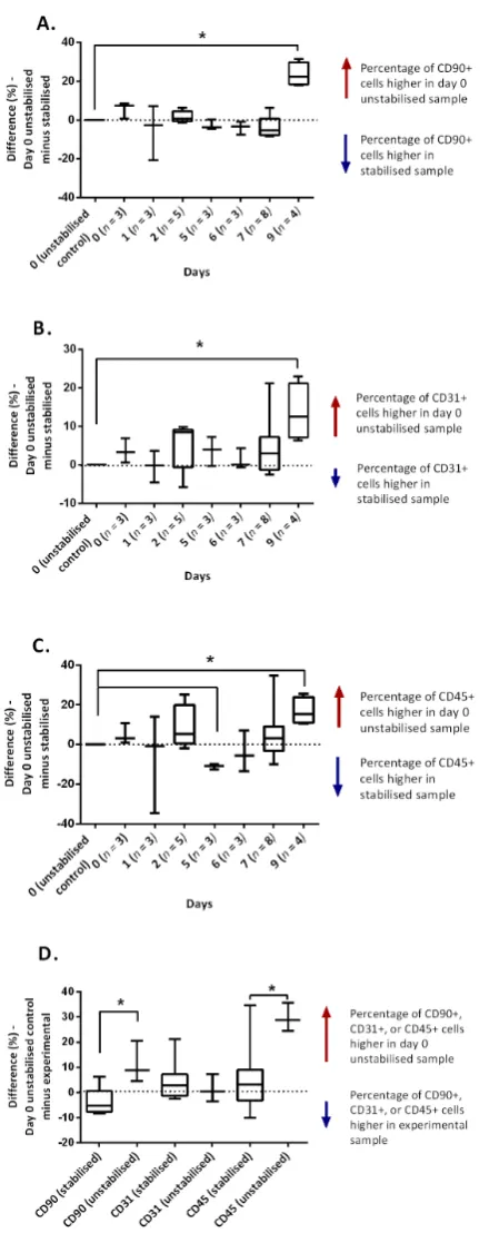
3.2. Monoclonal Antibody Staining for CD Surface Markers (Immunophenotyping)
tive cells in the Day 9 samples was significantly lower than that observed in the original unstabilised Day 0 samples, indicating a notable discrepancy at this time point between the Day 0 unstabilised and the Day 9 stabi-lised samples for each patient. The variation that is observed in the Day 1 data for CD90 (Figure 4) is a result of one sample that displayed a 20% increase of CD90+ cells after one day of stabilisation. Considerable biological variation in the proportion of CD31+ cells for each patient was evidenced in all samples.
There was no significant difference in the presence of the CD45 marker between the unstabilised and the sta-bilised samples for Day 1, 2, 6 and 7 (Figure 4). Interestingly, each patient sample stabilised for five days (n = 3) had a significantly higher proportion of CD45+ cells than its Day 0 unstabilised counterpart. In addition, consi-derable biological variation in the proportion of CD45+ cells was observed for every time point except for the Day 5 samples. The significant variation observed at the Day 1 stabilised comparison is a result of one sample, which appeared to increase the percentage of CD45+ cells by 35% after one day of stabilisation. Although this difference is quite large, the results for the stabilised counterpart are closer to the average proportion of CD45+ cells in adipose tissue (42.4% ± 10.4%) than the Day 0 samples.
Likewise to the results observed for the CD90 and CD31 markers, there was a significant difference in the amount of CD45+ cells observed after nine days of stabilisation when compared to the Day 0 unstabilised coun-terparts (Figure 4). The biological variation observed for the presence of CD45+ cells in the Day 0 unstabilised samples was notable, with the presence of CD45+ varying between 9% and 73% of the total population.
After extended storage without a stabilising reagent, there was a significant loss of CD90 and CD45 positive cells when compared to the stabilised counterpart, however the proportion of CD31 positive cells remained rela-tively consistent (Figure 4).
4. Discussion
Biological variation can be a confounding factor in patient to patient comparisons, especially so at the cellular level. For this reason, each patient served as their own control, wherein the results of all the cell samples were compared to the results of the corresponding Day 0, unstabilised aliquot. The biological variation of cell types in Day 0 samples was demonstrated by the widely varying proportion of CD45+ cells in the samples (between 9% and 73%, n = 29). The proportion of how many CD45+ cells (leukocytes) in blood, and likewise in a sample of SVF, can be affected by a range of factors such as the immune status of the patient on the day of the procedure, any exposure to pro-inflammatory stimuli from diet or environment, or the overall weight of a patient [16]-[18].
It was anticipated that the total nucleated cell count of the Day 0 samples would be the maximal cell count of all samples (unstabilised and stabilised) as storage time before analysis was minimal (up to 24 hrs, as opposed to one to nine days). However, the opposite proved to be true (Figure 3). There was a statistically significant crease in total cell number after stabilisation across all storage times compared to the Day 0 controls. The in-creased nucleated cell count after stabilisation is potentially a result of the disruption of cell clumping in the samples, which was not actively inhibited in the unstabilised samples. The impact of TransFix® on cell aggrega-tion has not been previously reported in the literature. The fixative properties of TransFix® reagent also appear to be able to stabilise cellular protein structures for immunophenotyping in white blood cells [11], and so it is likely that it also inhibits processes involved in cell division and protein synthesis.
We hypothesise that the stabilised cell counts in this data set are likely to be more accurate in the quantifica-tion of single cells in suspension because of the inhibiquantifica-tion of cellular processes that lead to cell loss such as cell clumping. Filtration of the sample to remove cell clumps prior to flow cytometry is essential, but this can result in a significant discrepancy between the total cell number that is counted and the actual cell number that is present in the sample. The use of TransFix® stabilisation may prevent cell aggregation, explaining the significant typical increase in total cell number in the samples after stabilisation and storage. Additionally, the TransFix® treated samples were stabilised immediately after SVF sample preparation, whilst unstabilised samples were stored at 4˚C for up to 24 hrs before assessment without the addition of inhibitors of cell division, apoptosis, the formation of clumps or cell-matrix aggregation. Storage of whole blood at 4˚C has been previously demonstrat-ed to negatively impact cell viability [19], cell population yield [20], and function [21]. The cell viability dropped consistently after stabilisation for seven days, in that the TransFix® samples are likely to have a viabili-ty that is 62.8% ± 16.5% of the Day 0 samples (Figure 3). As a result of this, an estimate of the original cell viability can be back-calculated from the stabilised data.
days post-collection in TransFix® reagent, with no statistical difference between the results attained post and pre-stabilisation (with the exception of the CD45 marker at five days) (Figure 4). The results at Day 5 do not appear to demonstrate the biological variation observed at each other time point, however further analysis would be required to test this hypothesis.
5. Conclusion
For post-hoc analysis of cell count and viability of nucleated cells in the stromal vascular fraction of digested adipose tissue, it is concluded that the sample remains stable in TransFix® reagent for up to seven days with the ability to provide an accurate total nucleated cell count and to back calculate the viability of the original sample. TransFix® reagent also appears to capable of preserving the CD90, CD31 and CD45 cellular marker on cells within an adipose-derived mixed cell population for up to seven days after stabilisation.
References
[1] Herrmann, R., Sturm, M., Shaw, K., Purtill, D., Cooney, J., Wright, M., et al. (2102) Mesenchymal Stromal Cell Therapy for Steroid-Refractory Acute and Chronic Graft versus Host Disease: A Phase 1 Study. International Journal of Hematology, 95, 182-188. http://dx.doi.org/10.1007/s12185-011-0989-2
[2] Oh, J.Y., Kim, M.K., Shin, M.S., Lee, H.J., Ko, J.H., Wee, W.R., et al. (2008) The Anti-Inflammatory and Anti-An- giogenic Role of Mesenchymal Stem Cells in Corneal Wound Healing Following Chemical Injury. Stem Cells, 26, 1047-1055. http://dx.doi.org/10.1634/stemcells.2007-0737
[3] Varma, M.J.O., Breuls, R.G., Schouten, T.E., Jurgens, W.J., Bontkes, H.J., Schuurhuis, G.J., et al. (2007) Phenotypical and Functional Characterization of Freshly Isolated Adipose Tissue-Derived Stem Cells. Stem Cells and Development,
16, 91-104. http://dx.doi.org/10.1089/scd.2006.0026
[4] De Ugarte, D., Morizono, K., Elbarbary, A., Alfonso, Z., Zuk, P., Zhu, M., et al. (2003) Comparison of Multi-Lineage Cells from Human Adipose Tissue and Bone Marrow. Cells, Tissues, Organs, 174, 101-109.
http://dx.doi.org/10.1159/000071150
[5] Riordan, N.H., Ichim, T.E., Min, W.P., Wang, H., Solano, F., Lara, F., et al. (2009) Non-Expanded Adipose Stromal Vascular Fraction Cell Therapy for Multiple Sclerosis. Journal of Translational Medicine, 7, 29.
http://dx.doi.org/10.1186/1479-5876-7-29
[6] Zimmerlin, L., Rubin, J.P., Pfeifer, M.E., Moore, L.R., Donnenberg, V.S. and Donnenberg, A.D. (2013) Human Adi-pose Stromal Vascular Cell Delivery in a Fibrin Spray. Cytotherapy, 15, 102-108.
http://dx.doi.org/10.1016/j.jcyt.2012.10.009
[7] Premaratne, G.U., Ma, L.P., Fujita, M., Lin, X., Bollano, E. and Fu, M. (2011) Stromal Vascular Fraction Transplanta-tion as an Alternative Therapy for Ischemic Heart Failure: Anti-Inflammatory Role. Journal of Cardiothoracic Surgery,
6, 43. http://dx.doi.org/10.1186/1749-8090-6-43
[8] Olson, W.C., Smolkin, M.E., Farris, E.M., Fink, R.J., Czarkowski, A.R., Fink, J.H., et al. (2011) Shipping Blood to a Central Laboratory in Multicenter Clinical Trials: Affect of Ambient Temperature on Specimen Temperature, and Ef-fects of Temperature on Mononuclear Cell Yield, Viability and Immunologic Function. Journal of Translational Medi-cine, 9, 26. http://dx.doi.org/10.1186/1479-5876-9-26
[9] Canonico, B., Zamai, L., Burattini, S., Granger, V., Mannello, F., Gobbi, P., et al. (2004) Evaluation of Leukocyte Sta-bilisation in TransFix-Treated Blood Samples by Flow Cytometry and Transmission Electron Microscopy. Journal of Immunological Methods, 295, 67-78. http://dx.doi.org/10.1016/j.jim.2004.09.013
[10] Jani, V., Janossy, G., Iqbal, A., Mhalu, F., Lyamuya, E., Biberfeld, G., et al. (2001) Affordable CD4+ T Cell Counts by Flow Cytometry. II. The Use of Fixed Whole Blood in Resource-Poor Settings. Journal of Immunological Methods,
257, 145-154. http://dx.doi.org/10.1016/S0022-1759(01)00458-6
[11] Barnett, D., Granger, V., Pockley, A., Saxton, J., Storie, I., Whitby, L., et al. (1999) TransFix: A Clinical Sample Sta-bilising Fluid for Use in Cellular Haematology and Immunology. Cytometry, 38, 88.
[12] Ng, A.A., Lee, B.T., Teo, T.S., Poidinger, M. and Connolly, J.E. (2012) Optimal Cellular Preservation for High Di-mensional Flow Cytometric Analysis of Multicentre Trials. Journal of Immunological Methods, 385, 79-89.
http://dx.doi.org/10.1016/j.jim.2012.08.010
[13] Blaber, S.P., Webster, R.A., Hill, C.J., Breen, E.J., Kuah, D., Vesey, G. and Herbert, B.R. (2012) Analysis of in Vitro Secretion Profiles from Adipose-Derived Cell Populations. Journal of Translational Medicine, 10, 172.
http://dx.doi.org/10.1186/1479-5876-10-172
http://dx.doi.org/10.1002/cyto.990080411
[15] Oldaker, T.A. (2007) Quality Control in Clinical Flow Cytometry. Clinics in Laboratory Medicine, 27, 671-685.
http://dx.doi.org/10.1016/j.cll.2007.05.009
[16] Field, J., Gougeon, R. and Marliss, E.B. (1991) Changes in Circulating Leukocytes and Mitogen Responses during Very-Low-Energy All-Protein Reducing Diets. American Journal of Clinical Nutrition, 54, 123-129.
[17] Kondo, T., Hayashi, M., Takeshita, K., Numaguchi, Y., Kobayashi, K., Iino, S., Inden, Y. and Murohara, T. (2004) Smoking Cessation Rapidly Increases Circulating Progenitor Cells in Peripheral Blood in Chronic Smokers. Arterio-sclerosis, Thrombosis, and Vascular Biology, 24, 1442-1447. http://dx.doi.org/10.1161/01.ATV.0000135655.52088.c5
[18] Potter, C.G., Potter, A.C., Hatton, C.S., Chapel, H.M., Anderson, M.J., Pattison, J.R., et al. (1987) Variation of Erythroid and Myeloid Precursors in the Marrow and Peripheral Blood of Volunteer Subjects Infected with Human Parvovirus (B19). Journal of Clinical Investigation, 79, 1486-1492. http://dx.doi.org/10.1172/JCI112978
[19] Ashmore, L.M., Shopp, G.M. and Edwards, B.S. (1989) Lymphocyte Subset Analysis by Flow Cytometry Comparison of Three Different Staining Techniques and Effects of Blood Storage. Journal of Immunological Methods, 118, 209- 215. http://dx.doi.org/10.1016/0022-1759(89)90008-2
[20] Garraud, O. and Moreau, T. (1984) Effect of Blood Storage on Lymphocyte Subpopulations. Journal of Immunological Methods, 75, 95-98. http://dx.doi.org/10.1016/0022-1759(84)90228-X