0095-1137/10/$12.00 doi:10.1128/JCM.01807-09
Copyright © 2010, American Society for Microbiology. All Rights Reserved.
Characterization of
Streptococcus milleri
Group Isolates from
Expectorated Sputum of Adult Patients with Cystic Fibrosis
䌤
Margot E. Grinwis,
1Christopher D. Sibley,
1Michael D. Parkins,
2,4Christina S. Eshaghurshan,
1Harvey R. Rabin,
2,4and Michael G. Surette
1,3*
Department of Microbiology and Infectious Diseases,1Department of Medicine,2Department of Biochemistry and Molecular Biology,3and
Adult Cystic Fibrosis Clinic,4University of Calgary, Calgary, Alberta, Canada T2N 4N1
Received 14 September 2009/Returned for modification 6 November 2009/Accepted 3 December 2009
With the recent insights into theStreptococcus millerigroup (SMG) as pulmonary pathogens in patients with cystic fibrosis (CF), we sought to characterize 128 isolates from the sputum of adults with CF, along with 45 isolates from patients with invasive diseases for comparison. The tests performed included Lancefield group-ing; tests for hemolysis; tests for the production of hyaluronidase, chondroitin sulfatase, DNase, proteases, and hydrogen peroxide; and PCR for the detection of the intermedilysin gene (ily). We also generated biochemical profiles with the Rapid ID Strep 32 API system and tested cell-free supernatants for the presence of the signal molecule autoinducer-2 (AI-2) using aVibrio harveyibioassay with a subset of CF strains. TheS. intermedius
isolates from both strain collections were similar, while theS. constellatus andS. anginosusisolates yielded several biotypes that differed in prevalence between the two strain collections. Beta-hemolytic, Lancefield group
CS. constellatuscomprised 74.4% of theS. constellatusisolates from patients with CF but only 13.3% of the
corresponding isolates from patients with invasive infections. This was the onlyS. constellatusbiotype associ-ated with pulmonary exacerbations. Hyaluronidase-positiveS. anginosuswas detected only among the isolates from patients with CF. Strain-to-strain variability in AI-2 expression was evident, with the mean values being the highest forS. anginosus, followed byS. constellatusand thenS. intermedius. Cluster analysis and 16S rRNA sequencing revealed that the species of SMG could be accurately determined with a minimum of three phenotypic tests: tests for the Lancefield group, hyaluronidase production, and chondroitin sulfatase produc-tion. Furthermore, isolates from patients with invasive infections clustered with isolates from the sputum of patients with CF, suggesting that the respiratory tract isolates were equally pathogenic.
Streptococcus intermedius, Streptococcus anginosus, and
Streptococcus constellatusare a group of organisms collectively
referred to as theStreptococcus millerigroup (SMG) or theS.
anginosusgroup (8). Members of the SMG are generally
con-sidered to be commensals of the oral cavity, gastrointestinal tract, and female urogenital tract in 15 to 30% of healthy individuals (22) but are clinically known for their association with purulent infections throughout the body (3). Recently, members of the SMG were found to be responsible for up to half of invasive pyogenic streptococcal infections in a large Canadian health region (13) and, afterStaphylococcus aureus, were the most common isolates in a microbiological analysis of skin and soft tissue infections (36). Members of the SMG are not widely recognized as pulmonary pathogens, but they have recently gained attention as such, particularly in those with cystic fibrosis (CF) (20, 32; C. D. Sibley, M. E. Grinwis, T. R. Field, M. D. Parkins, J. C. Norgaard, D. B. Gregson, H. R. Rabin, and M. G. Surette, submitted for publication). We have previously shown that the members of the SMG are linked with clinical deterioration in roughly 40% of hospital admissions in our population of adult patients with CF (32; Sibley et al., submitted). We have also shown that the members of the SMG
can easily be overlooked by standard clinical microbiology analysis with samples from patients with CF, even when it is the numerically dominant pathogen.
Characterization of members of the SMG has shown that these microorganisms are phenotypically diverse (1) and that several strains produce candidate virulence factors, such as hydrolytic enzymes and hemolysins (4, 11, 28, 42, 44). While less than half of the members of the SMG appear to be he-molytic on standard media (10, 22), Nagamune et al. found that virtually allS. intermediusstrains produce a potent human-specific cytoxin, intermedilysin, which shows little to no activity against the erythrocytes of other animals (18). Although the members of the SMG have been extensively characterized, there is little information on strains originating from patients with respiratory tract infections. Routine regimens for the cul-ture of specimens from patients with CF do not readily permit the growth of members of the SMG from sputum (9, 25, 32) and differentiation between members of the SMG and other streptococci from nonsterile sites is difficult (27), but the recent development of the semiselective medium McKay agar has led to the improved recovery of members of the SMG from spu-tum (32; Sibley et al., submitted). In the study described here, we characterized 128 SMG isolates from the sputum of pa-tients with CF along with 45 SMG isolates from papa-tients with invasive infections and 3 reference strains.
MATERIALS AND METHODS
Bacterial strains and growth conditions.All respiratory isolates from patients with CF were obtained from sputum samples collected in sterile containers
* Corresponding author. Mailing address: Department of Micro-biology and Infectious Diseases, University of Calgary, Faculty of Medicine, 3330 Hospital Drive NW, Calgary, Alberta, Canada T2N 4N1. Phone: (403) 220-2744 Fax: (403) 270-2772. E-mail: surette @ucalgary.ca.
䌤Published ahead of print on 9 December 2009.
395
on May 16, 2020 by guest
http://jcm.asm.org/
according to ethical guidelines for adult patients with CF attending the Southern Alberta Cystic Fibrosis Clinic. Ninety-nine strains were isolated on McKay agar as part of a 6-month surveillance study from September 2007 to March 2008 (Sibley et al., submitted). The remaining 29 isolates were collected between 2006 and 2008 and placed on McKay agar (32), brain heart infusion (BHI) agar (Difco), Trypticase soy agar (Difco) with 0.3% yeast extract (Bacto), Columbia blood agar (CBA; Difco) with 5% sheep blood (Med-Ox), Columbia-colistin-nalidixic acid (CNA) agar (BBL) with 5% sheep blood, BHI agar with 0.01 mg/ml colistin sulfate salt and 0.005 mg/ml oxilinic acid (Sigma-Aldrich, St. Louis, MO) (BHI-CO), chocolate agar, fastidious anaerobe agar (Becton Dickinson), and phenylethyl alcohol agar (BBL) at 37°C under CO2-enriched (5%) and anaerobic conditions. Forty-five isolates from patients with invasive infections (blood, em-pyema, unknown sources, brain abscess, liver abscess, and hip abscess) were obtained from Calgary Laboratory Services; 42 of these isolates were recovered on CBA. Strains from patients with invasive infections had been identified as members of the SMG according to standard phenotypic procedures, and the species had been determined by partial 16S rRNA gene PCR and sequencing. Reference strainsS. intermediusATCC 27335,S. constellatusATCC 27823, and
S. anginosusATCC 33397 were included in this study. All strains from patients with CF were identified by partial 16S rRNA gene PCR and sequencing, as described by Sibley et al. (32). The isolates were stored at⫺80°C with 15% glycerol or 10% skim milk as cryopreservatives.
Hemolysis and Lancefield grouping.Isolates were screened for hemolysis on CBA at 37°C with 5% CO2after 48 h of incubation. Lancefield grouping was performed with all isolates by use of a commercial kit (Oxoid, Nepean, Ontario, Canada).
Intermedilysin PCR.PCR for the intermedilysin gene (ily) was performed as described previously (19) by using Chelex-extracted DNA (Bio-Rad, Hercules, CA), as described previously (40).
Biochemical profiling.Biochemical profiles were determined with Rapid ID Strep 32 API strips (bioMe´rieux, Marcy l’Etoile, France), according to the manufacturer’s instructions, with the following adjustments: each inoculum was prepared from isolated colonies that had been subcultured onto BHI-CO agar and incubated for 72 h at 37°C with 5% CO2.
Arginine hydrolysis and VP tests.Arginine hydrolysis and Vogues-Proskauer (VP) tests were performed as described by Ruoff and Ferraro (26). Moeller decarboxylase broth base supplemented with arginine, MR-VP broth, alpha-naphthol, and 40% potassium hydroxide were purchased from Oxoid; creatine powder was purchased from Sigma-Aldrich.
Hydrogen peroxide assay.All strains were tested for hydrogen peroxide pro-duction by the Prussian blue plate assay described by Saito et al. (29). Briefly, isolates were patched onto Prussian blue agar, and the plates were incubated at 37°C with 5% CO2. The plates were checked for the development of the Prussian blue precipitate after 2 to 7 days. Dilutions of hydrogen peroxide were used as positive controls.
Determination of enzymatic activities.Hyaluronidase and chondroitin sulfa-tase activities were determined by the plate assays described by Smith and Willet (33), except that hyaluronic acid sodium salt from aStreptococcus equispecies (Fluka) was used instead of human umbilical potassium hyaluronidate and chon-droitin sulfate from whale and shark cartilage (sodium salt) (Sigma-Aldrich) was used instead of bovine nasal chondroitin sulfate. DNase activity was determined by the plate assay described by Porschen and Sonntag (23) with BHI agar as the basal medium, except that the yeast extract and dyes were omitted. DNase plates were flooded with 1 M HCl after 2 to 3 days of incubation and were observed for a zone of clearing around the growth. Protease activity was determined by the plate assay described by Lutticken et al. (15), except that 2% skim milk powder (Difco) was used in place of purified casein. The hyaluronic acid plates were incubated for 24 h, and the DNA and skim milk plates were incubated for 48 to 72 h at 37°C with 5% CO2. The chondroitin sulfate plates were incubated anaerobically for 7 days. AnS. intermediusisolate from this study was used as a positive control since that isolate yielded zones of clearing for all of the assays described above.
Cluster analysis.The results for all tests were recorded as either 0 (negative) or 1 (positive). Average-linkage hierarchal clustering was performed by using the Cluster program (version 3.0) and was viewed by use of the Java Treeview program (version 1.1.3) (7).
AI-2 expression assays.At 72 h, colonies of 27 strains from patients with CF (9S. intermedius, 9S. constellatus, and 9S. anginosusstrains) and 3 reference strains were used to inoculate 2 ml of BHI broth, and the broths were incubated for 48 h at 37°C with 5% CO2. Fifty microliters was transferred to 5 ml of Todd-Hewitt-yeast broth with 0.5% yeast extract (Bacto), and the broths were incubated for 24 h at 37°C with 5% CO2.The tubes were checked for turbidity as a confirmation of growth, 1 ml was transferred to a sterile 1.5-ml tube, and the
tube was centrifuged at maximum speed for 5 min. Four hundred microliters of the supernatant was transferred to a clean tube and was stored at⫺20°C until use. The detection of AI-2 in the supernatants was performed in triplicate as described by Surette and Bassler (37).Vibrio harveyiAI-2 reporter strain MM32 (luxSandluxNnegative) was maintained on Luria-marine agar (2) at 30°C. Sterile Todd-Hewitt-yeast broth was used as a negative control. Luciferase pro-duction (counts per second) was measured for 24 h by using a Wallac Victor2
. For statistical analysis, Tukey’s multiple-comparison test was performed by the use of Prism software (version 5.0; GraphPad Software, Inc.).
RESULTS
We characterized 128 isolates from the sputum of 47 pa-tients with CF recovered at times of both clinical stability and pulmonary exacerbation. We also examined 45 isolates from patients with invasive infections and 3 reference strains for comparison. Of the isolates from patients with CF, 22 from eight patients were cultured from hospital admission samples at levels ofⱖ107CFU/ml, which was greater than or equivalent
to the levels of the conventional CF pathogen present (
Pseudo-monas aeruginosain seven patients,Staphylococcus aureusin
one patient). This is a unique strain collection from a single patient population with no evidence of transmission between patients (unpublished data).
Table 1 shows the phenotypic characteristics of all isolates with respect to hemolysis, Lancefield grouping, the production of hydrolytic enzymes and hydrogen peroxide, and detection of the intermedilysin gene (ily). TheS. intermediusisolates from the collections of isolates from patients with CF and patients with invasive infections were highly similar. All strains were Lancefield nongroupable, and all except two strains produced all of the hydrolytic enzymes for which tests were conducted; the exceptions were two strains from patients with invasive infections, which lacked proteases. TheS. intermediusisolates were virtually devoid of-hemolysis on CBA (60/61, 98.4%), althoughilywas found exclusively in all of theS. intermedius isolates. Two of theS. intermediusisolates from patients with CF produced a rosy pigment on BHI-CO agar.
The majority ofS. constellatusisolates from patients with CF (32/43, 74.4%) were beta-hemolytic and Lancefield group C and produced all of the hydrolytic enzymes for which tests were conducted (Table 1). This was the onlyS. constellatusprofile that was linked to multiple pulmonary exacerbations in our collection. These strains resembleS. constellatussubsp.
phar-yngis, a variant ofS. constellatusof oral origin associated with
pharyngitis (43). Only 2 of 15 (13.3%) invasiveS. constellatus isolates shared this profile. All Lancefield group FS.
constel-latusisolates were beta-hemolytic and lacked chondroitin
sul-fatase and proteases. Likewise, all nongroupableS. constellatus isolates lacked chondroitin sulfatase, but 6/10 (60%) isolates from patients with CF and 4/7 (57.1%) isolates from patients with invasive infections were positive for proteases. The pro-tease-positive nongroupableS. constellatusisolates both from patients with CF and from patients with invasive infections tended to be nonhemolytic; only a single invasive isolate was both beta-hemolytic and protease positive. AllS. constellatus isolates were positive for hyaluronidase and DNase.
TheS. anginosusisolates from both strain collections
exhib-ited diverse phenotypes (Table 1). Only beta-hemolytic Lance-field group CS. anginosusisolates were positive for hyaluron-idase (n⫽5), and all of these strains were from patients with
on May 16, 2020 by guest
http://jcm.asm.org/
CF. Two of these strains were linked to separate exacerbations in a single patient. None of theS. anginosusisolates produced chondroitin sulfatase, and only a single isolate from a patient with CF failed to produce DNase. Ninety-four percent of theS.
anginosus(45/48) isolates from patients with CF were protease
positive, while 66.7% of theS. anginosus(6/9) isolates from patients with invasive infections were protease positive. Over-all, 10.2% (13/128) of the isolates from patients with CF were positive for hydrogen peroxide production, and most of these
wereS. anginosus(10/13, 76.9%); the remaining 3 isolates were
S. constellatus. Similarly, 6.3% of the isolates from patients
with invasive infections (two S. constellatus isolates, one S.
anginosusisolate) were hydrogen peroxide positive.
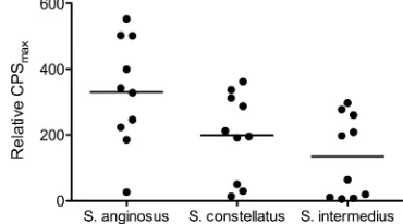
A subset of strains (nineS. anginosus, nineS. constellatus, and nine S. intermedius strains) of various phenotypes and from various patients with CF plus three reference strains were used in aVibrio harveyibioassay for detection of the quorum-sensing signal molecule autoinducer-2 (AI-2) in cell-free su-pernatants (37). The levels of detection were from 5- to 552-fold greater than the background levels, and variability was evident within each species (Fig. 1). The mean levels were the highest forS. anginosus(330-fold), followed byS. constellatus
(199-fold) andS. intermedius(134-fold). Only the values forS.
anginosusandS. intermediuswere significantly different (P⬍
0.05).
[image:3.585.326.511.556.659.2]In addition to the phenotypes described in Table 1, testing
TABLE 1. Phenotypic differentiation of theS. milleriisolates used in this study
Isolate source No. of isolates
No. of patients
No. of isolates positive for:
Hemolysis Lancefield group
Hyaluronidase production
Chondroitin sulfatase production
DNase production
Protease production
H2O2 production ily
a
S. intermedius
CF stable sputumb 31 21 1 1 NGc 1 1 1 1 0 1
30␣or␥ 30 NG 30 30 30 30 0 30
Exacerbation associated 6 3 6␣or␥ 6 NG 6 6 6 6 0 6
Invasived 24 24␣or␥ 24 NG 24 24 24 22 0 24
S. constellatus
CF stable sputum 30 11 22 19 C 19 19 19 19 0 0
1 F 1 0 1 0 1 0
2 NG 2 0 2 0 0 0
8␣or␥ 8 NG 8 0 8 6 2 0
Exacerbation associated 13 3 13 13 C 13 13 13 13 0 0
Invasive 15 11 2 C 2 2 2 2 0 0
6 F 6 0 6 0 0 0
3 NG 3 0 3 1 1 0
4␣or␥ 4 NG 4 0 4 3 1 0
S. anginosus
CF stable sputum 45 24 15 3 C 3 0 3 3 0 0
4 F 0 0 4 3 1 0
3 G 0 0 3 2 1 0
5 NG 0 0 5 4 1 0
30␣or␥ 6 C 0 0 6 6 1 0
17 F 0 0 17 17 3 0
7 NG 0 0 6 7 2 0
Exacerbation associated 3 2 2 2 C 2 0 2 2 0 0
1␣or␥ 1 F 0 0 1 1 1 0
Invasive 9 6 1 A 0 0 1 1 0 0
2 C 0 0 2 1 1 0
2 G 0 0 2 2 0 0
1 NG 0 0 1 0 0 0
3␣or␥ 2 F 0 0 2 2 0 0
1 NG 0 0 1 0 0 0
aily, intermedilysin gene, as determined by PCR.
bSputum samples were collected during periods of clinical stability. cNG, Lancefield nongroupable.
dSamples from patients with invasive infections.
FIG. 1. Detection of AI-2 in SMG isolates. As a measure of AI-2 expression, the maximum counts per second (CPSmax) were deter-mined for three replicates of each supernatant and the negative control (sterile Todd-Hewitt-yeast broth) during a 24-h assay period. Black circles, ratio of the average maximum counts per second for each supernatant to the average maximum counts per second for the neg-ative control; bars, mean for each species.
on May 16, 2020 by guest
http://jcm.asm.org/
with the Rapid API system was used to generate biochemical profiles for all strains. The statistical relationships between the phenotype profiles are depicted in Fig. 2. Data for tests in which less than 10% of the total strains were positive were
[image:4.585.71.518.64.621.2]excluded, as were data forily, which is unique toS. intermedius. We found that the SMG could be divided into two main groups on the basis of biochemical and enzymatic activities: the upper group consisted of less active strains of S. anginosus and S.
FIG. 2. Cluster analysis of phenotypic test results for the SMG. Blue,S. anginosus; red,S. constellatus; green,S. intermedius. Abbreviations and symbols: APPA, alanyl-phenylalanyl-proline arylamidase; GTA, glycyl-tryptophan arylamidase; stars, strains from patients with invasive infections; circles with dots in the center, reference strains, which clustered accordingly. The cluster is divided into two main groups: the upper group consists of less active strains, which showed fewer positive results than the lower, more active group. Subcluster designations are indicated on the right.
on May 16, 2020 by guest
http://jcm.asm.org/
constellatus(fewer numbers of positive results), while the lower group consisted of more active strains of all three species (greater numbers of positive results). Seven unique clusters were identified (Fig. 2). From these clusters, it is clear thatS.
anginosus and S. constellatus each comprised at least three
distinct biotypes, whileS. intermediusis more homogeneous. In two cases, clades that primarily consisted of invasive strains were formed. One was the active Lancefield group FS.
con-stellatus cluster, and the other was within theS. intermedius
cluster, distinguished from the other clades by arginine hydro-lysis and-glucosidase production. The beta-hemolytic, Lance-field group C, hyaluronidase-positiveS. anginosuscluster con-sisted solely of isolates from patients with CF. Each of the reference strains clustered accordingly.
Only 35% of the strains hydrolyzed arginine and 81% of the strains were VP test positive by use of the Rapid API strips (Fig. 2). Several reports have indicated that more than 90% of SMG isolates are positive both for arginine hydrolysis and by the VP test (1, 36, 44), which are considered to be of primary value in distinguishing the SMG from other streptococci (26). To confirm whether this was due to a deficiency in the Rapid API strips, we tested a subset of strains (111 strains from patients with CF, 37 strains from patients with invasive infec-tions) for arginine hydrolysis and positivity by the VP test by the method described by Ruoff and Ferraro (26). By this method, we found that 88% and 98% of the isolates from patients with CF were positive for arginine hydrolysis and by the VP test, respectively. The isolates from patients with inva-sive infections yielded similar results: 86% and 95% were pos-itive for arginine hydrolysis and by the VP test, respectively. Of the isolates in both groups that were arginine hydrolysis neg-ative (n⫽ 19), 89.5% (17/19) wereS. intermedius. Of theS.
intermedius isolates from patients with CF (n ⫽ 11), 81.8%
(9/11) were from patients who have hadS. intermedius -associ-ated exacerbations. Two of the arginine hydrolysis-negativeS.
intermediusinvasive isolates were also from a patient with CF.
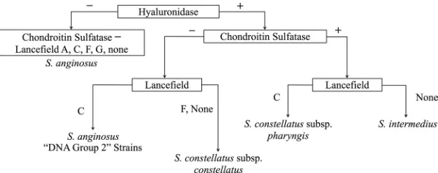
Furthermore, we found that the cluster diagram was surpris-ingly accurate for determination of the species of the SMG. Before the identification of 35 isolates from patients with in-vasive infections by the 16S rRNA PCR, we used the seven cluster designations to determine the species of the strains and did a blind comparison to the identifications obtained by the 16S rRNA PCR. We observed a complete correlation between the two identification methods and were able to compose a
minimal scheme that could accurately determine the species of all of the strains in our study (Fig. 3). This scheme depends on hyaluronidase production, chondroitin sulfatase production, and the Lancefield grouping.
DISCUSSION
Our knowledge of pathogenic microorganisms and their phenotypic traits predominantly stems from the analysis of strains from patients with acute infections, in which the patho-gen and the disease are short-lived. The airway of a patient with CF, however, presents a unique scenario because patho-gens that typically form acute infections have the opportunity to adapt to their host and establish chronic infections, which are also subject to the influence of antibiotic selective pres-sures. Thus, it is possible that the airways of patients with CF harbor unique biotypes of microorganisms that are different from their invasive counterparts causing acute disease. We sought to investigate this for the SMG by comparing strains from the airways of patients with CF to strains from patients with invasive infections. Additionally, our isolation and iden-tification techniques, which included a variety of types of me-dia, incubation conditions, and molecular methods, may have recovered biotypes either that fail to grow on the typically employed blood agar or that would be misidentified by stan-dard biochemical tests. Our novel collection of SMG strains from a total of 47 patients with CF contained 22 isolates from sputum linked to pulmonary exacerbation and 106 isolates from sputum during periods of clinical stability.
Because of their ability to break down components of host tissue, hydrolytic enzymes are presumed virulence determi-nants in the SMG. All of theS. intermediusandS. constellatus strains studied produced hyaluronidase, while only 10% ofS.
anginosusstrains produced hyaluronidase, consistent with the
findings of previous studies (11, 44). None of the hyaluroni-dase-positive S. anginosus isolates were from the group of patients with invasive infections, although they were similar to the DNA group 2 strains described by Whiley et al. (43).S.
anginosuswas rarely associated with pulmonary exacerbation
in this study, but hyaluronidase-positive S. anginosus was linked to two separate exacerbations in a single patient.
[image:5.585.137.448.68.191.2]The proportion of DNase-positive isolates was much higher in this study than in previous ones, in which no more than 75% of isolates produced detectable DNases (11, 24). Since the
FIG. 3. Proposed scheme for determination of the SMG species. The assignment of subtypes, namely,S. constellatussubsp.constellatus,S. constellatussubsp.pharyngis, and DNA group 2 strains, was based on the strain descriptions provided by Whiley et al. (43).
on May 16, 2020 by guest
http://jcm.asm.org/
strains from patients with invasive infections and patients with CF did not differ in this respect, it is possible that DNase is more widespread in the SMG than was previously thought, which would be consistent with the invasive nature of these pathogens (35, 41). It is interesting that the majority of strains also produced proteases, while none of the 72 SMG isolates tested by Ruoff and Ferraro (28) or the 86 isolates tested by Lutticken et al. (15) showed protease activity. This observation may be due to differences in the methodology used or the interpretation of the results, as the zones of clearing in this assay were not as vivid as those in the other plate assays used. In this study, many protease-negative strains belonged to the beta-hemolytic nongroupable and Lancefield group FS.
con-stellatusbiotypes, which were more common in the group of
strains from patients with invasive infections. Chondroitin sul-fatase activity was also more prevalent in the SMG than has previously been reported (11, 28) and was uniformly detected
in S. intermedius and beta-hemolytic Lancefield group C S.
constellatusisolates. These findings corroborate those found by
Takao and colleagues (38, 39), in that two types of hylauroni-dases exist within the SMG: those with chondroitin sulfatase activity, found only inS. intermediusandS. constellatussubsp.
pharyngis, and those without chondroitin sulfatase activity,
found inS. constellatussubsp.constellatusand reference strain MAS624, a beta-hemolytic Lancefield group C S. anginosus strain (43). Thus, the hyaluronidase and chondroitin sulfatase activities observed are likely due to a single enzyme with a broad substrate range.
Hydrogen peroxide production in the SMG was consistent with the findings described in previous reports (42, 44) and was most commonly associated with S. anginosus. In agreement with the findings of Nagamune et al. (19), the intermedilysin gene (ily) was found exclusively in all S. intermediusisolates. The heightened recovery of beta-hemolytic Lancefield group C
S. constellatusstrains, otherwise known asS. constellatussubsp.
pharyngis, from the sputum of patients with CF (43) is likely
attributable to the use of McKay agar in our studies. This biotype tended to grow poorly on CBA, the standard nonse-lective medium used for the primary isolation of streptococci from CF sputum and other specimens from non-CF patients, which might explain its low incidence in the collection of iso-lates from patients with invasive infections.
The quorum-sensing signal molecule AI-2 has been shown to influence virulence traits in several streptococcal species, in-cluding the SMG (16, 17, 21, 34). Unlike experiments involving
Streptococcus mutans orStreptococcus pneumoniae, in which
AI-2 was difficult to detect in culture supernatants (12, 17), AI-2 was readily detected in the supernatants of the SMG isolates. The range of levels observed highlights the strain-to-strain variability characteristic of AI-2 expression profiles. Since the SMG are commonly isolated from individuals with polymicrobial infections (3) and have been shown to act syn-ergistically with other organisms (30, 31), it is possible that AI-2 mediates synergistic virulence between species (5, 6).
Use of the combination of 16S rRNA PCR identification methods and phenotypic profiling affirms that the SMG is indeed composed of three distinct species, and these may be further divided into multiple subtypes, as suggested previously (43, 45). The phenotypic diversity and the overlapping traits of the SMG isolates have generally made determination of the
species of the strains a difficult task (27). Through substantial characterization of the SMG, an effective and reliable scheme which differentiates the three species on the basis of eight enzymatic tests has been developed (14, 44). We have found here that by the use of 16S rRNA PCR identification as the “gold standard” for comparison, tests for hyaluronidase pro-duction, chondroitin sulfatase propro-duction, and Lancefield group are sufficient for determination of the species of the members of the SMG and the subtype. Similarly, Takao et al. (39) found that the genes responsible for hyaluronidase and chondroitin sulfatase activity were useful for determination of the species of the members of the SMG by PCR, since their respective amplicons differed in size. We have yet to determine whether the scheme presented here can be used to distinguish the SMG from other streptococci or to what extent it holds true for the SMG.
This is the first study to provide a comprehensive character-ization of SMG isolates from patients with CF. Strains from patients with invasive infections and patients with CF alike are equipped with potential virulence factors, which may even be useful for species and subtype determination. We have shown here that most SMG biotypes are shared by strains from pa-tients with invasive infections and papa-tients with CF, but certain biotypes may be more prevalent and clinically relevant in pa-tients with CF, such as beta-hemolytic Lancefield group CS.
constellatusor arginine hydrolysis-negative S. intermedius. In
our previous studies, the only time that the members of the SMG were detected at elevated levels (ⱖ107CFU/ml) was at
hospital admission, and treatment directed toward the SMG resulted in clinical resolution (20, 32). Our strain collection is relatively small and is limited to one patient population, yet it provides the groundwork for resolving the complexity of SMG biotypes in chronic airway infections in patients with CF.
ACKNOWLEDGMENTS
We thank the Southern Alberta Cystic Fibrosis Clinic and CF pa-tients for their vital contributions to this study. We also thank Calgary Laboratory Services for the generous provision of strains.
This work was supported by a grant from the Canadian Cystic Fibrosis Foundation to M.G.S. M.G.S. is supported as an Alberta Heritage Foundation for Medical Research Scientist and Canada Re-search Chair in Microbial Gene Expression. M.E.G. is supported by studentships from the Canadian Institutes of Health Research, Cana-dian Cystic Fibrosis Foundation, and the Alberta Heritage Foundation for Medical Research.
REFERENCES
1.Ball, L. C., and M. T. Parker.1979. The cultural and biochemical characters ofStreptococcus milleristrains isolated from human sources. J. Hyg. (Lond.)
82:63–78.
2.Bassler, B. L., M. Wright, and M. R. Silverman.1994. Multiple signalling systems controlling expression of luminescence inVibrio harveyi: sequence and function of genes encoding a second sensory pathway. Mol. Microbiol.
13:273–286.
3.Belko, J., D. A. Goldmann, A. Macone, and A. K. M. Zaidi.2002. Clinically significant infections with organisms of theStreptococcus millerigroup. Pe-diatr. Infect. Dis. J.21:715–723.
4.Bridge, P. D., and P. H. Sneath.1983. Numerical taxonomy ofStreptococcus. J. Gen. Microbiol.129:565–597.
5.Duan, K., C. Dammel, J. Stein, H. Rabin, and M. G. Surette.2003. Modu-lation of Pseudomonas aeruginosa gene expression by host microflora through interspecies communication. Mol. Microbiol.50:1477–1491. 6.Duan, K., C. D. Sibley, C. J. Davidson, and M. G. Surette.2009. Chemical
interactions between organisms in microbial communities. Contrib. Micro-biol.16:1–17.
7.Eisen, M. B., P. T. Spellman, P. O. Brown, and D. Botstein.1998. Cluster analysis and display of genome-wide expression patterns. Proc. Natl. Acad. Sci. U. S. A.95:14863–14868.
on May 16, 2020 by guest
http://jcm.asm.org/
8.Facklam, R. R.1984. The major differences in the American and British
Streptococcustaxonomy schemes with special reference toStreptococcus mil-leri. Eur. J. Clin. Microbiol.3:91–93.
9.Harris, J. K., M. A. De Groote, S. D. Sagel, E. T. Zemanick, R. Kapsner, C. Penvari, H. Kaess, R. R. Deterding, F. J. Accurso, and N. R. Pace.2007. Molecular identification of bacteria in bronchoalveolar lavage fluid from children with cystic fibrosis. Proc. Natl. Acad. Sci. U. S. A.104:20529–20533. 10.Jacobs, J. A., C. S. Schot, and L. M. Schouls.2000. Haemolytic activity of the ‘Streptococcus millerigroup’ and relationship between haemolysis restricted to human red blood cells and pathogenicity inS. intermedius. J. Med. Mi-crobiol.49:55–62.
11.Jacobs, J. A., and E. E. Stobberingh.1995. Hydrolytic enzymes of Strepto-coccus anginosus,Streptococcus constellatusandStreptococcus intermediusin relation to infection. Eur. J. Clin. Microbiol. Infect. Dis.14:818–820. 12.Joyce, E. A., A. Kawale, S. Censini, C. C. Kim, A. Covacci, and S. Falkow.
2004. LuxS is required for persistent pneumococcal carriage and expression of virulence and biosynthesis genes. Infect. Immun.72:2964–2975. 13.Laupland, K. B., T. Ross, D. L. Church, and D. B. Gregson.2006.
Popula-tion-based surveillance of invasive pyogenic streptococcal infection in a large Canadian region. Clin. Microbiol. Infect.12:224–230.
14.Limia, A., T. Alarco´n, M. L. Jime´nez, and M. Lo´pez-Brea.2000. Comparison of three methods for identification ofStreptococcus millerigroup isolates to species level. Eur. J. Clin. Microbiol. Infect. Dis.19:128–131.
15.Lutticken, R., U. Wendorff, D. Lutticken, E. A. Johnson, and L. W. Wanna-maker.1978. Studies on streptococci resemblingStreptococcus milleriand on an associated surface-protein antigen. J. Med. Microbiol.11:419–431. 16.Lyon, W. R., J. C. Madden, J. C. Levin, J. L. Stein, and M. G. Caparon.2001.
Mutation ofluxSaffects growth and virulence factor expression in Strepto-coccus pyogenes. Mol. Microbiol.42:145–157.
17.Merritt, J., F. Qi, S. Goodman, and M. Anderson.2003. Mutation ofluxS
affects biofilm formation inStreptococcus mutans. Infect. Immun.71:1972– 1979.
18.Nagamune, H., C. Ohnishi, A. Katsuura, K. Fushitani, R. A. Whiley, A. Tsuji, and Y. Matsuda.1996. Intermedilysin, a novel cytotoxin specific for human cells secreted byStreptococcus intermediusUNS46 isolated from a human liver abscess. Infect. Immun.64:3093–3100.
19.Nagamune, H., R. A. Whiley, T. Goto, Y. Inai, T. Maeda, J. M. Hardie, and H. Kourai.2000. Distribution of the intermedilysin gene among the angino-sus group streptococci and correlation between intermedilysin production and deep-seated infection withStreptococcus intermedius. J. Clin. Microbiol.
38:220–226.
20.Parkins, M. D., C. D. Sibley, M. G. Surette, and H. R. Rabin.2008. The
Streptococcus millerigroup—an unrecognized cause of disease in cystic fi-brosis: a case series and literature review. Pediatr. Pulmonol.43:490–497. 21.Pecharki, D., F. C. Petersen, and A. A. Scheie.2008. LuxS and expression of
virulence factors inStreptococcus intermedius. Oral Microbiol. Immunol.
23:79–83.
22.Poole, P. M., and G. Wilson.1979. Occurrence and cultural features of
Streptococcus milleriin various body sites. J. Clin. Pathol.32:764–768. 23.Porschen, R. K., and S. Sonntag. 1974. Extracellular deoxyribonuclease
production by anaerobic bacteria. Appl. Microbiol.27:1031–1033. 24.Pulliam, L., R. K. Porschen, and W. K. Hadley.1980. Biochemical properties
of CO2-dependent streptococci. J. Clin. Microbiol.12:27–31.
25.Rogers, G. B., T. W. Daniels, A. Tuck, M. P. Carroll, G. J. Connett, G. J. David, and K. D. Bruce.2009. Studying bacteria in respiratory specimens by using conventional and molecular microbiological approaches. BMC Pulm. Med.9:14.
26.Ruoff, K. L., and M. J. Ferraro.1986. Presumptive identification of Strepto-coccus milleriin 5 h. J. Clin. Microbiol.24:495–497.
27.Ruoff, K. L.1988.Streptococcus anginosus(“Streptococcus milleri”): the un-recognized pathogen. Clin. Microbiol. Rev.1:102–108.
28.Ruoff, K. L., and M. J. Ferraro.1987. Hydrolytic enzymes of “Streptococcus milleri.” J. Clin. Microbiol.25:1645–1647.
29.Saito, M., M. Seki, K. Iida, H. Nakayama, and S. Yoshida.2007. A novel agar medium to detect hydrogen peroxide-producing bacteria based on the Prussian blue-forming reaction. Microbiol. Immunol.51:889–892. 30.Shinzato, T., and A. Saito.1994. A mechanism of pathogenicity of “
Strepto-coccus millerigroup” in pulmonary infection: synergy with an anaerobe. J. Med. Microbiol.40:118–123.
31.Shinzato, T., and A. Saito.1995. TheStreptococcus millerigroup as a cause of pulmonary infections. Clin. Infect. Dis.21(Suppl. 3):S238–S243. 32.Sibley, C. D., M. D. Parkins, K. Duan, J. C. Norgaard, H. R. Rabin, and
M. G. Surette.2008. A polymicrobial perspective of pulmonary exacerba-tions exposes an enigmatic pathogen in cystic fibrosis patients. Proc. Natl. Acad. Sci. U. S. A.105:15070–15075.
33.Smith, R. F., and N. P. Willett.1968. Rapid plate method for screening hyaluronidase and chondroitin sulfatase-producing microorganisms. Appl. Microbiol.16:1434–1436.
34.Stroeher, U. H., A. W. Paton, A. D. Ogunniyi, and J. C. Paton.2003. Muta-tion ofluxSofStreptococcus pneumoniaeaffects virulence in a mouse model. Infect. Immun.71:3206–3212.
35.Sumby, P., K. D. Barbian, D. J. Gardner, A. R. Whitney, D. M. Welty, R. D. Long, J. R. Bailey, M. J. Parnell, N. P. Hoe, G. G. Adams, F. R. Deleo, and J. M. Musser.2005. Extracellular deoxyribonuclease made by group A Strep-tococcusassists pathogenesis by enhancing evasion of the innate immune response. Proc. Natl. Acad. Sci. U. S. A.102:1679–1684.
36.Summanen, P. H., M.-C. Rowlinson, J. Wooton, and S. M. Finegold.2009. Evaluation of genotypic and phenotypic methods for differentiation of the members of the anginosus group streptococci. Eur. J. Clin. Microbiol. Infect. Dis.28:1123–1128.
37.Surette, M. G., and B. L. Bassler.1998. Quorum sensing inEscherichia coli
andSalmonella typhimurium. Proc. Natl. Acad. Sci. U. S. A.95:7046–7050. 38.Takao, A.2003. Cloning and expression of hyaluronate lyase genes of Strep-tococcus intermediusandStreptococcus constellatussubsp.constellatus. FEMS Microbiol. Lett.219:143–150.
39.Takao, A., H. Nagamune, and N. Maeda.2004. Identification of the angino-sus group within the genusStreptococcususing polymerase chain reaction. FEMS Microbiol. Lett.233:83–89.
40.Walsh, P. S., D. A. Metzger, and R. Higuchi.1991. Chelex 100 as a medium for simple extraction of DNA for PCR-based typing from forensic material. Biotechniques10:506–513.
41.Wartha, F., K. Beiter, S. Normark, and B. Henriques-Normark.2007. Neu-trophil extracellular traps: casting the NET over pathogenesis. Curr. Opin. Microbiol.10:52–56.
42.Whiley, R. A., and D. Beighton.1991. Emended descriptions and recognition of Streptococcus constellatus,Streptococcus intermedius, andStreptococcus anginosusas distinct species. Int. J. Syst. Bacteriol.41:1–5.
43.Whiley, R. A., L. M. Hall, J. M. Hardie, and D. Beighton.1999. A study of small-colony, beta-haemolytic, Lancefield group C streptococci within the anginosus group: description ofStreptococcus constellatussubsp.pharyngis
subsp. nov., associated with the human throat and pharyngitis. Int. J. Syst. Bacteriol.49(Pt 4):1443–1449.
44.Whiley, R. A., H. Fraser, J. M. Hardie, and D. Beighton.1990. Phenotypic differentiation ofStreptococcus intermedius,Streptococcus constellatus, and
Streptococcus anginosus strains within the “Streptococcus milleri group.” J. Clin. Microbiol.28:1497–1501.
45.Winstanley, T. G., J. T. Magee, D. I. Limb, J. M. Hindmarch, R. C. Spencer, R. A. Whiley, D. Beighton, and J. M. Hardie.1992. A numerical taxonomic study of theStreptococcus millerigroup based upon conventional phenotypic tests and pyrolysis mass spectrometry. J. Med. Microbiol.36:149–155.