Three-dimensional Image Segmentation using
Tissue-like P System
Salah I. Yahya
1,2,3, Rafaa I. Yahya
3,4, Bisan Al-Salibi
3, Ghada K. Al-Khafaji
3,5and
Siti Mariyam Shamsuddin
31Department of Software Engineering, Faculty of Engineering, Koya University, Danielle Mitterrand Boulevard, Koya KOY45, Kurdistan Region – F.R. Iraq 2Department of Computer Science and Engineering, School of Science and Engineering,
University of Kurdistan Hewler, Erbil, Kurdistan Region – F.R. Iraq
3UTM Big Data Center, Ibnu Sina Institute for Scientific and Industrial Research, Universiti Teknologi Malaysia, UTM Skudai, Malaysia
4Department of Computer, Collage of Science, University of Al-Mustansiriyah, Baghdad, F.R. Iraq
5Department of Computer, College of Science, University of Baghdad, Baghdad, F.R. Iraq
Abstract– Membrane computing (MC), which abstracts computational models from the structure and functioning of biological cells or population of cells in tissues, has served as a rich framework for handling many problems. Various types of P systems have been proposed in the literature to perform edge-based and region-based segmentation of two-dimensional digital images. However, less attention has been paid to the segmentation of three-dimensional (3D) medical images. Hence, the main contribution of this paper is to propose a tissue-like P system for segmenting 3D medical images. To the best of our knowledge, this is the first work that practically adapts MC for 3D images. Experimental results demonstrate the efficiency of the proposed approach in segmenting 3D images, and it has the potential to be used in real-world applications.
Index Terms–Membrane computing, Region-based image segmentation, Three-dimensional images, Tissue-like P systems.
I. Introduction
Membrane computing (MC) has emerged as a recent branch of natural computing, which is mainly based on
the assumption that the flow of metabolites within the
compartmental architecture and functioning of biological
cells can be interpreted as a flow of information for
computations (Paun, 2002). The computational devices in MC are known as P systems in honor of their initiator,
Paun and Rozenberg (2002). More importantly, P systems are massively parallel and distributed computing models
processing multisets of objects based on predefined evolution
rules in a nondeterministic manner. Due to the inherent parallelism, MC has the ability of solving NP-complete problems in polynomial or often in linear time and this feature has been shown extensively in the literature. The basic ingredients of a typical P system model consist of (1) membrane structure, (2) a set of evolution rules, and (3) multisets of objects. The main novelty of MC relies on the fact that membrane systems look at the whole cell as a
computing device. They are defined as systems consisting of
a hierarchical arrangement of membranes delimiting regions, which represent various compartments of a cell, and with each region having associated rules, which represent different biochemical processes taking place inside each compartment (Bernardini and Gheorghe, 2005). Basically, MC devices or P systems constitute three different models of computing devises depending on the particular features of the cell which are as follows: (1) Cell-like P systems (inspired from the structure of the cell), (2) tissue-like P systems (inspired from organization of cells in tissue), and (3) spiking neural-like membrane systems - inspired from the way neurons are linked in neural nets (Paun and Prez-Jimnez, 2006).
In this paper, tissue-like P system is used for segmentation of three-dimensional (3D) images. MC has applications in
many fields including digital image analysis (Alsalibi, et al.,
2015) and optimization (Alsalibi, et al., 2014; Alsalibi Venkat and Al-Betar, 2017).
This paper is organized as follows: The related work is
presented in Section II. Section III provides a basic definition
of tissue-like P system. Section IV describes the use of tssue-like P system for automatic region-based segmentation of 3D images as well as presents the methodology of segmentation with MC using P-lingua, and Section V concludes the paper and suggests some directions for the future work.
ARO-The Scientific Journal of Koya University Volume V, No. 2(2017), Article ID: ARO.10316, 8 pages DOI: 10.14500/aro.10316
Received 18 October 2017; Accepted 28 November 2017 Regular research paper: Published 08 December 2017
II. Related Work
The fundamental objective of digital image processing is to extract meaningful information from images without human assistance. Segmentation is an important task of image processing for satellite and medical images (Somasundaram and Alli, 2011). Technically speaking, in computer vision (Shapiro and Stockman, 2001), segmentation is the process of partitioning a digital image into multiple segments (sets of pixels), aiming to simplify or modify the representation of an image to be more expressive and easier to analyze and understand. Image segmentation is typically used to locate objects and boundaries (lines and curves) in images. More precisely, image segmentation is the process of assigning a label to every pixel in an image such that pixels with the same label share certain visual characteristics.
Recently, MC techniques have been used in solving problems arising from digital images. Image segmentation has been addressed extensively in the literature. For example, the authors (Christinal, et al., 2009; Yahya, et al., 2016) presented a tissue-like P systems to improve the standard edge-based segmentation method using MC rules. Christinal, et al. (2010) presented a MC rule framework to solve the threshold problem using cell-like P system, wherein the solution has been reached in linear time depending on the number of pixels of the input image. Similarly, Reina-Molina, et al. (2010) presented a new segmentation approach based on a tissue-like P system with the use of MC rules with multiple auxiliary cells for solving segmentation problem. Diaz-Pernil, Reina-Molina, and Carnero (2010) proposed a new software tool for performing a segmentation of two-dimensional (2D) digital images based on MC platform. However, they did not explain in detail the technical aspects of implementing the tool. The authors (Christinal, et al., 2011) developed a tissue-like P system (using MC rules) to design a region-based segmentation algorithm in a constant number of steps. In their approach, 4-adjacency relation between pixel’s neighborhoods has been adopted for 2D digital images where 6-adjacency relation between voxel neighborhoods has been used for 3D digital images. Sheeba, et al. (2011) proposed a tissue-like P system algorithm to enhance morphological segmentation methods in medical image. Christinal, et al. (2012) proposed bio-inspired MC through tissue-like P system (MC rules) to perform a parallel color segmentation of images using a threshold method. Yang, et al. (2013) proposed a novel membrane algorithm (tissue-like P system) to develop region-based segmentation. Along with this line, Peng, et al. (2012) proposed a novel threshold segmentation approach using cell-like P system (membrane algorithm) to improve threshold segmentation. Daz-Pernil, et al. (2013) presented a novel device architecture called CUDATM to implement tissue-like P system MC rules for segmentation of images with gradient-based edge detection. Furthermore, Peng, et al. (2014) proposed a novel segmentation by adaptive traditional region-based color segmentation method using tissue-like P system (membrane algorithm). Isawasan, et al. (2014) developed a tissue-like P system using MC rules to segment hexagonal images, wherein the segmentation has been done
in 7 steps. Peng, et al. (2014) proposed a novel method using cell-like P system (membrane algorithm) to solve the optimal multilevel thresholding problem. Yahya, et al. (2015) presented region-based segmentation with tissue-like P system rules
that implement a simple artificial image with a more detailed
illustration of how P system works furthermore, where different color relations have been explored to show the effect of color on the segmentation results. Interestingly, a new research line has been recently launched in which MC has been adopted to solve several problems relating to digital imagery. For example, region-based segmentation of 2D and 3D images have been investigated. However, the main drawback of their
approach is that the image has been manually codified in the
tissue simulator.
In the work of Christinal, et al. (2009), P systems have been linked to computational topology with digital images where this development paved way for a new and promising line of research. Christinal, et al. (2009) designed a collection of tissue-like P systems that used the communication rules of MC to perform edge-based segmentation. This communication entails the discovery of adequate different region boundaries among the input images. The experiment was conducted such
that the artificial 2D and 3D images, using 4-adjacency and
26-adjacency, respectively, have been employed. Experiments
show that results were obtained in a fixed number of 9
and 26 steps pertaining to 2D and 3D images, respectively. Furthermore, in the work of Christinal, et al. (2011), a tissue-like P system was proposed with the use of MC rules for the design of an edge-based segmentation algorithm in a constant number of steps. In their work, 4-adjacency relationships between neighboring pixels were adopted for 2D digital images.
They proved that only 9 steps were sufficient to get an
edge-based segmentation for a 2D image. In addition, 26-adjacency relationships between voxel neighborhoods were implemented for 3D digital images. They theoretically proved that 26 steps are required to get an edge-based segmentation for a 3D image. However, no practical examples have been provided. Meanwhile, the main weakness of their method is the fact that
the image has been manually codified in the tissue simulator. This leads to a lack of efficiency in favor of expressiveness.
For a comprehensive review of image segmentation using MC, interested readers are referred to Yahya, et al. (2017).
Thus, in this paper, we present a dynamic software using P-lingua for edge-based segmentation of 3D digital images based on the segmentation rules presented by Christinal, et al. (2011). Basically, edge-based segmentation is performed based on membrane computing and implemented in P-lingua with real medical images which are loaded automatically in
the system. P-lingua is an official programing language for
MC that offers a general syntactic framework that could
define a unified standard for MC, covering a broad variety of
models (Daz-Pernil, et al., 2009).
III. Tissue-like P System
a graph representing all the communication channels existing between interacting cells as can be seen in Fig 1. The set of interconnections is dynamic, and in the course of the evolution of the system, it can possibly change whosesoever a new communication way is established or another one is closed (Bianco, 2007). From the computational perspective, the essential characteristic of tissue-like P system is that membranes do not have electrical charges as in the cell-like P systems. Typically, the form of tissue-like P systems model with the input of degree q is a tuple.
Π=(Γ, Σ, ε, W1,…, Wq, (R1),…, (Rq), iΠ, oΠ)
Where
1. Γ is a finite alphabet, whose symbols are called objects;
2. Σ⊂Γ is the input alphabet;
3. ε⊆Γ is the list of objects in the environment, each one in
arbitrarily infinite copies;
4. µ is the membrane structure;
5. Mi is a set of strings over Γ representing the multisets of
objects associated with the membrane i, 1≤i≤q at the initial configuration;
6. Ri is a finite set of communication rules of the following
form: (i, u/v, j), for i, j∈ {0, 1, 2,…, q}, i = j, u, v∈Γ∗
7. oΠ∈ {0, 1, 2,…, q} refers to the output cell; 8. iΠ∈ {1, 2,…, q} refers to the input cell.
In the typical framework of MC, each cell is viewed as a computing unit working in a maximally parallel and nondeterministic way. The configuration is an instantaneous description of the P system at a particular moment, where a sequence of computation steps can be applied in a parallel manner to obtain a new configuration. A computation is said
to be successful if it halts, reaching a specific configuration
where no more rules can be applied to the current objects.
With a halting computation, the associated output can be codified by the content of the output membrane.
IV. Tissue-like P System for Automatic Segmentation of 3D Images
Edge-based approaches rely on common patterns in intensity values within a cluster of neighboring pixels. The cluster is referred to as the region, and the goal of segmentation algorithm is to group regions according to their anatomical
or functional roles (Siddiqui and Richhariya, 2013). The main aim of edge-based segmentation is to use image characteristics to map individual pixels in an input image to set of pixels called regions that might correspond to an object (Zhao, et al., 2008). Segmentation in digital imagery has several interesting features which makes it suitable for mechanisms inspired by nature. One of them is that it can be solved in a parallelized and localized manner. Regardless of how large is the picture, the segmentation process can be simultaneously performed in different local areas of the picture. Another interesting feature is that the basic required information can be easily encoded by different bioinspired representations.
In this paper, automatic edge-based segmentation will be applied to 3D images (.nrrd) rather than typical 2D (.png) images. To automatically load the images in the system, the segmentation rules will be written using P-lingua programing language. Furthermore, to deal with 3D images automatically, P- lingua will be linked to MATLAB to enable the processing of 3D images. P-LinguaCore4 library will
be also integrated to handle P-lingua input files and check
possible programing errors, both lexical/syntax and semantics (Frisco, et al., 2014). This library is used to implement MC with edge-based segmentation of 3D images.
Different from the 2D segmentation, in 3D segmentation, the input data will be voxels of the type C {i, j, k}, where c is the color of the voxel and i, j, and k are the three coordinates of that voxel. The edge-based segmentation of the 3D digital image problem can be formulated as follows: Given a digital 3D image with voxels of (possibly) different colors, obtain the boundaries of regions in that image. The solution of this problem can be described with several steps as follows:
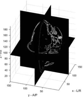
Fig. 3. Input three-dimensional (3D) image (3D slice) Fig. 4. Input three-dimensional image (horizontal slice)
• First marking voxels: Here, mark some of the voxels which
belong to a boundary between two regions (border voxels).
• Second marking voxels: We add voxels adjacent to two edge
voxels with the same color as them and adjacent to other voxel with a different color to the edge voxels.
• Output stage: The system sends to the environment all the
marked voxels. This stage works in parallel to the previous steps.
Another procedure of solving the 3D image segmentation problem is to convert the 3D image into a set of 2D slices. In this case, the 3D segmentation problem will be
simplified to the 2D segmentation case. After segmenting
all the slices, it will be combined and rendered back to the 3D shape. This procedure will be illustrated in the following section.
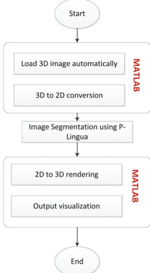
A. Methodology of the Proposed Work
As mentioned previously, the tissue-like P system rules will be implemented sing P-lingua to segment the 3D images. Fig. 2 shows the steps of performing the segmentation using P-lingua and MATLAB software.
B. Load the 3D Image Automatically
In the first step of the work, the 3D image with type (.nrrd)
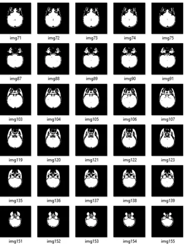
will be automatically loaded and processed by MATLAB software using “NRRDREAD” function. Examples of the 3D input image are shown in Figs. 3, and 4. Fig. 3 represents the vertical slice of the combined 3D image.
C. 3D to 2D Conversion
Here, the MATLAB processes the 3D image using “NRRDREAD” command which reads the image volume
and its associated metadata from the NRRD-format file specified by the FILENAME. In this case, the 3D image will
be converted into m 2D slices to simplify the segmentation process and to be able to use the P-lingua segmentation rules. Note that the number of slices m corresponds to the z
dimension of the 3D image. The result of this step is shown in Fig. 5.
D. Image Segmentation using P-Lingua
Typically, in the standard P-Lingua syntax format, the pixels should have color and coordinates. Basically, the color and the coordinates of each pixel will be read and converted to P-lingua syntax. The standard syntax is to indicate the color of the pixel followed by curly brackets that contain the coordinate of the associated pixel, which in turn will be
written into the.txt file. For example, in B{x,y}, x and y are
the coordinates of the black pixel in the image, whereas in
W{x,y}, x and y are the white pixel.
In this paper, the segmentation rules adopt the rules of MC used by Christinal, et al., 2011. The rules are written in the
P-lingua format and saved in a text file. After loading the images, the images will be automatically codified as an input and concatenated to the rules file to the final form .PLI file. This file is now ready to be executed.
If the image contains two colors which are white and black, then if the four adjacencies are considered, the segmentation rules will be as follows:
[Wi,j, Bi,j]1 ↔ [Wxi,j, Bi,j+1]0 [Wi,j, Bi,j-1]1 ↔ [Wxi,j, Bi,j-1]0 [Wi,j, Bi+1,j]1 ↔ [Wxi,j, Bi+1,j]0 [Wi,j, Bi-1,-j]1 ↔ [Wxi,j, Bi-1,j]0
[Wxi,j, Wi-1,j, Wxi-1,j+1, Bi,j+1]1 ↔ [Wxi,j, Wxi-1,j, Wxi-1,j+1, Bi,j+1]0 [Wxi,j, Wi,j+1, Wxi-1,j+1, Bi-1,j]1 ↔ [Wxi,j, Wxi,j+1, Wxi-1,j+1, Bi-1,j]0 Once the images have been segmented using MC into one of two cells, the output can be read from any of these cells as needed. There are two types of segmentation strategies regarding the philosophy of reading the output from a chosen cell. Cell one contains the input image that has had its boundary pixels sent to cell two so that it contains only the regions of the image. If the output is read from cell one, region-based segmentation is obtained. If the output is read from cell two, edge-based segmentation is obtained and visualized. Cell two contains the border and edge pixels.
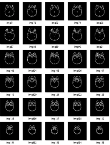
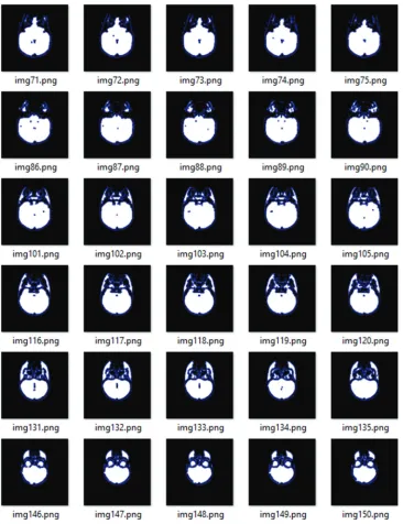
The result of segmentation of the 2D slices is shown in Fig. 6 for edge-based segmentation and Fig. 7 for region-based segmentation.
E. 2D to 3D Rendering
(black and white) images. The binarization will reduce not only the number of required rules but also the computational time required to achieve the segmentation.
Therefore, the input image will be converted into binary format with only black and white pixels to simplify the segmentation rules and speed up the execution. Binarization refers to the process in which each pixel in an image is converted into one bit with the value of one or zero
depending on the specified threshold value of all pixels.
If the pixel value is greater than the particular threshold value, then the pixel will be converted to one; otherwise, it will be zero. A binary image is a digital image with only two possible values for each pixel (black or white). In the proposed method, the average RGB value of each pixel is computed and then compared with the threshold value which is set to 128. If it is greater than the threshold, then the pixel will be converted to white; otherwise, it will be black.
Once all the 2D slices are being segmented using P-lingua, step-D, those images will be combined to form the segmented 3D image as shown in Figs. 8 and 9.
V. Conclusion
Segmentation has special characteristics which enables it to be suitable for methods of natural computing. However, for those techniques to be more effective and practical in real applications, a suitable software tool is of great importance. In this paper, MC software was presented as a tool for automatically segmenting 3D images using P-lingua programing language and MATLAB. The proposed software is able to deal with images of different sizes and has the potential of becoming a helping tool for dealing with large medical images. According to the literature of related work pertaining to image segmentation using MC, the majority of work has been focused on 2D images, but very less attention has been paid to 3D images. Hence, the contributions of the paper as can be summarized as follows: First, the input
image has been codified automatically by linking P-lingua
with the Java platform. Second, the proposed approach can deal with 3D images of different sizes. However, there are still some drawbacks of the proposed approach which need to be addressed thoroughly. One of the major limitations of our approach is that the proposed approach has been only simulated using a sequential architecture which in turn does not exploit the massive parallelism inherited in P systems. In our future work, to fully make use of the MC parallelism, a parallel architecture such as CUDA™ speedups over the serial implantation will be used to gain higher performance.
References
Alsalibi, B., Venkat, I., Subramanian, K.G., Lutfi, S. and Wilde, P.D., 2015. The
impact of bio-inspired approaches towards the advancement of face recognition. ACM Computing Surveys, 48(1), p.5.
Alsalibi, B., Venkat, I., Subramanian, K.G. and Christinal, H.A., 2014. A Bio-Inspired Software for Homology Groups of 2d Digital Images. Asian Conference on Membrane Computing ACMC 2014, Coimbatore, pp.1-4.
Fig. 8. The segmented three-dimensional image
Fig. 9. Horizontal slice of the segmented three-dimensional images
Alsalibi B., Venkat I. and Al-Betar M., 2017. A membrane-inspired bat algorithm to recognize faces in unconstrained scenarios. Engineering Applications of
Artificial Intelligence, 64, pp.242-260.
Bianco, L., 2007. Membrane Models of Biological Systems, (Doctoral Dissertation, Ph.D. Thesis, University of Verona). In: Bernardini, F. and Gheorghe, M., editors. 2005. Membrane Systems for Molecular Computing and Biological Modelling. University of Sheffield. Ph.D. Thesis.
Christinal, H.A., Daz-Pernil, D. and Jurado, P.R., 2009. Segmentation in 2D and 3D image using tissue-like P system. In: Progress in Pattern Recognition, Image Analysis, Computer Vision, and Applications. Springer. Berlin, Heidelberg. pp.169-176. Christinal, H.A., Daz-Pernil, D. and Real, P., 2011. Region-based segmentation of 2D and 3D images with tissue-like P systems. Pattern Recognition Letters, 32(16), pp.2206-2212.
Christinal, H.A., Diaz-Pernil, D., Gutierrez-Naranjo, M.A. and Perez-Jimenez, M.J., 2010. Thresholding of 2D images with cell-like P systems. Romanian Journal of Information Science and Technology (ROMJIST), 13(2), pp.131-140. Christinal, H.A., Diaz-Pernil, D., Jurado, P.R. and Selvan, S.E., 2012. Color Segmentation of 2D images with thresholding. Ecofriendly Computing and Communication Systems, 305, pp.162-169.
Daz-Pernil, D., Berciano, A., Pena-Cantillana, F. and GutiRrez-Naranjo, M.A., 2013. Segmenting images with gradient-based edge detection using membrane computing. Pattern Recognition Letters, 34(8), pp.846-855.
Daz-Pernil, D., Perez-Hurtado, I., Perez-Jimenez, M.J. and Riscos-Nunez, A., 2009. A P-lingua programming environment for membrane computing. Membrane Computing, 5391, pp.187-203.
Diaz-Pernil, D., Reina-Molina, R. and Carnero, J., 2010. A bio-inspired software for segmenting digital images. In: Bio-Inspired Computing: Theories and Applications (BIC-TA). 2010 IEEE 5th International IEEE Conference. Frisco, P., Gheorghe, M. and Prez-Jimnez, M.J., 2014. Applications of Membrane Computing in Systems and Synthetic Biology. Springer, Cham, Switzerland. Isawasan, P., Venkat, I., Subramani, K., Khader, A., Oman, O. and Christinal, H., 2014. Region-Based Segmentation of Hexagonal Digital Images using Membrane Computing. 2014 Asian Conference on Membrane Computing (ACMC). Martín-Vide, C., Paun, G., Paros, J. and Rodríguez-Patón, A., 2003. Tissue P systems. Theoretical Computer Science, 296(2), pp.295-326.
Paun, G., 2002. Membrane Computing. Springer, Heidelberg. pp.1-6. Paun, G. and Prez-Jimnez, M.J., 2006. Membrane computing: Brief introduction, recent results and applications. Biosystems, 85(1), pp.11-22.
Paun, G. and Rozenberg, G. 2002. A guide to membrane computing. Theoretical Computer Science, 287(1), pp.73-100.
Peng, H., Wang, J. and Prez-Jimnez, M.J., 2014. Optimal multi-level thresholding
with membrane computing. Digital Signal Processing, 37, pp.53-64.
Peng, H., Yang, Y., Zhang, J., Huang, X. and Wang, J., 2014. A region-based
color image segmentation method based on P systems. Romanian Journal of Information Science and Technology, 17(1), pp.63-75.
Shapiro, L. and Stockman, G.C., 2001. Computer Vision. 1st ed. Prentice Hall,
Pearson.
Sheeba, F., Thaburaj, R., Nagar, A.K. and Mammen, J.J., 2011. Segmentation of Peripheral Blood Smear Images Using Tissue-Like P Systems. Bio-Inspired Computing: Theories and Applications (BIC-TA), 2011 6th International
Conference.
Siddiqui, F.K. and Richhariya, V., 2013. An efficient image segmentation
approach through enhanced watershed algorithm. Computer Engineering and Intelligent Systems, 4(6), pp.1-7.
Somasundaram, P. and Alli, P., 2011. A review on recent research and implementation methodologies on medical image segmentation. Journal of Computer Science, 8(1), pp.170-174.
Yahya, R.I., Hasan, S., George, L.E. and Alsalibi, B. 2015. Membrane computing for 2D image segmentation. International Journal Advance Soft Computer Applications, 7(1), pp.35-50.
Yahya, R.I., Shamsuddin, S.M., Hasan, S. and Yahya, S.I., 2016. Tissue-like P
system for segmentation of 2D hexagonal images. ARO-The Scientific Journal
of Koya University, 4(1), pp.35-42.
Yahya, R.I., Shamsuddin, S.M., Yahya, S.I., Hasan, S., Al-Salibi, B. and Al-Khafaji, G.H., 2017. Image segmentation using membrane computing: A literature survey. Bio-inspired Computing Theories and Applications. Vol. 681. Springer, China. pp.314-335.
Yang, Y., Peng, H., Jiang, Y., Huang, X. and Zhang, J., 2013. A Region-based image segmentation method under P systems. Journal Information Computer Science, 10(10), pp.2943-2950.
Zhao, Y., Liu, J., Li, H. and Li, G., 2008. Improved Watershed Algorithm for
Dowels Image Segmentation. In: Intelligent Control and Automation.7th World Congress on WCICA 2008.
Reina-Molina, R., Carnero, J. and Diaz-Pernil, D., 2010. Image segmentation using tissue-like P systems with multiple auxiliary cells. Image-A, 1(3), pp.143-150.
Peng, H., Yang, Y., Zhang, J., Huang, X. and Wang, J., 2012. Image thresholding with cell-like P systems. In: Proceedings of the 10th