S H O R T G E N O M E R E P O R T
Open Access
High quality draft genomic sequence of
Flavihumibacter solisilvae
3-3
T
Gang Zhou
1, Chong Chen
1, Che Ok Jeon
2, Gejiao Wang
1*and Mingshun Li
1*Abstract
Flavihumibacter solisilvae3-3T(= KACC 17917T= JCM 19891T) represents a type strain of the genusFlavihumibacter within the familyChitinophagaceae. This strain can use various sole carbon sources, making it applicable in industry and bioremediation. In this study, the draft genomic information ofF. solisilvae3-3Tis described.F. solisilvae3-3T owns a genome size of 5.41 Mbp, 47 % GC content and a total of 4,698 genes, including 4,215 protein coding genes, 439 pseudo genes and 44 RNA encoding genes. Analysis of its genome reveals high correlation between the genotypes and the phenotypes.
Keywords:Flavihumibacter,Flavihumibacter solisilvae, Genomic information, Genotype, Phenotype
Introduction
The genus Flavihumibacter was established in 2010 [1] and comprises three recognized species,Flavihumibacter petaseusT41T[1],Flavihumibacter cheonanensisWS16T [2] and Flavihumibacter solisilvae 3-3T [3], that were isolated from a subtropical rainforest soil, a shallow stream sediment and a forest soil, respectively. The Fla-vihumibacter members are Gram-positive, rod-shaped, strictly aerobic, non-motile, yellow-pigmented bacteria. The strains all contain phosphatidylethanolamine (as the major polar lipid, menaquinone-7 as the major respira-tory quinine, iso-C15:0 and iso-C15:1G as the principal
fatty acids. In addition, the strains are oxidase- and catalase-positive and with a G + C content range of 45.9-49.5 mol% [1–3].
To the best of our knowledge, the genomic informa-tion ofFlavihumibactermembers still remains unknown. In this study, we present the draft genome information of F. solisilvae 3-3T. A polyphasic taxonomic study re-vealed that F. solisilvae 3-3T could utilize 33 kinds of sole carbon substrates, including 11 kinds of saccharides and 22 kinds of organic acids and amino acids [3]. Spe-cially, this strain could utilize aromatic compound
4-hydroxyphenylacetic acid as a sole carbon source making it applicable environmental bioremediation [4–6]. In addition, this strain could utilize quinic acid as a sole carbon. Quinic acid is the substrate used to synthesize aromatic amino acids (phenylalanine, tyrosine and tryp-tophan) via the shikimate pathway. These aromatic amino acids are very useful as food additives, sweetener and pharmaceutical intermediates [6, 7]. The genome analysis of F. solisilvae 3-3T will provide the genomic basis for better understanding these mechanisms and ap-plying the strain to industries and bioremediation more efficiently.
Organism information
Classification and features
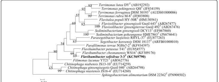
F. solisilvae3-3Twas isolated from forest soil of Bac Kan province in Vietnam [3]. The classification and features of F. solisilvae3-3Tare shown in Table 1. A maximum-likelihood tree was constructed based on the 16S rRNA gene sequences using MEGA 5.0 [8]. The bootstrap values were calculated based on 1,000 replications and distances were calculated in accordance with Kimura’s two-parameter method [9]. The phylogenetic tree showed that F. solisilvae 3-3T was clustered with the other Flavihumibacter members (Fig. 1).
Cells of F. solisilvae 3-3T (Fig. 2) are Gram-positive, aerobic, non-motile, and rod-shaped. Colony is yellow due to the production of flexirubin-type pigments [10]. F. solisilvae 3-3T grows well on NA and R2A agar * Correspondence:[email protected];[email protected]
1State Key Laboratory of Agricultural Microbiology, College of Life Sciences and Technology, Huazhong Agricultural University, Wuhan 430070, People’s Republic of China
Full list of author information is available at the end of the article
(optimum), but do not grow on LB or TSA agar [3]. It can hydrolyze aesulin, gelatin, casein and tyrosine [3]. F. solisilvae3-3Tcan also utilize various carbohydrate sub-strates (Table 1) and produces several glycosyl hydrolases, such as β-N-acetylhexosaminidase, α-galactosidase, β -glucosidase, β-galactosidase, α-fucosidase, α-mannosidase andα-glucosidase [3].
F. solisilvae 3-3T contains iso-C15:0, iso-C15:1G and
summed feature 3 (C16:1 ω6c/C16:1 ω7c) as the principal
fatty acids, MK-7 as the major respiratory quinine. The major polar lipids were PE, three unidentified aminolipids and three unidentified lipids [3].
Genome sequencing information
Genome project history
F. solisilvae 3-3T was selected for sequencing based on its taxonomic representativeness and the potential appli-cation in food industry and bioremediation. The genome ofF. solisilvae 3-3Twas sequenced at Wuhan Bio-Broad Co., Ltd, Hubei, China. This Whole Genome Shotgun project has been deposited at DDBJ/EMBL/GenBank under the accession JSVC00000000. The version described in this paper is version JSVC00000000.1. A summary of the genomic sequencing project information is given in Table 2.
Table 1Classification and general features ofF. solisilvae3-3Taccording to the MIGS recommendations [22]
MIGS ID Property Term Evidence codea
Classification DomainBacteria TAS [23]
PhylumBacteroidetes TAS [24,25]
ClassSphingobacteriia TAS [25,26]
OrderSphingobacteriales TAS [25,27]
FamilyChitinophagaceae TAS [28]
GenusFlavihumibacter TAS [1]
SpeciesFlavihumibacter solisilvae TAS [3]
Strain: 3-3T TAS [3]
Gram stain Positive TAS [3]
Cell shape Rod-shaped TAS [3]
Motility Non-motile TAS [3]
Sporulation Non-sporulating NAS
Temperature range 20–37 °C TAS [3]
Optimum temperature 28 °C TAS [3]
pH range; Optimum 5.5–9.5; 7.5 TAS [3]
Carbon source Sucrose, D-glucose, D-galactose, lactose, N-acetyl-glucosamine, L-arabinose, D-maltose, glycerol, dextrin, D-melibiose, glucuronamide, succinic acid mono-methyl ester, L-aspartic acid, D-galacturonic acid, D-glucosaminic
acid, D-glucuronic acid, malonic acid,β-hydroxybutyric acid, 4-hydroxyphenlyacetic acid, quinic acid, D-saccharic acid, bromosuccinic acid, succinamic acid,
L-pyroglutamic acid, urocanic acid,γ-aminobutyric acid, L-alanine, L-serine, D-alanine, L-histidine, L-alanyl-glycine, L-alaninamide and L-asparagine.
TAS [3]
GS-6 Habitat Forest soil TAS [3]
MIGS-6.3 Salinity 0–0.5 % NaCl (w/v) TAS [3]
MIGS-22 Oxygen requirement Aerobic TAS [3]
MIGS-15 Biotic relationship Free-living NAS
MIGS-14 Pathogenicity Non-pathogenic NAS
MIGS-4 Geographic location Bac Kan Province, Viet Nam TAS [3]
MIGS-5 Sample collection 2012 TAS [3]
MIGS-4.1 Latitude Not reported
MIGS-4.2 Longitude Not reported
MIGS-4.4 Altitude Not reported
a
Growth conditions and genomic DNA preparation F. solisilvae 3-3T was grown aerobically in 50 ml R2A broth at 28 °C for 24 h with 160 r/min shaking. About 20 mg cells were harvested by centrifugation and sus-pended in normal saline, and then lysed using lysozyme. The DNA was obtained using the QiAamp kit according to the manufacturer’s instruction (Qiagen, Germany).
Genome sequencing and assembly
The genome of F. solisilvae 3-3T was sequenced by Illumina Hiseq 2,000 technology [11] with Paired-End li-brary strategy (300 bp insert size). TruSeq DNA Sample Preparation Kits are used to prepare DNA libraries with insert sizes from 300–500 bp for single, paired-end, and multiplexed sequencing. The protocol supports shearing by either sonication or nebulization of 1μg of DNA [12]. The genome ofF. solisilvae3-3Tgenerated 7,041,525 reads totaling 1,422,388,050 bp data with an average coverage of
250 ×. Sequence assembly and quality assessment were performed using velvet v.1.2.10 [13] software. Finally, all reads were assembled into 75 contigs (> 200 bp) with a genome size of 5.41 Mbp.
Genome annotation
Genome annotation was performed through the NCBI Prokaryotic Genome Annotation Pipeline which com-bined the Best-Placed reference protein set and the gene caller GeneMarkS+. WebMGA-server [14] with E-value cutoff 1-e3was used to assess the COGs. The translated predicted CDSs were also used to search against the Pfam protein families database [15]. TMHMM Server v.2.0 [16], SignalP 4.1 Server [17] and CRISPRfinder pro-gram [18] were used to predict transmenbrane helices,
Fig. 1A 16S rRNA gene based ML phylogenetic tree showing the phylogenetic position ofF. solisilvae3-3T. Bootstrap values (>50 %) based on 1,000 replications are shown at branch nodes. Bar, 1 substitutions per 100 nucleotide positions.Sphingobacterium alimentariumDSM 22362Tis used as the outgroup. The GenBank accession numbers are shown in parentheses
Fig. 2A transmission electron micrograph ofF. solisilvae3-3Tcell.
The bar indicates 0.5μm
Table 2Project information ofF. solisilvae3-3T
MIGS ID Property Term
MIGS-31 Finishing quality High-quality draft
MIGS-28 Libraries used Illumina Paired-End library (300 bp insert size)
MIGS-29 Sequencing platforms Illumina Hiseq 2000
MIGS-31.2 Fold coverage 250 ×
MIGS-30 Assemblers velvet v.1.2.10
MIGS-32 Gene calling method GeneMarkS+
Locus Tag OI18
Genbank ID JSVC00000000
Genbank Date of Release 2015-01-05
BIOPROJECT PRJNA265817
MIGS-13 Source Material Identifier F. solisilvae3-3T
(= KACC 17917T= JCM 19891T)
signal peptides and CRISPRs in the genome, respectively. The metabolic pathway analysis were constructed using the KEGG (Kyoto Encyclopedia of Genes and Genomes) [19].
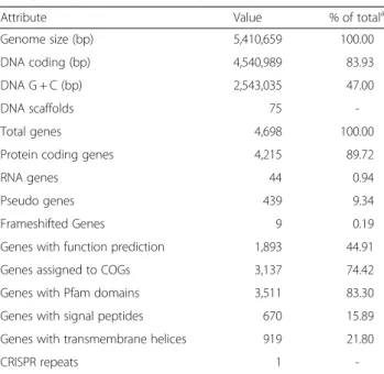
Genome properties
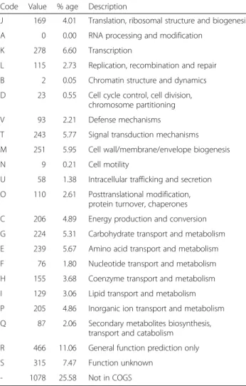
The daft genome size ofF. solisilvae3-3Tis 5,410,659 bp with 47 % GC content and contains 75 contigs. From a total of 4,698 genes, 4,215 (89.72 %) genes are protein coding genes, 439 (9.34 %) are pseudo genes and 44 (0.94 %) are RNA encoding genes. The genome properties and statistics are shown in Table 3 and Fig. 3. Altogether, 3,137 (74.42 %) protein coding genes are distributed into COG functional categories (Table 4).
Insights from the genome sequence
F. solisilvae 3-3Tcould grow on 33 kinds of sole carbon substrates including saccharides, organic acids and amino acids [3] (Table 1). Analysis of the genome reveals that this strain possesses putative enzymes for central carbohydrate metabolism to assimilate these carbon sources through
Table 3Genome statistics ofF. solisilvae3-3T
Attribute Value % of totala
Genome size (bp) 5,410,659 100.00
DNA coding (bp) 4,540,989 83.93
DNA G + C (bp) 2,543,035 47.00
DNA scaffolds 75
-Total genes 4,698 100.00
Protein coding genes 4,215 89.72
RNA genes 44 0.94
Pseudo genes 439 9.34
Frameshifted Genes 9 0.19
Genes with function prediction 1,893 44.91
Genes assigned to COGs 3,137 74.42
Genes with Pfam domains 3,511 83.30
Genes with signal peptides 670 15.89
Genes with transmembrane helices 919 21.80
CRISPR repeats 1
-a
The total is based on either the size of the genome in base pairs or the total number of protein coding genes in the annotated genome
Fig. 3A Graphical circular map ofF. solisilvae3-3Tgenome. From outside to center, ring 1, 4 show protein-coding genes colored by COG categories on
different metabolic pathways [20]. The putative enzymes that responsible to the utilization of 20 sole carbons were found in the genome (Table 5). All key enzymes in the Embden-Meyerhof-Parnas pathway (glucokinase, pyruvate kinase and 6-phosphofructokinase) and TCA cycle are present inF. solisilvae 3-3T. The key enzymes of Pentose Phosphate pathway (glucose-6-phosphate dehydrogenase, 6-phosphogluconolactonase and 6-phosphogluconate de-hydrogenase) were also found.
The presence of 4-hydroxyphenylpyruvate dioxygenase (KIC95062), homogentisate 1,2-dioxygenase (KIC93392) and other related enzymes suggests that 4-hydroxypheny-lacetic acid is degradable via homogentisic acid pathway [21]. In addition, the presence of 3-dehydroquinate dehy-dratase (KIC93382), shikimate dehydrogenase (KIC92987), shikimate kinase (KIC93265), 3-phosphoshikimate 1-car-boxyvinyltransferase (KIC94147) and chorismate synthase (KIC94148) indicates thatF. solisilvae3-3Tcould probably utilize quinic acid to synthesize the three aromatic amino
acids (tryptophan, tyrosine and phenylalanine) via shi-kimate pathway [7].
Conclusion
To the best of our knowledge, this report provides the first genomic information of the genusFlavihumibacter. Analysis of the genome shows high correlation between the genotypes and the phenotypes. The genome pos-sesses many key proteins of central carbohydrate metab-olism which provides the genomic basis to utilize the various carbon sources. In addition, analyzing its gen-ome indicates that this strain has potential application for the production of aromatic amino acids and for en-vironmental bioremediation.
Table 4Number of genes associated with general COG functional categories ofF. solisilvae3-3Tgenome Code Value % age Description
J 169 4.01 Translation, ribosomal structure and biogenesis
A 0 0.00 RNA processing and modification
K 278 6.60 Transcription
L 115 2.73 Replication, recombination and repair
B 2 0.05 Chromatin structure and dynamics
D 23 0.55 Cell cycle control, cell division, chromosome partitioning
V 93 2.21 Defense mechanisms
T 243 5.77 Signal transduction mechanisms
M 251 5.95 Cell wall/membrane/envelope biogenesis
N 9 0.21 Cell motility
U 58 1.38 Intracellular trafficking and secretion
O 110 2.61 Posttranslational modification, protein turnover, chaperones
C 206 4.89 Energy production and conversion
G 224 5.31 Carbohydrate transport and metabolism
E 239 5.67 Amino acid transport and metabolism
F 76 1.80 Nucleotide transport and metabolism
H 155 3.68 Coenzyme transport and metabolism
I 129 3.06 Lipid transport and metabolism
P 205 4.86 Inorganic ion transport and metabolism
Q 87 2.06 Secondary metabolites biosynthesis, transport and catabolism
R 466 11.06 General function prediction only
S 315 7.47 Function unknown
- 1078 25.58 Not in COGS
The total is based on the total number of protein coding genes in the genome
Table 5Putative enzymes responsible the utilization of different sole carbon sources in the genome ofF. solisilvae3-3T
Substrates Enzymes Accession
no.
Sucrose Alpha-glucosidase KIC96373
D-maltose Alpha-glucosidase KIC96373
D-glucose Glucokinase KIC93940
D-galactose Aldose epimerase KIC96300
Galactokinase KIC95381
Lactose Beta-galactosidase KIC94337
Glycerol Glycerol kinase KIC93992
Glycerol-3-phosphate dehydrogenase
KIC94583
N-acetyl-glucosamine β-N-acetylhexosaminidase KIC92674
L-arabinose Arabinose isomerase KIC96297
D-melibiose Alpha-galactosidase KIC96021
4-hydroxyphenlyacetic acid 4-hydroxyphenylpyruvate dioxygenase
KIC95062
Homogentisate 1,2-dioxygenase KIC93392
Quinic acid 3-dehydroquinate dehydratase KIC93382
Shikimate dehydrogenase KIC92987
Shikimate kinase KIC93265
3-phosphoshikimate 1-carboxyvinyltransferase
KIC94147
Chorismate synthase KIC94148
Urocanic acid Urocanate hydratase KIC93805
L-asparagine L-asparaginase KIC93060
L-aspartic acid Aspartate ammonia-lyase KIC93059
L-histidine Histidine ammonia-lyase KIC93735
L-serine Serine dehydratase KIC94326
L-alanine Alanine dehydrogenase KIC92870
D-alanine D-alanine–D-alanine ligase KIC93315
D-glucuronic acid Glucuronate isomerase KIC95816
Abbreviations
TCA:Tricarboxylic acid cycle; PE: Phosphatidylethanolamine; MK-7: Menaquinone-7; PGAP: Prokaryotic genome annotation pipeline.
Competing interests
The authors declare that they have no competing interests.
Authors’contributions
GZ performed laboratory experiments, analyzed the data and wrote the draft manuscript. CC and COJ cultured samples, analyzed the data and revised the manuscript. ML performed the genome comparison and revised the manuscript. GW organized the study and revised the manuscript. All authors read and approved the final manuscript.
Acknowledgment
This work was supported by the National Natural Science Foundation of China (31470227).
Author details
1State Key Laboratory of Agricultural Microbiology, College of Life Sciences and Technology, Huazhong Agricultural University, Wuhan 430070, People’s Republic of China.2Department of Life Science, Chung-Ang University, Seoul 156-756, Republic of Korea.
Received: 10 February 2015 Accepted: 8 July 2015
References
1. Zhang NN, Qu JH, Yuan HL, Sun YM, Yang JS.Flavihumibacter petaseusgen. nov., sp. nov., isolated from soil of a subtropical rainforest. Int J Syst Evol Microbiol. 2010;60:1609–12. doi:10.1099/ijs.0.011957-0.
2. Kim WH, Lee S, Ahn TY.Flavihumibacter cheonanensissp. nov., isolated from sediment of a shallow stream. Int J Syst Evol Microbiol. 2014;64:3235–9. doi:10.1099/ijs.0.063370-0.
3. Lee HJ, Jeong SE, Cho MS, Kim SH, Lee SS, Lee BH, et al.Flavihumibacter solisilvaesp. nov., isolated from forest soil. Int J Syst Evol Microbiol. 2014;64:2897–901. doi:10.1099/ijs.0.063669-0.
4. Martin M, Cibello A, Fernandez J, Ferrer E, Garrido-Pertierra A. Catabolism of 3- and 4-hydroxyphenylacetic acid byKelbsiella pneumoniae. J Gen Microbiol. 1991;137:621–8. doi:10.1099/00221287-137-3-621. 5. Méndez V, Agulló L, González M, Seeger M. The homogentisate and
homoprotocatechuate central pathways are involved in 3- and 4-hydroxyphenylacetate degradation byBurkholderia xenovoransLB400. PLoS One. 2011;6:e17583. doi:10.1371/journal.pone.0017583. 6. Koma D, Yamanaka H, Moriyoshi K, Ohmoto T, Sakai K. Production of
aromatic compounds by metabolically engineeredEscherichia coliwith an expanded shikimate pathway. Appl Environ Microbiol. 2012;78:6203–16. doi:10.1128/AEM.01148-12.
7. Guo J, Carrington Y, Alber A, Ehlting J. Molecular characterization of quinate and shikimate metabolism inPopulus trichocarpa. J Biol Chem.
2014;289:23846–58. doi:10.1074/jbc.M114.558536.
8. Tamura K, Peterson D, Peterson N, Stecher G, Nei M, Kumar S. MEGA5: molecular evolutionary genetics analysis using maximum likelihood, evolutionary distance, and maximum parsimony methods. Mol Biol Evol. 2011;28:2731–9. doi:10.1093/molbev/msr121.
9. Kimura M. A simple method for estimating evolutionary rates of base substitutions through comparative studies of nucleotide sequences. J Mol Evol. 1980;16:111–20.
10. Bernardet JF, Nakagawa Y, Holmes B. Proposed minimal standards for describing new taxa of the familyFlavobacteriaceaeand emended description of the family. Int J Syst Evol Microbiol. 2002;52:1049–70. doi:10.1099/ijs.0.02136-0.
11. Bennett S. Solexa Ltd. Pharmacogenomics. 2004;5:433–8. doi:10.1517/ 14622416.5.4.433.
12. Illumina official website. [www.illumina.com.cn]
13. Zerbino DR, Birney E. Velvet: algorithms for de novo short read assembly using de Bruijn graphs. Genome Res. 2008;18:821–9. doi:10.1101/gr.074492.107. 14. Wu S, Zhu ZW, Fu L, Niu BF, Li WZ. WebMGA: a customizable web server for
fast metagenomic sequence analysis. BMC Genomics. 2011;12:444. doi:10.1186/1471-2164-12-444.
15. The Pfam protein families database. [http://pfam.xfam.org/search]
16. Krogh A, Larsson B, Heijne GV, Sonnhammer ELL. Predicting transmembrane protein topology with a hidden Markov model: application to complete genomes. J Mol Biol. 2001;305:567–80. doi:10.1006/jmbi.2000.4315. 17. Petersen TN, Brunak S, Heijine GV, Nielsen H. SignalP 4.0: discriminating
signal peptides from transmembrane regions. Nat Methods. 2011;8:785–6. doi:10.1038/nmeth.1701.
18. Grissa I, Vergnaud G, Pourcel C. CRISPRFinder: a web tool to identify clustered regularly interspaced short palindromic repeats. Nucleic Acids Res. 2007;35:52–7. doi:10.1093/nar/gkm360.
19. Kanehisa M, Goto S, Sato Y, Kawashima M, Furumichi M, Tanabe M. Data, information, knowledge and principle: back to metabolism in KEGG. Nucleic Acids Res. 2014;42:D199–205. doi:10.1093/nar/gkt1076.
20. Justice NB, Norman A, Brown CT, Singh A, Thomas BC, Banfield JF. Comparison of environmental and isolateSulfobacillusgenomes reveals diverse carbon, sulfur, nitrogen, and hydrogen metabolisms. BMC Genomics. 2014;15:1107. doi:10.1186/1471-2164-15-1107.
21. van den Tweel WJJ, Smits JP, de Bont JAM. Catabolism of dl-α -phenylhydracrylic, phenylacetic and 3- and 4-hydroxyphenylacetic acid via homogentisic acid in aFlavobacteriumsp. Arch Microbiol. 1988;149:207–13. doi:10.1007/BF00422006.
22. Field D, Garrity G, Gray T, Morrison N, Selengut J, Sterk P, et al. The minimum information about a genome sequence (MIGS) specification. Nat Biotechnol. 2008;26:541–7. doi:10.1038/nbt1360.
23. Woese CR, Kandler O, Weelis ML. Towards a natural system of organisms. Proposal for the domainsArchaea,BacteriaandEucarya. Proc Natl Acad Sci U S A. 1990;87:4576–4579. doi:10.1073/pnas.87.12.4576 [PubMed]. 24. Krieg NR, Ludwig W, Euzéby J, Whitman WB. Phylum XIV. Bacteroidetes phyl.
nov. In: Krieg NR, Staley JT, Brown DR, Hedlund BP, Paster BJ, Ward NL, Ludwig W, Whitman WB (eds), Bergey's Manual of Systematic Bacteriology, Volume 4, 2nd ed. Springer, New York, 2011, p. 25.
25. List Editor. Validation List No. 143. Int J Syst Evol Microbiol 2012; 62:1-4. doi:10.1099/ijs.0.039487-0 [PubMed].
26. Kämpfer P. Class III.Sphingobacteriiaclass. nov. In: Krieg NR, Stately JT, Brown DR, Hedlund BP, Paster BJ, Ward NL, Ludwing W, Whitman WB (eds), Bergey’s Manual of Systematic Bacteriology, Volume 4, 2nd ed. New York: Springer; 2011. p. 330.
27. Kämpfer P. Order I.Sphingobacterialesord. nov. In: Krieg NR, Staley JT, Brown DR, Hedlund BP, Paster BJ, Ward NL, Ludwig W, Whitman WB (eds), Bergey's Manual of Systematic Bacteriology, Volume 4, 2nd ed. New York: Springer; 2011. p. 330
28. Kämpfer P, Lodders N, Falsen E.Hydrotalea flavagen. nov., sp. nov., a new member of the phylumBacteroidetesand allocation of the genera
Chitinophaga,Sediminibacterium,Lacibacter,Flavihumibacter,Flavisolibacter,
Niabella,Niastella,Segetibacter,Parasegetibacter,Terrimonas,Ferruginibacter,
FilimonasandHydrotaleato the familyChitinophagaceaefam. nov. Int J Syst Evol Microbiol 2011; 61:518-523. doi:10.1099/ijs.0.023002-0 [PubMed]. 29. Ashburner M, Ball CA, Blake JA, Botstein D, Butler H, Cherry JM, et al. Gene
ontology: tool for the unification of biology. The Gene Ontology Con-sortium. Nat Genet. 2000;25:25–9. doi:10.1038/75556.
Submit your next manuscript to BioMed Central and take full advantage of:
• Convenient online submission
• Thorough peer review
• No space constraints or color figure charges
• Immediate publication on acceptance
• Inclusion in PubMed, CAS, Scopus and Google Scholar
• Research which is freely available for redistribution
![Table 1 Classification and general features of F. solisilvae 3-3 T according to the MIGS recommendations [22]](https://thumb-us.123doks.com/thumbv2/123dok_us/669385.2065838/2.892.87.808.146.801/table-classification-general-features-solisilvae-according-migs-recommendations.webp)