Open Access
Research
Thumb force deficit after lower median nerve block
Zong-Ming Li*, Daniel A Harkness and Robert J Goitz
Address: Hand Research Laboratory, Departments of Orthopaedic Surgery and Bioengineering, University of Pittsburgh, PA 15213 USA
Email: Zong-Ming Li* - [email protected]; Daniel A Harkness - [email protected]; Robert J Goitz - [email protected] * Corresponding author
ThumbHandForceMedian nerve block
Abstract
Purpose: The purpose of this study was to characterize thumb motor dysfunction resulting from simulated lower median nerve lesions at the wrist.
Methods: Bupivacaine hydrochloride was injected into the carpal tunnel of six healthy subjects to locally anesthetize the median nerve. Motor function was subsequently evaluated by measuring maximal force production in all directions within the transverse plane perpendicular to the longitudinal axis of the thumb. Force envelopes were constructed using these measured multidirectional forces.
Results: Blockage of the median nerve resulted in decreased force magnitudes and thus smaller force envelopes. The average force decrease around the force envelope was 27.9%. A maximum decrease of 42.4% occurred in a direction combining abduction and slight flexion, while a minimum decrease of 10.5% occurred in a direction combining adduction and slight flexion. Relative decreases in adduction, extension, abduction, and flexion were 17.3%, 21.2%, 41.2% and 33.5%, respectively. Areas enclosed by pre- and post-block force envelopes were 20628 ± 7747 N.N, and 10700 ± 4474 N.N, respectively, representing an average decrease of 48.1%. Relative decreases in the adduction, extension, abduction, and flexion quadrant areas were 31.5%, 42.3%, 60.9%, and 52.3%, respectively.
Conclusion: Lower median nerve lesion, simulated by a nerve block at the wrist, compromise normal motor function of the thumb. A median nerve block results in force deficits in all directions, with the most severe impairment in abduction and flexion. From our results, such a means of motor function assessment can potentially be applied to functionally evaluate peripheral neuropathies.
Introduction
The thumb has unique anatomical and biomechanical characteristics that are required to perform many manipu-lative tasks. Thumb motor dysfunction resulting from neuromuscular and musculoskeletal pathologies severely hinders the performance of these daily tasks. Clinical
treatment, prevention protocols, and rehabilitation effi-cacy requires a thorough understanding of thumb motor capabilities, as well as its associated functional deficit. Investigations of underlying pathological mechanism of the thumb help advance clinical treatments such as Published: 19 October 2004
Journal of NeuroEngineering and Rehabilitation 2004, 1:3 doi:10.1186/1743-0003-1-3
Received: 30 August 2004 Accepted: 19 October 2004
This article is available from: http://www.jneuroengrehab.com/content/1/1/3
© 2004 Li et al; licensee BioMed Central Ltd.
Journal of NeuroEngineering and Rehabilitation 2004, 1:3 http://www.jneuroengrehab.com/content/1/1/3
tendon transfers [1], functional electrical stimulation [2] and plasticity suppression [3].
Measurement of strength during maximum voluntary contraction is a simple and direct means of assessing neu-romuscular function. Popular instruments used for quan-titative assessment of thumb strength are pinch dynamometers. The pinch output, however, provides lim-ited information about thumb motor function in that it offers a single generic force in one specific direction. Each muscle/tendon within the thumb has a distinct anatomi-cal origin and insertion, suggesting its external force potential in a particular direction [4-6]. Hence, evaluation of strengths in multiple directions offers insight concern-ing the motor capacity of individual muscles. Force pro-duction of a digit has been measured in various directions such as flexion/extension [7,8], abduction/adduction [9-14], or in combined directions [15,16]. Bourbonnais et al. developed an apparatus to measure thumb force produc-tion in eight direcproduc-tions in the transverse plane of the thumb and investigated force dependence on the direc-tion of effort [15]. Yokogawa and Hara measured index fingertip forces in various directions within the flexion/ extension plane [8]. Recently, we developed experimental apparatuses to measure multi-directional forces of a digit in its transverse plane [17-19]. From these multi-direc-tional forces we constructed force envelopes representa-tive of the characteristic force output pattern of a digit [17-19].
Disorders resulting from traumatic injuries to and various diseases of these nerves are common in clinical practice. Clinical manifestations of hand dysfunction are distinc-tive depending on the nerve involved. For example, thenar atrophy is a major clinical observation affecting thumb function at the later stages of compression neuropathy of the median nerve. Several studies have been conducted to investigate the effects of simulated peripheral neuropa-thies using local anesthetization [5,16,20,21]. Kozin et al. [21] studied the effects of median and ulnar nerve blocks on grip and pinch strength and showed significant decreases following nerve blockage [21]. Boatright and Kiebzak [20] investigated the effects of median nerve block on thumb abduction strength. Kaufman et al. [5] measured isometric thumb forces in eight directions together with electromyographic signals of thumb mus-cles after block of the median nerve. Labosky and Waggy [22] studied the strength related to grip, pinch, thumb adduction, thumb abduction, and finger flexion after radial nerve block [22]. Kuxhaus studied the three dimen-sional feasible force set at the thumb-tip before and after ulnar nerve block and reported this to be a reproducible and sensitive means to detect impairment.
The purpose of this study was to utilize our developed apparatus and protocols to investigate the effects of lower median nerve lesion on thumb motor function. The lesion was simulated by blocking the median nerve at the wrist using an anesthetic. We hypothesized that a median nerve block would cause (1) a decrease in force produc-tion, which would be direction-dependent with the most severe reduction in the abduction direction, and (2) a decrease in the force envelope area and force quadrant area, with the greatest decrease in the abduction quadrant.
Methods
Subjects
Six healthy male subjects (mean age: 26.9 ± 5.1 years) par-ticipated in this study. The subjects had no previous his-tory of neuromuscular or musculoskeletal disorders of the upper extremities. Each subject signed an informed con-sent form approved by the Institutional Review Board prior to participating in the experiment.
Median nerve block
Injections were performed under aseptic conditions while the subjects sat with the forearm supinated and the wrist slightly extended. After the skin at the palmer area of the wrist was cleaned with alcohol, 4 mL of 0.5% bupivacaine hydrochloride (Astra Pharmaceuticals, Westborough, MA, USA) was injected into the carpal tunnel with a sterile 25-gauge short-bevel needle. The needle was inserted through the transverse carpal ligament in line with the radial bor-der of the fourth digit slightly ulnar to the palmaris longus tendon at the level of the distal wrist crease. Forty minutes was allowed for the median nerve block to reach complete effectiveness [23] and was verified using the Semmes-Weinstein monofilament test. The average monofilament score was 2.85 across the five digits before nerve block. About 40 minutes after nerve injection, little sensory impairment occurred in the ulnar distribution (score = 3.22), while the sensory score in the median distribution was greater than 6.15. The effects of nerve block lasted more than 6 hours with all subjects regaining normal hand function within 12 hours.
Testing apparatus
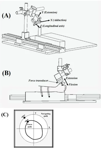
Schematic of experimental setup to measure thumb force production in the transverse plane
Figure 1
Journal of NeuroEngineering and Rehabilitation 2004, 1:3 http://www.jneuroengrehab.com/content/1/1/3
using a custom adapter. The ring served as a connection anchor for the transducer and the digit. The force trans-ducer and ring attachment were positioned in a desired orientation using an aluminum slide rail, tubing, and lockable mounting clamps (80/20 Inc., Columbia City, IN, USA). The slide rail was secured to an aluminum base plate. Foam padded wooden blocks with two locking straps secured the arm to the base plate.
The analog outputs from the transducer were digitized using a 16-bit analog-to-digital converter (PCI-6031, National Instruments, TX, USA). The X (abduction/adduc-tion) and Y (flexion/extension) force components in the transverse plane were displayed on the screen while the subject performed a force production task. The resolu-tions of the force transducer in its axial (flexion/exten-sion) and horizontal (abduction/adduction) directions were 0.16 N and 0.08 N, respectively. A personal compu-ter equipped with LabVIEW (National Instrument, TX, USA) was used for force data acquisition, display, and processing.
Experimental procedures
Each subject was tested before and after median nerve block. The nerve block procedures were performed imme-diately after the completion of the first testing session. Post-block testing started after the verification of complete median nerve block, approximately 40 minutes after the injection. During each test, the subject was seated in a chair adjacent to the testing station modified with a wooden board to align their back vertically throughout the trials. The subjects rested their forearm on padded wooden blocks positioning their shoulder in approxi-mately 60° of frontal plane abduction. Nylon straps fitted with plastic snap locking mechanisms secured the forearm and minimized the intervention of the elbow and shoul-der during thumb force application. Subjects grasped a vertical dowel secured to the distal end of the wooden blocks in a midprone position. Formable thermoplastic braces were used to fix the elbow in 90° of flexion, and the wrist in 20° of extension and 0° of ulnar deviation. A metallic brace was used to fix the interphalangeal joint of the thumb in full extension. The aluminum ring was placed around the middle of the proximal phalanx and oriented to accommodate comfortable thumb position with the metacarpophalangeal joint flexed approximately 15°. Prior to testing, a line was drawn on the proximal phalange at the midpoint between the interphalangeal and metacarpophalangeal joints. The alignment of the ring with the circumferential line standardized the loca-tion of force applicaloca-tion within and between subjects. As force application was at the middle of the proximal pha-lanx, mechanical action pertains to both the metacar-pophalangeal and carpometacarpal joints. We chose the terminology of flexion/extension and
adduction/adduc-tion based on the mechanical acadduction/adduc-tion with respect to the metacarpophalangeal joint. With the thumb in the ring (Figure 1B), extension and flexion occurred in parallel with the palm, and abduction and adduction occurred in a plane perpendicular to the palm.
Each subject performed 15 circumferential MVC trials with randomized starting directions (Figure 1C). The sub-ject was allotted 15 seconds to complete each circumfer-ential trial, and was instructed to use the entire time allotted to traverse the perimeter of the ring once. A dot generated on the computer screen was programmed to traverse a circle within 15 seconds to provide the subject with directional feedback of their force application. Sub-jects were given 60 seconds of rest between each circum-ferential trial. Each subject was familiarized with the task with a few practice trials. Data were collected from each subject at 100 samples per second producing a total of 22,500 pairs of force components from the 15 circumfer-ential trials. Our previous study [19] indicated that the testing protocol did not cause noticeable fatigue.
Force envelope and quadrants
Data from multiple circumferential trials were accumu-lated to construct a force envelope. The procedures to gen-erate a force envelope were as follows:
Division of force envelope into extension, abduction, flexion, and adduction quadrants
Figure 2
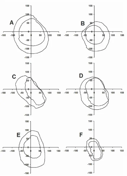
Force envelopes before and after median nerve block of subjects A, B, C, D, E, and F
Figure 3
Journal of NeuroEngineering and Rehabilitation 2004, 1:3 http://www.jneuroengrehab.com/content/1/1/3
Cartesian force coordinates (Xi, Yi) were transformed into polar coordinates (Rα, α), where Rαwas the force
magni-tude at an angular position α. Each α was rounded to the nearest integer ranging from 0 to 359 degrees.
The maximum, Fα, was determined from a string of N data
points along each radial line defined by α. At the comple-tion of the 15 trials, there were, on average, N = 63 data points on each radial line of α based on the distribution off the 22,500 data points around 360°.
A moving average with an interval of 10° was applied to the maximal series data Fα(α = 0, 1, 2,..., 359) to obtain
filtered maximal forces. These forces formed a force envelope.
The area formed by a force envelope was divided into adduction-extension, extension-abduction, abduction-flexion, and flexion-adduction quadrants by radial lines oriented at 0°, 90°, 180°, and 270° A quadrant force was represented using the mean magnitude of the forces in that quadrant. The areas of the entire envelope and each quadrant were calculated by summing the areas of indi-vidual arc sections formed by the polar coordinates of the force envelope. (Figure 2).
Statistical Analyses
One- and two-factor repeated measures analyses of vari-ance (ANOVA) were used to analyze outcome measures. The independent variables were testing SESSION (n = 2, i.e., pre- and post-block), force DIRECTION (n = 16), and force QUADRANT (n = 4), with SESSION as a repeated variable. Dependent variables were directional force, indi-vidual quadrant area and force envelope area. Statistical analyses were performed using SPSS 11 (SPSS Inc., Illi-nois) with statistical significance set at α = 0.05.
Results
Force envelope and directional forces
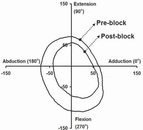
Figure 3 shows the force envelopes produced by each sub-ject (A to F) before and after median nerve block. The post-block force envelope was inside the pre-block envelope for each subject, indicating a decrease in force magnitude in all directions after nerve block. Figure 4 shows the average pre- and post-block force envelope across all subjects. Force magnitudes were significantly reduced after nerve block (p < 0.001) resulting in signifi-cantly smaller force envelopes. The average decrease across all directions was 27.9%. A maximum decrease of 42.4% occurred at 199°, corresponding to a combined direction of abduction and slight flexion, while a mini-mum decrease of 10.5% occurred at 328° corresponding to a combined direction of adduction and slight flexion. Relative decreases at 0° (adduction), 90° (extension),
180° (abduction), and 270° (flexion) directions were 17.3%, 21.2%, 41.2% and 33.5%, respectively.
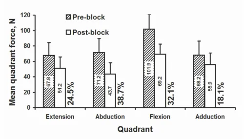
A single force in each quadrant was represented using the mean magnitude of the forces in that quadrant (see description in the Methods). The average quadrant forces were significantly decreased after nerve block (p < 0.001; Figure 5). The amount of decrease was also different between quadrants (p < 0.005). Relative decreases in mean quadrant forces were 24.5%, 38.7%, 32.1%, and 18.1% for extension, abduction, flexion, and adduction, respectively. The maximal decreases in mean quadrant force, 38.7%, occurred in the abduction quadrant.
Force envelope areas and quadrant areas
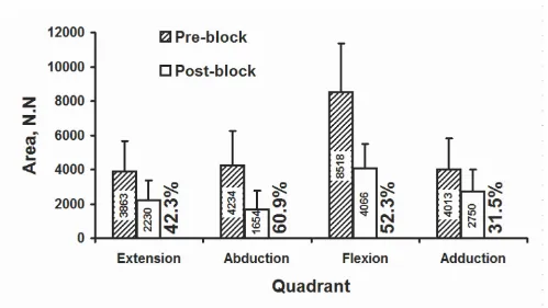
Areas enclosed by the post-block envelopes were signifi-cantly smaller than the pre-block envelopes (p < 0.001; Figure 4). Post-block force envelope area, 10700 ± 4474 N.N, was 51.9% of pre-block force envelope area, 20628 ± 7747 N.N. Quadrant area decreased significantly (p < 0.001; Figure 6). The maximal percentage decrease in area after nerve block was 60.9% in the abduction quadrant, followed by a 52.3% area decrease in the flexion quadrant.
Discussion
In this study we simulated a lower median nerve lesion and evaluated the resultant thumb motor function deficit.
Average force envelopes produced by the thumb before and after median nerve block
Figure 4
Our internal control via pre- and post-block design offered a particular advantage of investigating the mechanical role of muscles innervated by a targeted nerve. The testing and analytical methods employed have pro-vided advanced quantification of thumb motor function. The results have confirmed our initial hypotheses that greatest force decreases occurred in directions related to abduction, and that the post-block thumb force envelope area was smaller than the pre-block force envelope area.
Preferential force attenuation in the quadrants of abduc-tion and flexion after median nerve block are in agreement with anatomical and neuromuscular features of the thumb. The median nerve innervates the abductor pollicis brevis, the opponens pollicis and superficial head of the flexor pollicis brevis, all of which contribute to the abduction and flexion of the thumb [4]; therefore, dener-vation of these muscles after median nerve block would cause the greatest force deficit related to median nerve function [5]. Additionally, as force application moved towards adduction, the force deficit decreased as neu-romuscular control shifted from the median nerve to the
ulnar nerve via the first dorsal interosseous and adductor pollicis brevis. Force deficit in extension was also comparably small as extension forces are mainly pro-duced by the extensors pollicis brevis and longus originat-ing in the forearm.
Our reported force decreases following a median nerve block (40.9% in abduction, 34.1% in flexion) were smaller than those reported in the literature. Kozin et al. [21] reported a 60% decrease in pinch strength after a median nerve block using mepivicaine hydrochloride [21]. Boatright and Kiebzak [20] reported an approximate 70% decrease in thumb abduction strength after median nerve block using Lidocaine [20]. Kaufman et al. [5] stated that a median nerve block with Lidocaine almost com-pletely diminished force production in the abduction direction [5]. The discrepancy may be due to the anesthetic used and strength testing method. Although the sensory block appeared to be complete for each method, the motor capabilities of the muscles associated with the median nerve might or might not be completely eliminated. Such a result is largely dependent on a
partic-Average force magnitude, N, in individual quadrants
Figure 5
Journal of NeuroEngineering and Rehabilitation 2004, 1:3 http://www.jneuroengrehab.com/content/1/1/3
ular anesthetic, its concentration and dosage, as well as the efficacy of the injection technique at immersing the nerve. The methods of strength testing may also help explain the different magnitudes of strength deficit after the nerve block. All previous results were based on forces obtained in discrete direction(s), and focused exertions, while the current study utilized a method of force produc-tion in a continuous, circumferential and dynamic man-ner. Furthermore, thumb motor performance can be maintained despite the absence of certain individual mus-cles. For example, Britto and Elliot reported that the loss of abductor pollicis longus and extensor pollicis brevis in their two patients did not show functional compromise of strength and grip strength [24]. In a broader sense, the neuromuscular system has remarkable capabilities to accomplish the same motor function goal using different effectors and different goals using the same effectors, a phenomenon so called "motor equivalence" [25].
An unexpected finding from this study was that the force deficit occurred in all directions (Figure 4). In other words, the median nerve block caused reduced force pro-duction by those muscles not associated with the median
nerve. Several potential explanations exist to describe such a phenomenon. First, the injection into the carpal tunnel at the wrist, although localized, potentially diffused into the intrinsic fascia of the hand partially compromising function of the ulnar nerve, which inner-vates the adductor pollicis. Although Semmes-Weinstein monofilament testing confirmed the continued sensation of the digits within the ulnar nerve distribution, it is not inconceivable that the injection could have contaminated the ulnar innervated muscles, the first dorsal interosseous and deep head of the flexor pollicis brevis [20]. Secondly, thumb force in any direction is produced by synergistic activation of the many intrinsic muscles, and as a result, the muscular deficiency associated with one direction may hinder the force production in other directions by other muscles [5,22]. For example, Kaufman et al. demonstrated that thumb muscles not innervated by the median nerve displayed lower electromyographical activation and shifted the direction of maximum activation after a median nerve block [5]. Labosky and Waggy showed that a radial nerve block caused a 53% decrease in thumb abduction strength because of the lack of stabilization of the radial innervated extensor muscles [22].
Area (N-N) of individual force quadrants, and percentage decrease after nerve block
Figure 6
Consequently, deficiency of median innervated muscles inherently limits force production in other directions as neuromuscular switching is necessary to produce force in changing directions.
The median innervated muscles are the dominant abduc-tors of the thumb metacarpophalangeal and carpometa-carpal joint. The more than 50% residual abduction force found in this study suggests that the injection did not totally block the motor function of these muscles, even though a complete sensory loss was verified. This concurs with clinical observations of median compression neu-ropathy. Individuals with carpal tunnel syndrome com-plain of sensory dysfunction early in the disease process (at the beginning), while motor signs of thenar wasting and thumb weakness occur as the disease advances. The concept that the motor deficit is more resistant to periph-eral median neuropathy than sensory loss has been well documented [23,26,27]. Butterworth et al. studied the temporal effects on sensory and motor blockade after injection of bupivacaine or mepivacaine, and found that sensory loss was complete but about a 20% compound motor action potential remained after 40 minutes [23].
In conclusion, we have incorporated a method for assess-ing thumb motor deficit based on strength measurement with a standard local anesthetic to investigate the effects of a simulated median neuropathy on thumb motor function. Median nerve block results in force deficits in all directions, with the most severe impairment in abduction and flexion. Future endeavors using this methodology can potentially further elucidate underlying pathomecha-nisms of peripheral neuropathies in all digits of the hand.
Acknowledgements
This work was partially supported by the Aircast Foundation and the Whitaker Foundation. The authors thank Robert A. Kaufmann for helping perform anesthetic injections.
References
1. Cooney WP: Tendon transfer for median nerve palsy.Hand Clin
1988, 4:155-165.
2. Lauer RT, Kilgore KL, Peckham PH, Bhadra N, Keith MW: The func-tion of the finger intrinsic muscles in response to electrical stimulation.IEEE Trans Rehabil Eng 1999, 7:19-26.
3. Autti-Ramo I, Larsen A, Taimo A, von Wendt L: Management of the upper limb with botulinum toxin type A in children with spastic type cerebral palsy and acquired brain injury: clinical implications.Eur J Neurol 2001, 8(Suppl 5):136-144.
4. Smutz WP, Kongsayreepong A, Hughes RE, Niebur G, Cooney WP, An KN: Mechanical advantage of the thumb muscles.J Biomech
1998, 31:565-570.
5. Kaufman KR, An KN, Litchy WJ, Cooney WP, Chao EY: In-vivo function of the thumb muscles.Clin Biomech (Bristol, Avon) 1999,
14:141-150.
6. Valero-Cuevas FJ, Towles JD, Hentz VR: Quantification of finger-tip force reduction in the forefinger following simulated paralysis of extensor and intrinsic muscles.J Biomech 2000,
33:1601-1609.
7. Kilgore KL, Lauer RT, Peckham PH: A transducer for the meas-urement of finger joint moments.IEEE Trans Rehabil Eng 1998,
6:424-429.
8. Yokogawa R, Hara K: Measurement of distribution of maxi-mum index-fingertip force in all directions at fingertip in flexion/extension plane.J Biomech Eng 2002, 124:302-307. 9. Belanger AY, Noel G: Force-generating capacity of thumb
adductor muscles in the parallel and perpendicular plane of adduction.J Orthop Sports Phys Ther 1995, 21:139-146.
10. Boatright JR, Kiebzak GM, O'Neil DM, Peindl RD: Measurement of thumb abduction strength: normative data and a compari-son with grip and pinch strength. J Hand Surg [Am] 1997,
22:843-848.
11. Byers GJ, Goldstein BS, Sanders JE: An electromechanical testing device for assessment of hand motor function.IEEE Trans Reha-bil Eng 1998, 6:88-94.
12. Ditor D, Hicks A: The optimal joint angle for adductor pollicis force production in men and women.Can J Appl Physiol 1999,
24:570-580.
13. Liu F, Carlson L, Watson HK: Quantitative abductor pollicis brevis strength testing: reliability and normative values.J Hand Surg [Am] 2000, 25:752-759.
14. Schreuders TA, Roebroeck M, van der Kar TJ, Soeters JN, Hovius SE, Stam HJ: Strength of the intrinsic muscles of the hand meas-ured with a hand-held dynamometer: reliability in patients with ulnar and median nerve paralysis.J Hand Surg [Br] 2000,
25:560-565.
15. Bourbonnais D, Forget R, Carrier L, Lepage Y: Multidirectional analysis of maximal voluntary contractions of the thumb.J Hand Ther 1993, 6:313-318.
16. Kuxhaus L: Changes in thumb 3D force production with selec-tive paralysis. In M.S Thesis Cornell University, Department of Mechanical Engineering; 2003.
17. Li ZM, Goitz RJ: Biomechanical evaluation of the motor func-tion of the thumb.Technol Health Care 2003, 11:233-243. 18. Li ZM, Pfaeffle HJ, Sotereanos DG, Goitz RJ, Woo SL:
Multi-direc-tional strength and force envelope of the index finger.Clin Biomech 2003, 18:908-915.
19. Li ZM, Harkness DA: Circumferential force production of the thumb.Med Eng Phys 2004, 26:663-670.
20. Boatright JR, Kiebzak GM: The effects of low median nerve block on thumb abduction strength.J Hand Surg [Am] 1997,
22:849-852.
21. Kozin SH, Porter S, Clark P, Thoder JJ: The contribution of the intrinsic muscles to grip and pinch strength.J Hand Surg [Am]
1999, 24:64-72.
22. Labosky DA, Waggy CA: Apparent weakness of median and ulnar motors in radial nerve palsy.J Hand Surg [Am] 1986,
11:528-533.
23. Butterworth J, Ririe DG, Thompson RB, Walker FO, Jackson D, James RL: Differential onset of median nerve block: rand-omized, double-blind comparison of mepivacaine and bupi-vacaine in healthy volunteers.Br J Anaesth 1998, 81:515-521. 24. Britto JA, Elliot D: Thumb function without the abductor
polli-cis longus and extensor pollipolli-cis brevis.J Hand Surg [Br] 2002,
27:274-277.
25. Rosenbaum DA: In Human motor control London: Academic Press; 1991:314.
26. Gelberman RH, Hergenroeder PT, Hargens AR, Lundborg GN, Ake-son WH: The carpal tunnel syndrome. A study of carpal canal pressures.J Bone Joint Surg Am 1981, 63:380-383.
27. Mazur A: Role of thenar electromyography in the evaluation of carpal tunnel syndrome.Phys Med Rehabil Clin N Am 1998,