Original article
1: Parasitology & Medical Mycology Department,School of Medicine, Mashhad University of Medical Sciences Mashhad, Iran
*Corresponding author: Seyyed Ali Akbar Shamsian; Tel: +98 511- 7112410; Fax: +98 511- 7112616; E-mail: [email protected]
Received: Nov 28, 2012; Accepted: Feb 13, 2013
Identification of Leishmania species causing
cutaneous leishmaniasis using Random Amplified
Polymorphic DNA (RAPD-PCR) in Kharve, Iran
Fatemeh Saadabadi
1, Masoud Mohajery
1, Elham Poostchi
1,
Seyyed Ali Akbar Shamsian*
1Abstract
Background: Leishmaniasis, especially cutaneous leishmaniasis, is considered an important health problem in many parts of Iran including Kharve, Khorasan Razavi province. Cutaneous
leishmaniasis is caused by various species of Leishmania, each having a different secondary host.
Thus, identifying the parasites’ specie is of paramount importance for containment strategy
planning. The morphological differentiation of Leishmania species is not possible, rendering the
molecular methods as the sole means to this purpose. Therefore, to identify the causative agent of cutaneous leishmaniasis in Kharve, Random Amplified Polymorphic DNA-PCR (RAPD-PCR) was used.
Methods: The disease was first confirmed by direct smears. Samples were gathered from 22 patients with established cutaneous leishmaniasis. The samples were immediately cultured in NNN medium, followed by sub-culture in RPMI-1640. Afterwards, DNA was extracted and amplified using RAPD-PCR. Electrophoresis patterns from each isolate were compared with reference strains
of Leishmania major (L. major)and Leishmania tropica (L. tropica).
Results: The results of this study indicated that the parasite causing cutaneous leishmaniasis in
Kharve is L. tropica.
Conclusion: It seems that L. tropica is the only causative agent of cutaneous leishmaniasis in
Kharve, and RAPD-PCR is a suitable tool for Leishmania characterization in epidemiological
studies.
Keywords: Leishmania major, Leishmania tropica, RAPD-PCR, Khorasan, Kharve
Introduction
Cutaneous leishmaniasis is the most common form of leishmaniasis, with about 1.5 million new cases every year worldwide, mostly reported from Afghanistan, Iran, Iraq, Algeria, Saudi Arabia, Peru, and Pakistan (1). The surveys demonstrate that cases of leishmaniasis are increasing worldwide, mainly due to environmental changes (2). Irregular buildings in towns and collection of
domestic garbage in suburbs (3), plus migration of susceptible populations could also be causative factors. Poverty and malnutrition play important roles in increased morbidity of the disease (2).
Depending on the species causing the disease, different preventive measures are taken against
leishmaniasis (3). All Leishmania species have
the same morphology; hence, they are not distinguishable microscopically (4). Before the
Rep. Biochem. Mol. Biol, Vol. 1, No. 2, Apr 2013 70
advancement of new methods, characterization
and isolation of Leishmania parasites had been
only possible through painstaking procedures
(5). Applying iso-enzyme characteristics
simplified the diagnosis to some extent; however, these methods were accompanied by
problematic difficulties (6). Since the
introduction of molecular techniques however, several novel methods have been introduced to parasitology, such as the use of DNA probes, polymerase chain reaction-restriction fragment length polymorphism (PCR-RFLP), and random amplified polymorphic DNA-polymerase chain reaction (RAPD-PCR) (3, 7-8). Being more accurate and user-friendly than previous methods, RAPD-PCR was utilized in the present study.
Previous attempts to classify the
Leishmania parasite in Razavi Khorasan
Province (9) were based on clinical and epidemiological signs. It was previously thought that only dry cutaneous leishmaniasis was present in Mashad; however, recent molecular studies revealed the existence of both dry and wet forms. Kharve, as a leishmaniasis hot spot, has been neglected in studies thus far; therefore, this study was
conducted to investigate Leishmania species
occurring in Kharve, using RAPD-PCR.
Materials and Methods
In the present study, patients with suspicious cutaneous lesions referred to the health center laboratory of Kharve, between 15 Sep 2006 and 15 Sep 2007 were included in the primary examination. After the acquisition of written informed consent, all patients underwent direct smear testing to confirm the diagnoses of leishmaniasis. Cases with established
cutaneous leishmaniasis (n=22), were
recruited for species characterization by RAPD-PCR.
Aspirations were obtained from lesions on all subjects and cultured on Novy-MacNeal-Nicole (NNN) medium for 48 to 72 h followed by subculture in RPMI-1640 medium supplemented with 10% inactivated fetal calf serum to obtain a dense parasite population.
Genomic DNA preparation: Parasite DNA was isolated after the parasite concentration
reached 1x 10⁶/mL. Cells were pelleted in a
centrifuge at 3000 x g for 10 min at 4 °C, then washed using cold phosphate-buffered saline (PBS), pH 7.2, and the pellet was dissolved in 500 µl of lysis buffer (50mM NaCl, 50mM EDTA, 1% SDS, 50mM Tris-HCl, pH 8.0). Proteinase K, with final concentration of 100µg/mL was added, and the solution was incubated at 55 °C overnight. The resulting lysate was extracted with phenol followed by 1:1 phenol-chloroform, and finally with 100% chloroform.
The extracted material was precipitated with cold ethanol and the obtained DNA was dissolved in distilled water at 10 ng/mL.
RAPD-PCR: RAPD-PCR for each 20 µl reaction was performed with 1 µl of genomic DNA at 10 ng/µl, 2 µl 10X PCR buffer, 0.6 µl 50 mM
MgCl2, 0.6 µl 10 mM dNTPs, 1 µl 10 mM
primer, 2 µl of 0.5 unit/µl polymerase, and 12.8 µl distilled water. Thermal parameters were 45 cycles, a single denaturation step of 94 ºC for 3 minutes and afterwards a cycle of 94 ºC for one minute, 37 ºC for 2 minutes and 72 ºC for 2 minutes. This was followed by final extension cycle at 72 °C for 10 min. Eight µl of the PCR products were electrophoresed on 1.2% agarose gels containing ethidium bromide for 4 hours at 50 V in TAE buffer, and gel photos were prepared in a trans-illuminator . Four primers designated AB1-07, A4, 327, and 329 (Table 1) were utilized
to characterize the Leishmania species (8, 10).
RAPD-PCR results from subject’s samples were
compared to products obtained from Leishmania
major (MHOM/IR/75/ER) and Leishmania
tropica (MHOM/IR/99) (Figs. 1-4).
Table 1: Primers used in this study.
Sequence (5’-3’) Primer GGTGACGCAG AB1-07 ATACGGCGTC 327 AATCGGGCTG A4 GCGAACCTCC 329
Rep. Biochem. Mol. Biol, Vol. 1, No. 2, Apr 2013 71
Results
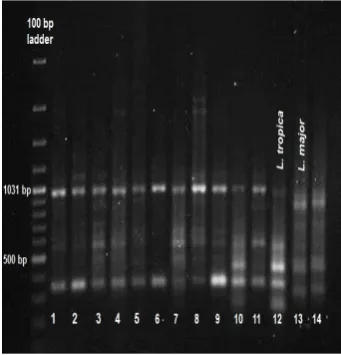
RAPD-PCR with each primer resulted in multiple bands on electrophoresis (Figs. 1-4). The number of bands in each strain varied and ranged from 250 to 2,500 base pairs (bps). Primer AB1-07 amplified two major PCR products of about 950 and 350 bps
from L. tropica and three major products of about
850, 450, and 300 bps from L. major (Fig. 1).
Primer 327 amplified four major products of about
1,900, 1,200, 950, and 350 bps from L. tropica, and
three major products of about 1,400, 1,000, and 600
bps from L. major (Fig. 2). Primer 329 amplified
two major products of about 850 and 450 bps from
L. tropica and two major products of about 750 and
650 bps from L. major (Fig. 3). Primer A4
amplified three major products of about 700, 550,
and 250 bps from L. tropica and three major
products of about 1,350, 850, and 200 bps from L.
major (Fig. 4). All 22 samples collected from
subjects in Kharve with cutaneous leishmaniasis
were diagnosed by RAPD-PCR as L. tropica.
Fig. 1: The profiles obtained from the RAPD-PCR with primer AB1-07; First lane: 100 bp ladder; Lanes 1-11: cutaneous leishmaniasis subject samples, lane 12: L. tropica, Lanes 13 and 14: L. major.
Fig. 2: The profiles obtained from the RAPD-PCR with primer 327; First lane: 100 bp ladder; Lanes 1-10: cutaneous leishmaniasis subject samples, lane 11: L. tropica, Lane 12: L. major.
Fig. 3: The profiles obtained from the RAPD-PCR with primer 329; Lanes 1-14: cutaneous leishmaniasis subject samples; L. major lane: L. major; L. tropica lane: L. tropica; 100 bp ladder lane: 100 bp ladder.
Fig. 4: The profiles obtained from the RAPD-PCR with primer A4; First lane: 100 bp ladder; Lanes 1-3, 5-7, 9-13: cutaneous leishmaniasis subject samples, lane 4: L. tropica, Lane 8: 100 bp ladder; Lane 14: L. major. 1
1 1 3 1
Rep. Biochem. Mol. Biol, Vol. 1, No. 2, Apr 2013 72
Discussion
According to the World Health Organization (WHO), leishmaniasis is one of the six most common infectious diseases in the world, and cases are increasing (2). Research about different aspects of this ailment is necessary (11).
Cutaneous leishmaniasis poses a critical health problem across Iran (12-14). Previous studies conducted in Khorasan province
reported both dry leishmaniasis, caused by L.
tropica, and wet leishmaniasis, caused by L.
major (3-4, 8). Dry leishmaniasis is observed
in many cities of Iran including Mashhad, Neishabur, and Sabzevar, whereas the wet form is reported to be endemic to Sarakhs, Lotf Abad, and Esfarayen by (Unpublished).
Kharv, a city in Neishabur, Khorasan Razavi Province, is one of the principal foci of leishmaniasis. In the past years, population growth and overall financial improvements in residential regions in arid lands have led to an increase in this disease in this region despite preventive measures (3). Therefore, to plan more effective preventive strategies, epidemiological studies and species identification using precise
molecular methods are of utmost importance. RAPD-PCR was used in this study to identify
the Leishmania species present in the area. A
great advantage of the primers used is that they are species-specific. In addition, this method is both cost- and time-efficient (15). The possibility of evaluating several loci simultaneously, the high number of markers, and the low amount of DNA sample needed have added to the popularity of this method.
Of the 22 the study subjects, most had one or two lesions and many were afflicted with the disease for more than two months. Because the
causative agent in all 22 specimens was L.
tropica, we conclude that, similar to Neishabur,
the cutaneous leishmaniasis type in Kharve has not changed and is the dry type; whereas in Mashhad, a modification in the epidemiologic pattern occurred, because both dry and wet cutaneous leishmaniasis have been reported.
Acknowledgements
This study has been supported by the Research Council of Mashhad University of Medical Sciences (MUMS), Mashhad, Iran.
References
1. Vidyashankar C, Agrawal A. Leishmaniasis EMedicine. 2006: Available from:
http://www.emedicine.com/ped/topic1292.htm.
2. Khan SJ, Muneeb S. Cutaneous leishmaniasis in Pakistan. Dermatol Online J. 2005;11(1):4.
3. Mohajery M, Hatam GR, Javaheri A. Isolation of Leishmania major by isoenzyme electrophoresis in Mashhad. Med J Mashhad Univ Med Sc. 2005;48(88):177-84.
4. Mahmoodi MR, Mohajery M, Tavakkol Afshari J, Shakeri MT, Yazdan Panah MJ, Berenji F, et al. Molecular identification of Leishmania species causing cutaneous leishmaniasis in Mashhad, Iran. Jundishapur Journal of Microbiology. 2010;3(4):195-200.
5. Klaus SN, Frankenburg S, Ingber A. Epidemiology of cutaneous leishmaniasis. Clin Dermatol. 1999 May-Jun;17(3):257-60.
6. Motazedian H, Noyes H, Maingon R. Leishmania and Sauroleishmania: the use of random amplified polymorphic DNA for the identification of parasites from vertebrates and invertebrates. Exp Parasitol. 1996 Jun;83(1):150-4.
7. Mauricio IL, Howard MK, Stothard JR, Miles MA. Genomic diversity in the Leishmania donovani complex. Parasitology. 1999 Sep;119 ( Pt 3):237-46.
8. Hajjaran H, Mohebali M, Alimoradi S, Abaei MR, Edrissian GH. Isolation and characterization of pathogenic Leishmania turanica from Nesokia indica (Rodentia, Muridae) by PCR-RFLP and ITS1 sequencing in Iran. Trans R Soc Trop Med Hyg. 2009 Nov;103(11):1177-9. 9. Rassi Y, Javadian E, Nadim A, Rafizadeh S, Zahraii A,
Azizi K, et al. Phlebotomus perfiliewi transcaucasicus, a vector of Leishmania infantum in northwestern Iran. J Med Entomol. 2009 Sep;46(5):1094-8.
10. Zemanova E, Jirku M, Mauricio IL, Miles MA, Lukes J. Genetic polymorphism within the leishmania donovani complex: correlation with geographic origin. Am J Trop Med Hyg. 2004 Jun;70(6):613-7.
11. Rafati S, Salmanian AH, Hashemi K, Schaff C, Belli S, Fasel N. Identification of Leishmania major cysteine proteinases as targets of the immune response in humans. Mol Biochem Parasitol. 2001 Mar;113(1):35-43.
12. Rahbarian N, Mesgarian A, Mahmoudi Rad M, Hajaran H, Shahbazi F, Mesgarian Z, et al. Identification of leishmania species isolated from human cutaneous leishmaniasis using PCR method. J Res Health Sci. 2009;9(2):48-51.
13. Motazedian H, Noamanpoor B, Ardehali S. Characterization of Leishmania parasites isolated from provinces of the Islamic Republic of Iran. East Mediterr Health J. 2002 Mar-May;8(2-3):338-44.
Rep. Biochem. Mol. Biol, Vol. 1, No. 2, Apr 2013 73 14. Motazedian H, Karamian M, Noyes HA, Ardehali S. DNA
extraction and amplification of leishmania from archived, Giemsa-stained slides, for the diagnosis of cutaneous Leishmaniasis by PCR. Ann Trop Med Parasitol. 2002 Jan;96(1):31-4.
15. Craft KJ, Owens JD, Ashley MV. Application of plant DNA markers in forensic botany: genetic comparison of Quercus evidence leaves to crime scene trees using microsatellites. Forensic Sci Int. 2007 Jan 5;165(1):64-70.