Copyright © 2001, American Society for Microbiology. All Rights Reserved.
Characterization of
Chlamydia trachomatis omp1
Genotypes
among Sexually Transmitted Disease Patients in Sweden
MARGARETHA JURSTRAND,1* LARS FALK,2HANS FREDLUND,1MARGRET LINDBERG,2PER OLCE´N,1
SO¨ REN ANDERSSON,1KENNETH PERSSON,3JAN ALBERT,4ANDANDERS BA¨ CKMAN1
Department of Clinical Microbiology and Immunology1and Outpatient Sexually Transmitted Disease Clinic,
Department of Dermatovenereology,2O¨ rebro Medical Centre Hospital, SE-70185 O¨rebro, Department of
Clinical Microbiology, Malmo¨ University Hospital, SE-20502 Malmo¨,3and Division of Virology, Department of
Microbiology, Pathology and Immunology, Karolinska Institutet, Huddinge University Hospital, SE-14186 Huddinge/
Stockholm,4Sweden
Received 16 April 2001/Returned for modification 15 June 2001/Accepted 12 August 2001
A method for detection and genotyping of genital Chlamydia trachomatis infections based on omp1 gene amplification and sequencing was developed. DNA was extracted from urogenital or urine samples using a Chelex-based method, and an approximately 1,100-bp-long fragment from theomp1gene was directly amplified and sequenced. Genotyping was performed by BLAST similarity search, and phylogenetic tree analysis was used to illustrate the evolutionary relationships between clinical isolates and reference strains. The method was used to determine the genotypes ofC. trachomatisin 237 positive urogenital and/or urine specimens collected at a Swedish sexually transmitted disease clinic during 1 year. The most common genotypes corresponded to serotypes E (47%) and F (17%). Theomp1 gene was highly conserved for genotype E (106 of 112 samples without any mutation) and F (41 of 42 samples without any mutation) strains but appear slightly less conserved for genotypes G (nⴝ6) and H (nⴝ6), where the sequences displayed one to four nucleotide substitutions relative to the reference sequence. Genotyping of samples collected at the follow-up visit indicated that two patients had become reinfected, while three other patients suffered treatment failure or reinfection. One woman appeared to have a mixed infection with two differentC. trachomatisstrains. Thisomp1genotyping method had a high reproducibility and could be used for epidemiological characterization of sexually transmitted
Chla-mydiainfections.
Sexually transmitted infection withChlamydia trachomatisis a common treatable urogenital infection in young adults world-wide and is associated with a spectrum of clinical diseases, including urethritis and epididymitis among men and cervicitis, salpingitis, and pelvic inflammatory disease among women (4, 10, 21). In Sweden, reporting gonorrhea and syphilis has been mandatory under the Communicable Diseases Act since 1919, and reporting genital infection with C. trachomatishas been mandatory since April 1988. The incidence of genital chlamyd-ial infections declined in all Swedish counties until 1996. It has been suggested that this decline was due to contact tracing, screening, and treatment of asymptomatic men and women (10), but data from the Swedish Institute for Infectious Disease Control show that genital infections withC. trachomatisare now again increasing. The incidence was 172 cases per 100,000 inhab-itants in 1998 and 217 cases per 100,000 inhabinhab-itants in 2000 (27). Characterization ofC. trachomatisstrains can provide valu-able information about the variants circulating in the commu-nity, and with better knowledge of the epidemiology of
Chla-mydiainfection, efforts against spread can probably be more
effective. Serotyping with monoclonal antibodies recognizing antigenic determinants located on the major outer membrane protein (MOMP) is the reference method for typingC.
tracho-matisisolates (12, 29, 30). The MOMP is the immunodominant
antigen ofC. trachomatisand contains four variable domains (VDI to VDIV) that are flanked and interspaced by five con-stant domains (30). In order to study the epidemiology ofC.
trachomatisinfections, new methods—such as PCR, restriction
fragment length polymorphism, and sequencing of theomp1
gene, which encodes the MOMP protein—have recently been described (7, 9, 11, 12, 17, 20, 23, 25).
The aim of the present study was to establish a PCR method for genetic characterization of clinicalC. trachomatisisolates in a Swedish population by sequence analysis of theomp1gene.
MATERIALS AND METHODS
Clinical samples and strains.Urogenital and/or urine samples for diagnosis of C. trachomatiswere prospectively obtained from all new attendees (n⫽2,195) of the Outpatient Sexually Transmitted Disease (STD) Clinic, O¨ rebro Medical Centre Hospital, O¨ rebro, Sweden, during 1 year (1 March 1999 to 29 February 2000). The mean age for men (n⫽1,141) was 28.5 (range, 14 to 68) years, and the mean age for women (n⫽1,054) was 25.7 (range, 13 to 59) years.
Urethral or endocervical specimens for tissue culture were obtained from males and females, respectively, using sterile Dacron swabs. Swabs were placed into transport medium containing sucrose-phosphate buffer, 5% fetal bovine serum, and antibiotics (2SP medium) and were directly transported to the lab-oratory and stored at⫺70°C until processed for tissue culture. At the same examination, first-void urine samples (5 to 10 ml) from both men and women were collected and stored at 2 to 8°C in a sterile screw-cap plastic tube. The first 779 urine samples were tested by theChlamydia trachomatis Amplicor PCR (Roche Diagnostic Systems, Inc., Branchburg, N.J.), and the remaining samples were tested by the COBAS AmplicorChlamydia trachomatisTest (Roche Diag-nostic Systems) due to changed diagDiag-nostic screening PCR methods during the period of the study. AllC. trachomatis-positive patients were treated with ap-propriate antibiotics and requested to come for a checkup visit 4 to 5 weeks after the initial sample was obtained.
* Corresponding author. Mailing address: Department of Clinical Microbiology and Immunology, O¨ rebro Medical Centre Hospital, SE-70185 O¨ rebro, Sweden. Phone: 46-19-6021520. Fax: 46-19-127416. E-mail: [email protected].
3915
on May 15, 2020 by guest
http://jcm.asm.org/
A total of 240 specimens were found to beC. trachomatispositive by tissue culture and/or by the Amplicor PCR or COBAS Amplicor test. Two samples were lost, while 238 were stored at⫺20°C until used in the study. The first choice for theomp1PCR was the urogenital specimen (n⫽190), and the second choice was the urine sample (n⫽48), if the urogenital sample was negative or not obtained. OneC. trachomatis-negative patient sample per everyC. trachomatis-positive sample was randomly selected each day, and these were used as negative controls in the study. Twenty-four patients who were epidemiologically highly suspected of having aC. trachomatisinfection, but whose diagnostic tests forC. trachomatiswere negative, were also included in the study.
The followingC. trachomatisreference strains were used for optimization of PCR and DNA sequencing: serotypes A/HAR-1/OT, B/TW-5, Ba/AP-2/OT, C/UW-1/OT, D/ICCAL-8/ON, E/DK-20/ON, F/MRC-301/GU, G/IOL-238/R, H/UW-4/GCx, I/UW-12/GU, J/UW-36/GCx, K/UW-31/GCx, L1/440 Bu, L2/434 Bu, and L3/404 Bu, as well asChlamydia pneumoniaestrain IOL-207 and Chla-mydia psittacistrain 6BC. AC. trachomatisserotype E strain, used as a control in the diagnostic tissue culture and PCR analyses above (provided from Depart-ment of Microbiology, Halmstad Hospital, Halmstad, Sweden), was used as a positive control in each PCR run. All the reference strains were originally from the Institute of Ophthalmology, London, United Kingdom.
Isolation of DNA.DNA was isolated directly from the clinical samples and the reference strains using Chelex 100 resin (catalog no. CA 94547; Bio-Rad Labo-ratories) (28). A volume of 100l from the clinical specimens (in 2SP medium) for tissue culture or a pellet from 1,000l of urine was washed in distilled water for 30 min at room temperature and microcentrifuged for 5 min at 18,000⫻g. The pellet was resuspended to a final volume of 200l in distilled water and mixed with 2l of 10-mg/ml proteinase K (catalog no. P6556; Sigma) and incubated for 1 h at 37°C. The tubes were then microcentrifuged for 5 min at 18,000⫻g, and the pellet was resuspended in 200l of 5% Chelex 100 resin, thoroughly mixed and incubated at 56°C for 30 min, mixed again, and incubated in boiling water for 8 min. The cell debris was pelleted by centrifugation at 10,000⫻gfor 3 min, and the supernatant containing the DNA was withdrawn and stored at 4°C until used.
omp1PCR.The optimizedomp1PCR was carried out as follows. The DNA preparation (10l) was added to the PCR mixture (40l) containing 1.5 U of AmpliTaqGold DNA polymerase (PE Biosystems, Branchburg, N.J.), 1⫻PCR buffer (PE Biosystems), 2.0 mM MgCl2, a 200M concentration of each
de-oxynucleoside triphosphate (PE Biosystems), and a 1.0M concentration of each primer (Scandinavian Gene Synthesis AB, Ko¨ping, Sweden): primer P1
(5⬘-ATG AAA AAA CTC TTG AAA TCG G-3⬘) (7) and primer OMP 2 (5⬘-ACT GTA ACT GCG TAT TTG TCT G-3⬘) (designed fromC. trachomatis strains sequenced in reference 25). The PCR mixture was overlaid with 50l of mineral oil and was run in aGeneAmpPCR System 9600 (PE Biosystems) at 94°C for 10 min followed by 40 cycles of 94°C for 30 s, 55°C for 30 s, and 72°C for 1.5 min. At the end of the final cycle, an extension at 72°C for 7 min was included before storage at 4°C. A positive control (DNA ofC. trachomatis serotype E) and a negative reagent control (distilled water), as well as the randomly selected negative sample controls, were included in each PCR run. The amplification product, which consisted of approximately 1,100 bp from theomp1 gene, was visualized after electrophoresis through a 1.5% agarose gel containing ethidium bromide. The DNA Molecular Weight Marker VI (Boehringer Mann-heim, GmbH, MannMann-heim, Germany) was included in each electrophoresis.
DNA sequencing.The PCR products were purified using the High Pure PCR product purification kit (Boehringer Mannheim, Indianapolis, Ind.) according to the manufacturer’s instructions. Sufficient amounts of purified DNA (1 to 4l) were mixed with 2l of a 50M concentration of one of the primers, S1 (5⬘-TTG AGT TCT GCT TCC TCC T-3⬘) or OMP 2 (5⬘-ACT GTA ACT GCG TAT TTG TCT G-3⬘) (Scandinavian Gene Synthesis AB), in separate reaction mixtures and sequenced with the ABI PRISM BigDye Terminator Cycle Sequencing Ready Reaction kit (PE Biosystems, Warrington, United Kingdom). The DyeEx Spin kit (catalog no. 63106; Qiagen, Hilden, Germany) was used to remove the dye terminators in the cycle sequencing reaction mixtures according to the manufac-turer’s instructions. The sequence of theomp1gene was determined by using an ABI PRISM 310 Genetic Analyser (PE Biosystems). Each PCR product was sequenced twice in each direction, and this gave an overlap of about 200 bp in the middle of theomp1gene.
BLAST and phylogenetic analyses.In a first analysis, the individual consensus sequence (about 1,100 nucleotides) of the clinical isolates was determined by comparison toomp1nucleotide sequences of known serovars ofC. trachomatis strains using the BLAST search tool at the National Center for Biotechnology Information (http://www.ncbi.nlm.nih.gov).
Phylogenetic tree analyses were used to illustrate the evolutionary relation-ships between clinical isolates and the following reference strains ofC.
tracho-matisobtained from GenBank (accession numbers in parentheses): A/SA1/OT (M58938), Ba/AP-2 (AF063194), B/TW-5 (M33636), C/TW-3/OT (M17343), D/B-120 (X62918), D/B-185 (X62919), D/IC-CAL-8 (X62920), E/DK-20/ON (X52557), F/MRC-301/GU (X52080), G/UW57/Cx (AF063199), H/Wash (X16007), I/UW-12 (AF063200), J/UW36/Cx (AF063202), K/UW31/Cx (AF063204), L1/440 Bu (M36533), L2/434 Bu (M14738), and L3/404 Bu (X55700). MOMP sequences representingC. psittaci (AF131889),C. pneu-moniae(L25436), and a murine variant ofC. trachomatis(MoPn [M64171]) were used as outgroup sequences to root the tree. The sequences were manually aligned using BioEdit (version 5.0.0) software. Preliminary phylogenetic trees with all sequences were constructed using the DNADIST and NEIGHBOR programs in the PHYLIP (version 3.52c) package (6). A final tree with selected sequences was constructed using a parallelized version of DNAml (5, 6; A. Holmberg et al., unpublished data). Bootstrapping was performed using the SEQBOOT, DNADIST, NEIGHBOR, and CONSENSE programs in the PHYLIP (version 3.52c) package (6). All programs were run under Linux on a custom-built Beowulf cluster, consisting of one master and four slaves (all five were 350-MHz 586 AMD-K6 PCs).
Ethics.The research ethics committee at O¨ rebro County Council, O¨rebro, Sweden, approved the study.
RESULTS
omp1PCR.All tested prototype isolates of serovars A to L3
ofC. trachomatiswere successfully amplified in theomp1PCR,
whereas theC. pneumoniaeand C. psittacistrains were PCR negative. Theomp1PCR showed high concordance with the diagnostic tests forC. trachomatis, and out of 238C.
tracho-matis-positive clinical samples analyzed with the diagnostic
tests, 235 were found to be positive in theomp1PCR. Conse-quently, three clinical samples were found to be negative in the
omp1PCR and cell culture but positive in the Amplicor PCR. Retesting with the COBAS Amplicor test showed that two of the three samples were negative, and thus these were con-sidered false positives in the Amplicor PCR. The remaining COBAS Amplicor-positive sample was repeatedly negative in theomp1PCR.
Samples from 2 of the 24C. trachomatis-exposed but -neg-ative partners were found to be positive in the omp1PCR. When these two samples were retested with the COBAS Am-plicor test, one was positive and the other was negative.
All clinical samples included in the study as negative con-trols (one per positive sample) were negative in theomp1PCR. Sequence and phylogenetic analyses. Optimization of the DNA sequence analysis was performed using reference isolates
forC. trachomatisserovars A, B, Ba, C, D, E, F, G, H, I, J, K,
L1, L2, and L3. All isolates were successfully amplified and sequenced.
Sequence analysis of the omp1gene from amplified DNA from the 237 clinical strains revealed that the most prevalent genotypes corresponded toC. trachomatisserovar E (47.3%), followed by F, K, D/B-120, D, J, Ia, D/B-185, G, H, and 1 strain of Ba. All nucleotide sequences were easy to read and interpret when compared by BLAST similarity search. The results are summarized in Table 1, and the evolutionary relationships between clinical isolates and reference strains are shown in Fig. 1.
The phylogenetic analysis of theomp1gene showed that the serovars ofC. trachomatisare segregated into three main clus-ters. Two clusters were characterized by small genetic distances within each cluster; one consisted of genotypes F and G, and the other consisted of A, I, J, H, C, K, and L3. The third cluster consisted of genotypes B, Ba, D, D/B-120, D/B-185, E, L1, and L2 and displayed larger genetic distances (Fig. 1).
on May 15, 2020 by guest
http://jcm.asm.org/
Detailed analysis revealed that there were limited sequence differences within genotypes (Table 2 and Fig. 1). However, some differences from the reference sequences were observed, and these are listed in Table 2. Some of these sequence vari-ants were observed in a single sample, whereas others were observed in several samples. Notably, all of the sequences within genotypes G and H differed from the respective refer-ence sequrefer-ence by one or more nucleotides (Table 2). Some of these nucleotide substitutions resulted in amino acid replace-ments and could thus potentially alter the function and anti-genicity of the MOMP. For genotypes D, D/B-120, D/B-185, E, F, J, and K a majority of theomp1gene sequences from the clinical samples had sequences that were identical to those reported for the respective reference isolates. However, in genotypes E, J, and K several clinical isolates displayed iden-tical nucleotide differences relative to the reference sequence. Thus, 4 of 113 genotype E strains displayed an A3G substi-tution at position 420, and 3 strains displayed a G3A substi-tution at position 997 (compared to E/DK-20/ON [19]). The latter substitution resulted in an alanine-to-threonine amino acid replacement, whereas the former was silent. Similarly, 4 of 10 genotype J sequences displayed a silent C3T substitution at position 369 (compared to J/UW36), and 3 of 21 genotype K sequences displayed a silent C3T substitution at position 132 (compared to K/UW 31/Cx).
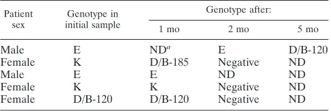
As shown in Table 3, three women and two men were found to beC. trachomatispositive at follow-up visits 1 to 5 months after treatment of their initial infections. Theomp1sequence of isolates obtained from the initial sample was compared to those obtained from the follow-up samples. The sequence analysis suggested that a reinfection had occurred in two of these five individuals. Thus, one woman was found to haveC.
trachomatis genotype K sequence in the initial endocervical
sample and a genotype D/B-185 sequence 1 month later. Sim-ilarly, in the initial urethral specimen from one man and also in the second sample (received 2 months later) a genotype E sequence was found, while the third urethral sample, 5 months after the initial sample, contained genotype D/B 120. The
remaining three individuals showed no change in genotype, which suggests either treatment failure or reinfection with the same genotype. Further on, one woman showed evidence of double infection, since she was found to haveC. trachomatis
genotype E in an endocervical sample and genotype F in the urine sample (Table 1).
The epidemiological information suggested that eight pa-tients had been infected abroad. One woman and one man, both infected with genotype E, were likely to have acquired their infections in Bulgaria. Like most other genotype E se-TABLE 1. omp1genotype distribution of 237 urogenital
C. trachomatisstrains isolated in O¨ rebro, Sweden, from March 1999 to February 2000
Genotype
No. of persons infected with genotype Men
(n⫽138) (Womenn⫽99) (nTotal⫽237) %a
Ba 1 0 1 0.4
D 9 3 12 5.1
D/B-120 8 6 14 5.9
D/B-185 3 3 6 2.5
E 71 41 112 47.3
F 24 17 41 17.3
G 2 4 6 2.5
H 3 3 6 2.5
Ia 4 3 7 3.0
J 6 4 10 4.2
K 7 14 21 8.9
Mixed (E⫹F) 0 1 1 0.4
aValues are percentages of total numbers ofC. trachomatis-positive specimens
(n⫽237).
FIG. 1. Phylogenetic tree showing the evolutionary relationship be-tween theC. trachomatisnucleotide sequences of theomp1gene from one representative of each genetic variant in the clinical material and 18 serovars of C. trachomatis available from GenBank. Sequences representingC. psittaci,C. pneumoniae, and a murine variant ofC. trachomatis(MoPn) were used as outgroups to root the tree. The tree was calculated by the maximum-likelihood method (transition/trans-version ratio, 1.4; global rearrangement option on) from an alignment of 1,032 aligned nucleotides. Relevant bootstrap values (as a percent-age of 1,000 replicates) are also given. Reference strains are shown in boldface text. The clinical strains (n⫽237) are illustrated with geno-type and number of mutations per number of samples (within paren-theses). Footnote letters: a, genotype D with one mutation at position 331 compared to D/IC-CAL-8; b, genotype D with one mutation at position 1092 compared to D/IC-CAL-8; c, genotype D/B120 with one mutation at position 975 compared to the strain X62918 (GenBank); d, genotype D/B120 with one mutation at position 1045 compared to the strain X62918 (GenBank); e, genotype E with one mutation at position 420 compared to E/DK20/ON; f, genotype E with one mutation at position 997 compared to E/DK20/ON.
on May 15, 2020 by guest
http://jcm.asm.org/
[image:3.587.302.538.74.413.2]quences these twoomp1gene sequences were identical to that of the reference strain. The other five patients infected abroad included one genotype Ia infection from the United States, one genotype F infection from Greece, one genotype D/B-120 in-fection from Austria, one genotype D inin-fection from Switzer-land, one genotype D infection from Norway, and one geno-type Ba infection from Thailand. Interestingly, this genogeno-type Ba infection was the only genotype Ba sample in the entire study.
DISCUSSION
In this study we have established a method for PCR ampli-fication and sequence analysis of theomp1gene ofC.
tracho-matisfrom clinical specimens. Ouromp1PCR worked
success-fully with both urogenital samples and urine samples and showed no cross-reactions withC. pneumoniaeorC. psittacior other false-positive reactions. All but 1 of 238 samples that were positive by the diagnosticChlamydiatests were positive in theomp1PCR. One reason for this false-negative result could be that theomp1gene is only present in one copy per organ-ism, whereas the plasmid that is targeted by the diagnostic tests is present in approximately 10 copies per organism (13, 18, 24). However, theomp1PCR was also positive in 2 of 24 diagnostic test negative samples from individuals epidemiologically highly suspected of having aC. trachomatisinfection. One of these two samples was positive after retesting by the diagnostic test,
perhaps due to the freeze-thawing, as recently described (3). The remaining sample, which was found positive in theomp1
PCR only, could contain a plasmid-free variant ofC.
tracho-matis(1).
C. trachomatis in these Swedish clinical isolates was most
frequently genotyped as E (47%) or F (17%). Also relatively prevalent wereomp1 genotypes D, D/B-120, J, and K (5 to 10%), whereas the remaining genotypes (Ba, D/B-185, G, H, and Ia) were more infrequently encountered. The genotype distribution in our 237 clinical specimens was broadly similar to the serovar distributions reported previously (15, 20). There was a high level of conservation of theomp1gene in infections caused by genotype E, of which 106 out of 112 had 100% similarity to the strain E/DK-20/ON. These findings are in agreement with those of Rodriguez et al. (22), who reported a high level of conservation of E strains from different geo-graphic origins. However, we found minor sequence variations within the different genotypes in our clinical material. Some of these genetic variants were detected only in a single sample, which makes it difficult to exclude the possibility that they represent sequencing artifacts, but most of them were inde-pendently detected in two or more samples, which strongly indicates that they were accurate. Thus, 4 of 10 clinical geno-type J strains were found to have one identical substitution, but at a different position than that described by Morre´ et al. (14). Similarly, five of six of genotype G sequences had identical substitutions that were different from the substitutions re-ported previously (14, 16), and all six genotype H sequences harbored two identical mutations. It has been speculated that suchomp1genovariants occur as a result of point mutations and recombination events selected by immune pressure (8). However, several of the nucleotide substitutions that we de-tected were synonymous (i.e., silent), which suggests that they were evolutionarily neutral.
The phylogenetic tree analysis of all the genotype sequences found indicates that there is no simple correlation between the disease manifestation and omp1 gene phylogeny. The omp1
[image:4.587.47.542.84.265.2]sequences of genotype H, I, J, and K, which were represented in our material from patients with symptomatic or subclinical TABLE 2. Mutations found in 237 clinical specimens compared to strains in GenBank
Genotype No.a No. of
mutations
Mutation Amino acid
change accession no.GenBank Nucleotide Position
D 1 (12) 1 G3A 331 R3H X62920
D 1 1 G3A 1092 A3T X62920
D/B-120 1 (14) 1 A3T 975 Silent X62918
D/B-120 2 1 T3C 1045 Silent X62918
E 4 (113) 1 A3G 420 Silent X52557
E 3 1 G3A 997 A3T X52557
F 1 (42) 1 A3T 1409 Silent X52080
J 4 (10) 1 C3T 369 Silent AF063202
K 3 (21) 1 C3T 132 Silent AF063204
H 6 (6) 2 A3G 440 D3S X16007
C3T 1018 Silent X16007
G 1 (6) 1 T3G 1003 S3A AF063199
G 5 4 T3A 228 Silent AF063199
G3A 487 G3S AF063199
G3C 700 E3Q AF063199
T3A 1003 S3T AF063199
aNumber of clinical strains with mutations. The values in parentheses are the total numbers of strains of the same genotype.
TABLE 3. Patients stillChlamydiapositive at follow-up visit after initial antibiotic treatment
Patient
sex initial sampleGenotype in
Genotype after:
1 mo 2 mo 5 mo
Male E NDa E D/B-120
Female K D/B-185 Negative ND
Male E E ND ND
Female K K Negative ND
Female D/B-120 D/B-120 Negative ND
aND, not done (diagnostic samples not obtained).
on May 15, 2020 by guest
http://jcm.asm.org/
[image:4.587.43.285.638.719.2]genital infections, were closely related to each other. However, they were also closely related to genotypes A and C, which are associated with trachoma, and genotype L3, which is associated with lymphogranuloma venerum. These observations are broadly the same as those reported by Stothard et al. when they comparedomp1sequences from clinical strains from the United States to sequences registered in GenBank (26). Few studies have addressed the possible correlation between spe-cific serovars or genotypes and disease manifestations and se-verity of disease. However, in a recent Finnish serological study it was stated that serovar G could be associated with subsequent development of cervical squamous cell carcinoma (2). This finding is interesting and requires further study, es-pecially due to the fact that some genotype G sequences in our study differ from the prototype sequence by three amino acids (Table 2).
Genotyping was also useful in the follow-up of the Chlamy-dia-infected patients, and five patients were stillC. trachomatis
positive at the follow-up visit. In two of these patients, the initial sample and follow-up sample showed different omp1
genotypes, which indicates that new sexual partners had rein-fected these patients. In the other three patients the genotypes were identical in the initial and follow-up samples, suggesting treatment failure or reinfection with the same genotype. Fur-thermore, one woman was found to have a mixed infection with two genotypes ofC. trachomatis, and she had had two sexual partners, each carrying one of these different genotypes. In conclusion, we have established a sensitive and relatively simple method for the genotyping ofC. trachomatisstrains in clinical samples based on sequencing of theomp1gene. Ge-notypes E and F dominated in our Swedish material, and the individual sequences were stable and showed limited variation. This omp1 genotyping method provided interesting results concerning double infections and reinfections and could be useful for epidemiological characterization of circulating C.
trachomatisstrains in the community.
ACKNOWLEDGMENTS
We thank the staff at the Outpatient Sexually Transmitted Disease Clinic and at the Department of Clinical Microbiology and Immunol-ogy for all effort and positive attitude shown during the study.
This work was supported by grants from the O¨ rebro Medical Re-search Foundation, O¨ rebro Medical Centre Hospital, and The Na-tional Institute for Public Health, Stockholm, Sweden.
REFERENCES
1.An, Q., G. Radcliffe, R. Vassallo, D. Buxton, W. J. O’Brien, D. A. Pelletier, W. G. Weisburg, J. D. Klinger, and D. M. Olive.1992. Infection with a plasmid-free variantChlamydiarelated toChlamydia trachomatisidentified by using multiple assays for nucleic acid detection. J. Clin. Microbiol.30:
2814–2821.
2.Antilla, T., P. Saikku, P. Koskela, A. Bloigu, J. Dillner, I. Ika¨heimo, E. Jellum, M. Lehtinen, P. Lenner, T. Hakulinen, A. Na¨rva¨nen, E. Pukkala, S. Thoresen, L. Youngman, and J. Paavonen.2001. Serotypes ofChlamydia trachomatisand risk for development of cervical squamous cell carcinoma. JAMA285:47–51.
3.Berg, E. S., G. Ånestad, H. Moi, G. Storvold, and K. Skaug.1997. False-negative results of a ligase chain reaction assay to detectChlamydia tracho-matisdue to inhibitors in urine. Eur. J. Clin. Microbiol. Infect. Dis.16:727– 731.
4.Cates, W. J., Jr., and J. N. Wassenheit.1991. Genital chlamydial infections: epidemiology and reproductive sequelae. Am. J. Obstet. Gynecol.164:1771– 1781.
5.Ceron, C., J. Dopazo, E. L. Zapata, J. M. Carazo, and O. Trelles.1998. Parallel implementation for DNAml program on message-passing
architec-tures. Parallel Comput.24:701–716.
6.Felsenstein, J. 1993. PHYLIP: phylogenetic inference package, version 3.52.c. University of Washington, Seattle.
7.Frost, E. H., S. Deslandes, S. Veilleux, and D. Bourgaux-Ramoisy.1991. TypingChlamydia trachomatisby detection of restriction fragment length polymorphism in the gene encoding the major outer membrane protein. J. Infect. Dis.163:1103–1107.
8.Hayes, L. J., P. Yearsley, J. D. Treharne, R. A. Ballard, G. H. Fehler, and M. E. Ward.1994. Evidence for naturally occurring recombination in the gene encoding the major outer membrane protein of lymphogranuloma venerum isolates ofChlamydia trachomatis.Infect. Immun.62:5659–5663. 9.Kaltenboeck, B., K. G. Kousoulas, and J. Storz.1992. Two-step polymerase
chain reactions and restriction endonuclease analyses detect and differenti-ateompADNA ofChlamydiaspp. J. Clin. Microbiol.30:1098–1104. 10.Kamwendo, F., L. Forslin, L. Bodin, and D. Danielsson.1998. Programmes
to reduce pelvic inflammatory disease—the Swedish experience. Lancet
351(Suppl. 3):25–28.
11.Lampe, M. F., K. G. Wong, and W. E. Stamm.1995. Sequence conservation in the major outer membrane protein gene amongChlamydia trachomatis strains isolated from the upper and lower urogenital tract. J. Infect. Dis.
172:589–592.
12.Lampe, M. F., R. J. Suchland, and W. E. Stamm.1993. Nucleotide sequence of the variable domains within the major outer membrane protein gene from serovariants ofChlamydia trachomatis. Infect. Immun.61:213–219. 13.Mahony, J. B., K. E. Luinstra, J. W. Sellors, and M. A. Chernesky.1993.
Comparison of plasmid- and chromosome-based polymerase chain reaction assays for detectingChlamydia trachomatisnucleic acids. J. Clin. Microbiol.
31:1753–1758.
14.Morre´, S. A., J. M. Ossewaarde, J. Lan, G. J. J. van Doornum, J. M. M. Walboomers, D. M. MacLaren, C. J. L. M. Meijier, and A. J. C. van den Brule.1998. Serotyping and genotyping of genitalChlamydia trachomatis isolates reveal variants of serovars Ba, G, and J as confirmed byomp1 nucleotide sequence analysis. J. Clin. Microbiol.36:345–351.
15.Morre´, S. A., R. Moesvan, I. Valkengoed, J. P. Boeke, J. T. M. van Eijk, C. J. L. Meijer, and A. J. C. van den Brule.1998. Genotyping ofChlamydia trachomatisin urine specimens will facilitate large epidemiological studies. J. Clin. Microbiol.36:3077–3078.
16.Norum Pedersen, L., O. H. Kjaer, J. Kjolseth Moller, T. Falck Orntoft, and L. Ostergaard.2000. High-resolution genotyping ofChlamydia trachomatis from recurrent urogenital infections. J. Clin. Microbiol.38:3068–3071. 17.Ostergaard, L.1999. Diagnosis of urogenitalChlamydia trachomatis
infec-tion by use of DNA amplificainfec-tion. APMIS107(Suppl. 89):5–36.
18.Palmer, L., and S. Falkow.1986. A common plasmid ofChlamydia tracho-matis. Plasmid16:52–62.
19.Peterson, E. M., B. A. Markhoff, and L. M. de la Maza.1990. The major outer membrane protein nucleotide sequence ofChlamydia trachomatis, serovar E. Nucleic Acids Res.18:3414.
20.Poole, E., and I. Lamont.1992.Chlamydia trachomatisserovar differentiation by direct sequence analyses of the variable segment 4 region of the major outer membrane protein gene. Infect. Immun.60:1089–1094.
21.Quinn, T C., C. Gaydos, M. Shepherd, L. Bobo, E. W. Hook III, R. Viscidi, and A. Rompalo.1996. Epidemiologic and microbiologic correlates of Chla-mydia trachomatisinfection in sexual partnerships. JAMA276:1737–1742. 22.Rodriguez, P., B. de Barbeyrac, K. Persson, B. Dutilh, and C. Bebear.1993.
Evaluation of molecular typing for epidemiological study ofChlamydia tra-chomatisgenital infections. J. Clin. Microbiol.31:2238–2240.
23.Sayada, C., E. Denamur, and J. Elion.1992. Complete sequence of the major outer membrane protein-encoding gene ofChlamydia trachomatisserovar Da. Gene120:129–130.
24.Sriprakash, K. S., and E. S. Macavoy.1987. Characterization and sequence of a plasmid from the trachoma biovar ofChlamydia trachomatis. Plasmid
18:205–214.
25.Stephens, R. S., R. Sanchez-Pescador, E. A. Wagar, C. Inouye, and M. S. Urdea.1987. Diversity ofChlamydia trachomatismajor outer membrane protein genes. J. Bacteriol.169:3879–3885.
26.Stothard, D. R., G. Boguslawski, and R. B. Jones.1998. Phylogenic analyses of theChlamydia trachomatismajor outer membrane protein and examina-tion of potential pathogenic determinants. Infect. Immun.66:3618–3625. 27.Swedish Institute for Infectious Disease Control. 2000. Annual report.
Swedish Institute for Infectious Disease Control, Stockholm, Sweden. 28.Walsh, S. P., D. A. Metzger, and R. Higuchi.1991. Chelex 100 as a medium
for simple extraction of DNA for PCR-based typing from forensic material. BioTechniques10:506–513.
29.Wang, S. P., C. C. Kuo, R. C. Barnes, R. S. Stephens, and J. T. Grayson.
1985. Immunotyping ofChlamydia trachomatiswith monoclonal antibodies. J. Infect. Dis.152:791–800.
30.Yuan, Y., Y. X. Zhang, N. G. Watkins, and H. D. Caldwell.1989. Nucleotide and deduced amino acid sequences for the four variable domains of the major outer membrane proteins of the 15Chlamydia trachomatisserovars. Infect. Immun.57:1040–1049.