C A S E R E P O R T
Open Access
Primary extramedullary plasmacytoma with
diffuse lymph node involvement: a case
report and review of the literature
Leonard Naymagon
1and Maher Abdul-Hay
2*Abstract
Background:Primary plasmacytomas are localized proliferations of clonal plasma cells occurring in the absence of a systemic plasma cell dyscrasia such as multiple myeloma. Primary plasmacytomas most commonly manifest as solitary lesions of the bone or of the upper aerodigestive tract. Presentation in a lymph node is very uncommon and can often be initially mistaken for lymphoma. Because they are local phenomena, primary plasmacytomas are managed with local therapies such as radiation or, less commonly, excision. Multifocal presentations are rare and are often not amenable to local treatment modalities, thus requiring systemic therapies. Because of their rarity, standardized treatment guidelines are not established, and treatment paradigms borrow heavily from those employed in multiple myeloma. Multifocal presentation in lymph nodes is nearly unheard of with only seven such cases reported in the existing literature, only four of which were diffuse enough to require systemic therapy. Here we describe the most diffuse and widely distributed instance of primary lymph node plasmacytoma yet reported and present a description of its successful treatment with systemic therapy.
Case presentation:A 71-year-old Asian man presented with progressive fatigue in the setting of diffuse hypermetabolic lymphadenopathy throughout his chest, abdomen, and pelvis. A diagnosis of lymphoma was initially suspected; however, a lymph node biopsy was consistent with plasmacytoma. A bone marrow biopsy was unremarkable, and no monoclonal protein was identified, establishing a diagnosis of primary extramedullary plasmacytomas of the lymph nodes. He was treated with a myeloma-like regimen consisting of four cycles of bortezomib/dexamethasone followed by two cycles of thalidomide/prednisone with improvement in symptoms and near complete resolution of prior hypermetabolic lymphadenopathy. He remains in remission over 18 months following completion of therapy.
Conclusion:This case report and accompanying literature review highlight the exceedingly rare and easily misclassified entity of primary plasmacytoma of diffuse lymph nodes. Importantly, we demonstrate that this entity may be treated with, and demonstrate excellent response to, systemic therapies often employed in multiple myeloma.
Keywords:Primary plasmacytoma, Plasma cell dyscrasia, Multiple myeloma, Lymph nodes, Bortezomib, Thalidomide
Introduction
Plasma cell neoplasms are characterized by the uncontrolled proliferation of malignant plasma cell clones. The most com-mon plasma cell malignancy is multiple myeloma (MM), which is characterized by a significant burden (> 10%) of clonal plasma cells within the bone marrow, monoclonal protein in serum and/or urine, and systemic symptoms (most commonly hypercalcemia, renal insufficiency, anemia,
and/or bone lesions) [1,2]. Plasmacytomas are clonal prolif-erations of plasma cells that are cytologically and immuno-phenotypically indistinguishable from MM; however, they demonstrate strictly local growth, without bone marrow or broader systemic involvement [3]. The most common mani-festation of primary plasmacytoma is as a single lesion of bone (solitary osseous plasmacytoma) [2]. Primary plasmacy-tomas not involving bone (extramedullary plasmacyplasmacy-tomas) typically manifest as lesions of the upper aerodigestive tract [4, 5]. Primary lymph node plasmacytomas are exceedingly rare with fewer than 40 cases having been described in the literature to date [6, 7]. The vast majority of lymph node
© The Author(s). 2019Open AccessThis article is distributed under the terms of the Creative Commons Attribution 4.0
International License (http://creativecommons.org/licenses/by/4.0/), which permits unrestricted use, distribution, and reproduction in any medium, provided you give appropriate credit to the original author(s) and the source, provide a link to the Creative Commons license, and indicate if changes were made. The Creative Commons Public Domain Dedication waiver (http://creativecommons.org/publicdomain/zero/1.0/) applies to the data made available in this article, unless otherwise stated.
* Correspondence:[email protected]
2New York University School of Medicine and New York University Perlmutter
plasmacytomas are solitary lesions, with only seven docu-mented cases involving multiple nodes.
Here we describe a highly atypical case of primary plasmacytoma with extensive lymph node involvement. This case demonstrates the most diffuse and widely distributed instance of primary plasmacytoma yet de-scribed, with enlarged hypermetabolic lymph nodes evi-dent throughout the neck, chest, abdomen, and pelvis. In fact, the dramatic lymphadenopathy at presentation initially prompted suspicion for lymphoma, and the eventual diagnosis of a plasma cell neoplasm was quite a surprise. Due to its remarkable dissemination this case could not be treated with the usual local modalities (namely radiation therapy) employed in the overwhelm-ing majority of patients with primary plasmacytoma. As such, systemic therapy had to be employed. As data for systemic treatment of primary plasmacytoma is minimal, we borrowed heavily from myeloma treatment para-digms. In fact, this is the first reported case describing the use of modern-style myeloma therapy for the treat-ment of diffuse primary plasmacytoma. The success of such therapy in this case demonstrates that diffuse pri-mary plasmacytomas not amenable to local therapies may be treated safely and effectively with drugs com-monly employed in MM, such as proteasome inhibitors and immunomodulatory agents.
Case presentation
Our patient is a 71-year-old Asian man with a history of rheumatoid arthritis (RA), type II diabetes, chronic kid-ney disease (CKD), and Hodgkin’s lymphoma, which was stage IV, diagnosed 6 years prior to presentation, status post six cycles of adriamycin/bleomycin/vinblastine/ dacarbazine (ABVD), with sustained complete remission; he presented with progressive fatigue and malaise of 2 months’duration. He was a former tobacco smoker with a 30 pack year history; however, he had quit smoking 20 years before presentation. He had no history of alcohol or illicit substance use. He was retired from a previous career as a farmer. He had no contributory family his-tory. His vital signs were within normal limits with temperature of 36.3 °C (97.3 °F), blood pressure of 128/ 78 mmHg, and a heart rate of 70 beats per minute. An examination was most notable for new cervical lymph-adenopathy. Other examination findings included finger deformities attributed to his RA and normal neurological examination. A laboratory evaluation was notable for pancytopenia with leukocyte count of 2.6 × 10⁹/L, plate-let count of 50 × 109/L, and hemoglobin of 11 mg/dL. His chemistry studies including calcium, creatinine, and liver function test were within normal limits. Positron emission tomography (PET) computed tomography (CT) demonstrated multiple enlarged hypermetabolic lymph nodes throughout his neck, chest, abdomen, and
pelvis, the largest measuring 2.2 cm × 1.2 cm. This prompted concern for recurrence of Hodgkin’s lymph-oma or de novo development of non-Hodgkin’s lymph-oma. A tissue biopsy of an enlarged hypermetabolic left inguinal lymph node was obtained, with pathology not-able for sheets of CD138+, MUM1+, MYD88-, and lambda restricted plasma cells, consistent with a plasma cell neoplasm. A bone marrow biopsy performed shortly afterward demonstrated largely unremarkable trilineage hematopoiesis without increased plasma cells, and with no monoclonal plasma cells detected. Our patient dem-onstrated normal renal function and electrolytes. Serum albumin, serum protein, and serum protein gap were within normal limits. His plasma and urine were nega-tive for monoclonal protein, with normal findings on serum protein electrophoresis (SPEP), urine protein electrophoresis (UPEP), serum immunofixation, serum free light chain assay, and urine free light chain assay. Based on the above data, a diagnosis of multiple diffuse extramedullary plasmacytomas of the lymph nodes was established. On diagnosis our patient was on metformin 1000 mg daily, Januvia (sitagliptin) 100 mg daily, glipizide 5 mg daily, Nexium (esomeprazole) 40 mg daily, Vesicare (solifenacin) 5 mg daily, folic acid 1 mg daily, nebivolol 10 mg daily, and tamsulosin 0.4 mg daily.
Treatment was promptly initiated with subcutaneously administered bortezomib 1.5 mg/m2 and orally adminis-tered dexamethasone 40 mg weekly for 3 weeks on and 1 week off. Our patient’s platelet count improved consider-ably following initiation of treatment, allowing for the addition of thalidomide. He received four cycles of bor-tezomib/dexamethasone followed by two continuous 28-day cycles of thalidomide 100 mg orally administered with prednisone. Thalidomide was chosen rather than lenalidomide due to our patient’s CKD and some persist-ent thrombocytopenia. His initial prespersist-enting symptoms improved throughout the course of therapy. An interval PET-CT was then done to assess response, and demon-strated near complete resolution of prior hypermetabolic lymphadenopathy with no new areas of fluorodeoxyglu-cose (FDG) avidity noted. He now remains in remission over 18 months following completion of therapy.
Discussion
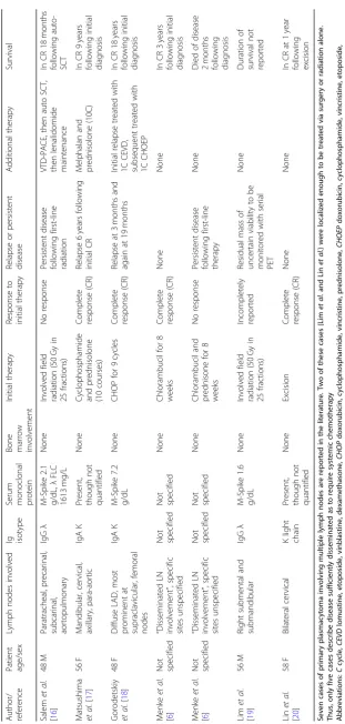
Table 1 Reported cases of primary plasmacytoma with multiple lymph node involvement Autho r/ referenc e Patient age/sex Lym ph nod es involved Ig isotype Serum monoc lonal protein Bone mar row involvement Initial therapy Re sponse to initial therapy Relap se or persistent disea se Add itional the rapy Survival Salem et al . [ 16 ] 48 M Para trache al, preca rinal, sub carinal, ao rtopulmonary IgG λ M-Spik e 2.1 g/dL, λ FLC 1613 mg /L None Involved field radiation (50 Gy in 25 fractions) No response Persi stent disea se followi ng first-line radiation VTD-P ACE, then auto SCT, then le nalidomide main tenanc e In CR 18 mont hs followi ng auto-SCT
Matsushima etal
[image:3.595.139.451.80.734.2]systemic treatments such as chemotherapy and/or im-mune therapy [11,12]. Given these radical differences in treatment, it is of utmost importance to unequivocally dis-tinguish between these two entities. In fact, a rigorous workup is required to definitively rule out systemic in-volvement and establish a diagnosis of primary plasmacy-toma [13–15].
The remarkably diffuse anatomic dissemination noted in this case is highly atypical of primary plasmacytoma and the primary diagnostic challenge lay in excluding MM. In fact, primary plasmacytomas of multiple lymph nodes is such a rare entity (only seven cases in the exist-ing literature, see Table1), that a diagnosis of underlying MM seemed likely [6,16–30]. Thus, great care and rigor had to be taken to definitely rule out underlying mye-loma, and establish with certainty a diagnosis of diffuse lymph node plasmacytoma [31–33]; to do so required demonstration of an uninvolved bone marrow, absence of monoclonal immunoglobulins or light chains (both serum and urine), and absence of any of the clinical sequels of myeloma [34,35]. It was only after frank mye-loma had been thoroughly and exhaustively ruled out as above that our patient could be deemed to have diffuse primary plasmacytoma of lymph nodes [36].
Once the above diagnostic challenge was settled, there then emerged the therapeutic challenge. Solitary primary plasmacytoma is the only curable plasma cell disorder, and the treatment of choice is radiation therapy [14]. Treatment paradigms are less well established for those with multiple primary plasmacytomas. Certainly, local radiation may be an option for those with relatively few lesions where radiation fields and cumulative dosage would not be prohibitive. However, those with many dif-fuse lesions cannot feasibly be treated with purely local modalities. Such cases must then be treated with sys-temic chemotherapy. Given the rarity of multiple diffuse primary plasmacytomas, there is lack of disease-specific data, either prospective or retrospective, to guide ther-apy. As such, treatment paradigms must borrow heavily from those established for MM.
As the disease in this case was far too widespread to benefit from standard treatments for localized plasmacy-toma (that is, radiation), our patient required systemic therapy. Due to the extreme paucity of data describing systemic therapy for primary plasmacytoma, treatment had to be planned largely without well-established prece-dent. Given the anatomically diffuse nature of the dis-ease, it was deemed prudent to pursue a myeloma-like regimen. Bortezomib and dexamethasone were started initially. Thalidomide was initially withheld to avoid thrombocytopenia, however, it was added following platelet response to initial treatment (as above, thalido-mide was chosen rather than lenalidothalido-mide due to our patient’s CKD and some persistent thrombocytopenia).
Interval PET-CT after four cycles of bortezomib/dexa-methasone followed by two cycles of thalidomide/pred-nisone demonstrated near complete resolution of prior hypermetabolic lymphadenopathy with no new areas of FDG avidity noted. Our patient remains in remission over 18 months following completion of therapy. Given this excellent response to initial therapy, and the absence of bone marrow involvement and monoclonal protein at diagnosis, his prognosis is felt to be relatively good, and the likelihood of progression to myeloma is felt to be relatively low [8–13].
Conclusion
Solitary plasmacytomas, which typically manifest in bone or the upper aerodigestive tract and demonstrate no con-current evidence of systemic myeloma, may be treated with purely local modalities such as involved field radi-ation. Multifocal plasmacytomas raise greater concern for concurrent occult myeloma; however, once systemic dis-ease is ruled out, they too may be treated with local mo-dalities if their extent and distribution are amenable. Multifocal primary plasmacytomas of lymph nodes are exceedingly rare and may be initially mistaken for lymph-oma. Although systemic myeloma should be diligently ruled out, treatment may require the use of myeloma-like therapeutic regimens if local modalities are not anatomic-ally feasible. Responses to myeloma-like regimens may be rapid, deep, and perhaps curative; however, a risk of future progression to systemic myeloma remains, particularly in cases with evidence of marrow involvement or monoclo-nal protein. It is not known why some plasma cell dyscra-sias such as MM manifest systemically while others such as primary plasmacytomas manifest locally. A better un-derstanding of the biologic mechanisms underlying these differences may potentially yield therapeutic insights for myeloma, as future treatments may aim to target those cellular elements most associated with diffuse or meta-static phenotypes.
Abbreviations
ABVD:Adriamycin/bleomycin/vinblastine/dacarbazine; CKD: Chronic kidney disease; CT: Computed tomography; FDG: Fluorodeoxyglucose; MM: Multiple myeloma; PET: Positron emission tomography; RA: Rheumatoid arthritis; SPEP: Serum protein electrophoresis; UPEP: Urine protein electrophoresis
Acknowledgements
We would like to thank the patient for allowing us to publish this case report.
Funding
No funding was received for this study.
Availability of data and materials
Authors’contributions
All authors (LN and MA-H) contributed equally and approved the final manuscript.
Ethics approval and consent to participate
No ethics committee approval is required at our institution for a case report involving a single patient. The patient consented to receiving all above therapies and all consents were documented in his medical record.
Consent for publication
Written informed consent was obtained from the patient for publication of this case report and any accompanying images. A copy of the written consent is available for review by the Editor-in-Chief of this journal.
Competing interests
The author Maher Abdul-Hay declares conflict of interest: Takeda honorarium and advisory board.
Publisher’s Note
Springer Nature remains neutral with regard to jurisdictional claims in published maps and institutional affiliations.
Author details
1New York University School of Medicine and Mount Sinai School of
Medicine, New York, USA.2New York University School of Medicine and New York University Perlmutter Cancer Center, 240 East 38th street, 19Floor, New York, NY 10016, USA.
Received: 14 August 2018 Accepted: 15 April 2019
References
1. Rajkumar SV, Kumar S. Multiple Myeloma: Diagnosis and Treatment. Mayo
Clin Proc. 2016;91(1):101–19.
2. Dores GM, Landgren O, McGlynn KA, Curtis RE, Linet MS, Devesa SS.
Plasmacytoma of bone, extramedullary plasmacytoma, and multiple myeloma: incidence and survival in the United States, 1992-2004. Br J
Haematol. 2009;144(1):86–94.
3. Tohda S. Overview of lymphoid neoplasms in the fourth edition of the
WHO classification. Rinsho Byori. 2012;60(6):560–4.
4. International Myeloma Working G. Criteria for the classification of
monoclonal gammopathies, multiple myeloma and related disorders: a report of the International Myeloma Working Group. Br J Haematol. 2003;
121(5):749–57.
5. Gerry D, Lentsch EJ. Epidemiologic evidence of superior outcomes for
extramedullary plasmacytoma of the head and neck. Otolaryngol Head
Neck Surg. 2013;148(6):974–81.
6. Menke DM, Horny HP, Griesser H,et al.Primary lymph node plasmacytomas
(plasmacytic lymphomas). Am J Clin Pathol. 2001;115(1):119–26.
7. Kilciksiz S, Karakoyun-Celik O, Agaoglu FY, Haydaroglu A. A review for
solitary plasmacytoma of bone and extramedullary plasmacytoma. ScientificWorldJ. 2012;2012:895765.
8. Rajkumar SV, Dimopoulos MA, Palumbo A,et al.International Myeloma
Working Group updated criteria for the diagnosis of multiple myeloma.
Lancet Oncol. 2014;15(12):e538–48.
9. Hughes M, Doig A, Soutar R. Solitary plasmacytoma and multiple myeloma:
adhesion molecule and chemokine receptor expression patterns. Br J
Haematol. 2007;137(5):486–7.
10. Soutar R, Lucraft H, Jackson G,et al.Guidelines on the diagnosis and
management of solitary plasmacytoma of bone and solitary extramedullary
plasmacytoma. Br J Haematol. 2004;124(6):717–26.
11. Rajkumar SV, Dispenzieri A, Kyle RA. Monoclonal gammopathy of
undetermined significance, Waldenstrom macroglobulinemia, AL amyloidosis, and related plasma cell disorders: diagnosis and treatment.
Mayo Clin Proc. 2006;81(5):693–703.
12. Kumar S, Fonseca R, Dispenzieri A,et al.Prognostic value of angiogenesis in
solitary bone plasmacytoma. Blood. 2003;101(5):1715–7.
13. Dimopoulos MA, Goldstein J, Fuller L, Delasalle K, Alexanian R. Curability of
solitary bone plasmacytoma. J Clin Oncol. 1992;10(4):587–90.
14. Creach KM, Foote RL, Neben-Wittich MA, Kyle RA. Radiotherapy for
extramedullary plasmacytoma of the head and neck. Int J Radiat Oncol Biol
Phys. 2009;73(3):789–94.
15. Dimopoulos MA, Moulopoulos LA, Maniatis A, Alexanian R. Solitary
plasmacytoma of bone and asymptomatic multiple myeloma. Blood. 2000;
96(6):2037–44.
16. Salem KZ, Nishihori T, Kharfan-Dabaja MA, Horna P, Alsina M. Primary
plasmacytoma involving mediastinal lymph nodes: A diagnostic mimicry of primary mediastinal lymphoma. Hematol Oncol Stem Cell Ther. 2016;9(1):
26–9.
17. Matsushima T, Murakami H, Tamura J, Sawamura M, Naruse T, Tsuchiya J.
Primary plasmacytoma of generalized lymph nodes: a long survivor. Am J
Hematol. 1993;43(3):237–9.
18. Gorodetskiy VR, Probatova NA, Vasilyev VI. Unusual course of generalized
lymph node primary plasmacytoma in a patient with Sjogren's syndrome: a case report. J Med Case Rep. 2017;11(1):116.
19. Lim YH, Park SK, Oh HS,et al.A case of primary plasmacytoma of lymph
nodes. Korean J Intern Med. 2005;20(2):183–6.
20. Lin BT, Weiss LM. Primary plasmacytoma of lymph nodes. Hum Pathol. 1997;
28(9):1083–90.
21. Addis BJ, Isaacson P, Billings JA. Plasmacytoma of lymph nodes. Cancer.
1980;46(2):340–6.
22. Andersen PE. Extramedullary plasmocytomas. Acta Radiol. 1949;32(5–6):365–74.
23. Gaston EA, Dollinger MR, Strong EW, Hajdu SI. Primary plasmacytoma of
lymph nodes. Lymphology. 1969;2(1):7–15.
24. Namikawa S, Hata H, Niinomi F, Yuasa H, Kubo K. Successfully treated case
of solitary extramedullary plasmacytoma occurring in the mediastinal lymph
node. Kyobu Geka. 1972;25(5):357–62.
25. Nelson MG, Lyons AR. Plasmacytoma of lymph glands. Cancer. 1957;10(6):
1275–80.
26. Ohmori T, Tabel R, Arita N,et al.Solitary plasmacytoma of the
submandibular lymph node with stromal amyloid deposits. Acta Pathol Jpn.
1983;33(5):999–1007.
27. Storch H, Puschel W, Raabe F, Schmidt F. Primary lymph node
plasmocytoma of IgD-class. Haematologia (Budap). 1986;19(3):219–25.
28. Tanimura A, Koziro M, Miyoshi T. Primary extramedullary plasmacytoma of
submaxillary lymph node. Acta Pathol Jpn. 1980;30(2):309–14.
29. Wiltshaw E. The natural history of extramedullary plasmacytoma and its
relation to solitary myeloma of bone and myelomatosis. Medicine
(Baltimore). 1976;55(3):217–38.
30. Woodruff RK, Whittle JM, Malpas JS. Solitary plasmacytoma. I: Extramedullary
soft tissue plasmacytoma. Cancer. 1979;43(6):2340–3.
31. Zhang YQJ, Qiu L. Multiple Solitary Plasmacytoma: Characteristics, Response
To Therapy, Survival In Nine Patients From a Single Institute. Blood. 2013;
122(21):5383. Accessed 08 Oct 2018. Retrieved fromhttp://www.
bloodjournal.org/content/122/21/5383
32. San-Miguel JF, Mateos MV. Can multiple myeloma become a curable
disease? Haematologica. 2011;96(9):1246–8.
33. Thumallapally N, Meshref A, Mousa M, Terjanian T. Solitary plasmacytoma:
population-based analysis of survival trends and effect of various treatment modalities in the USA. BMC Cancer. 2017;17(1):13.
34. Di Micco P, Di Micco B. Up-date on solitary plasmacytoma and its main
differences with multiple myeloma. Exp Oncol. 2005;27(1):7–12.
35. Rajkumar SV. Updated Diagnostic Criteria and Staging System for Multiple
Myeloma. Am Soc Clin Oncol Educ Book. 2016;35:e418–23.
36. Dimopoulos M, Kyle R, Fermand JP,et al.Consensus recommendations for
standard investigative workup: report of the International Myeloma