Determinants from Positive Blood Culture Broths by Use of the
Verigene Gram-Negative Blood Culture Multiplex Microarray-Based
Molecular Assay
Nathan A. Ledeboer,aBert K. Lopansri,bNeelam Dhiman,cRobert Cavagnolo,cKaren C. Carroll,dPaul Granato,e
Richard Thomson, Jr.,fSusan M. Butler-Wu,gHeather Berger,gLinoj Samuel,hPreeti Pancholi,iLettie Swyers,iGlen T. Hansen,j Nam K. Tran,kChristopher R. Polage,kKenneth S. Thomson,lNancy D. Hanson,lRichard Winegar,m Blake W. Buchana The Medical College of Wisconsin, Milwaukee, Wisconsin, USAa
; Intermountain Medical Center, Murray, Utah, USAb
; med fusion, Lewisville, Texas, USAc
; Johns Hopkins, Baltimore, Maryland, USAd
; Laboratory Alliance of Central New York, LLC Hospital, Fayetteville, New York, USAe
; NorthShore University Health System Evanston, Evanston, Illinois, USAf
; University of Washington, Seattle, Washington, USAg
; Henry Ford Hospital, Detroit, Michigan, USAh
; The Ohio State University Wexner Medical Center, Columbus, Ohio, USAi
; Hennepin County Medical Center, Minneapolis, Minnesota, USAj
; University of California-Davis Medical Center, Sacramento, California, USAk ; Creighton University School of Medicine, Omaha, Nebraska, USAl
; MRIGlobal, Kansas City, Missouri, USAm
Bloodstream infection is a serious condition associated with significant morbidity and mortality. The outcome of these infec-tions can be positively affected by the early implementation of effective antibiotic therapy based on the identification of the in-fecting organism and genetic markers associated with antibiotic resistance. In this study, we evaluated the microarray-based Verigene Gram-negative blood culture (BC-GN) assay in the identification of 8 genus or species targets and 6 genetic resistance determinants in positive blood culture broths. A total of 1,847 blood cultures containing Gram-negative organisms were tested using the BC-GN assay. This comprised 729 prospective fresh, 781 prospective or retrospective frozen, and 337 simulated cul-tures representing 7 types of aerobic culture media. The results were compared to those with standard bacterial culture and bio-chemical identification with nucleic acid sequence confirmation of the resistance determinants. Among monomicrobial cultures, the positive percent agreement (PPA) of the BC-GN assay with the reference method was as follows;Escherichia coli, 100%; Kleb-siella pneumoniae, 92.9%;Klebsiella oxytoca, 95.5%;Enterobacterspp., 99.3%;Pseudomonas aeruginosa, 98.9%;Proteusspp., 100%;Acinetobacterspp., 98.4%; andCitrobacterspp., 100%. All organism identification targets demonstrated>99.5% negative percent agreement (NPA) with the reference method. Of note, 25/26 cultures containingK. pneumoniaethat were reported as not detected by the BC-GN assay were subsequently identified asKlebsiella variicola. The PPA for identification of resistance determinants was as follows;blaCTX-M, 98.9%;blaKPC, 100%;blaNDM, 96.2%;blaOXA, 94.3%;blaVIM, 100%; andblaIMP, 100%. All resistance determinant targets demonstrated>99.9% NPA. Among polymicrobial specimens, the BC-GN assay correctly identi-fied at least one organism in 95.4% of the broths and correctly identiidenti-fied all organisms present in 54.5% of the broths. The sam-ple-to-result processing and automated reading of the detection microarray results enables results within 2 h of culture positivity.
B
loodstream infection (BSI) is a serious and life-threatening condition that has been associated with 25% to 80% mortality (1,2). The outcome of BSI can be dependent on host factors, such as underlying comorbidities, and microbiological factors, includ-ing the type of infectinclud-ing organism and its susceptibility to antibi-otics. It is estimated that up to 30% of hospital-acquired BSI are attributable to Gram-negative organisms (3). Infections caused by these bacteria, particularly when acquired in the hospital, have been associated with 15% to 29% increased crude mortality rates compared with those of the case controls (4,5). This is particularly true for infections with multidrug-resistant organisms, including those harboring extended-spectrum-lactamases (ESBLs) or car-bapenemases, which have been associated with prolonged hospital stay and increased 30-day mortality (6,7).Perhaps the most important intervention in the manage-ment of BSI is the early implemanage-mentation of effective antibiotic therapy (8,9). In one study, failure to administer effective an-tibiotics within 24 h of sepsis onset resulted in a two-day-lon-ger hospital stay, which was statistically significant, and was associated with a higher mortality rate (10). In a similar study,
ineffective antimicrobial therapy was associated with a statisti-cally significant 33% increase in mortality (8). Of note, both studies reported that up to one-third of patients initially re-ceived antibiotics that were ineffective against the infecting
Received3 March 2015Returned for modification2 April 2015 Accepted14 May 2015
Accepted manuscript posted online20 May 2015
CitationLedeboer NA, Lopansri BK, Dhiman N, Cavagnolo R, Carroll KC, Granato P, Thomson R, Jr, Butler-Wu SM, Berger H, Samuel L, Pancholi P, Swyers L, Hansen GT, Tran NK, Polage CR, Thomson KS, Hanson ND, Winegar R, Buchan BW. 2015. Identification of Gram-negative bacteria and genetic resistance determinants from positive blood culture broths by use of the Verigene Gram-negative blood culture multiplex microarray-based molecular assay. J Clin Microbiol 53:2460 –2472.doi:10.1128/JCM.00581-15.
Editor:P. Bourbeau
Address correspondence to Blake W. Buchan, [email protected].
Copyright © 2015, American Society for Microbiology. All Rights Reserved.
doi:10.1128/JCM.00581-15
on May 16, 2020 by guest
http://jcm.asm.org/
organism. These data underscore the importance of early or-ganism identification and, when possible, the detection of ge-netic markers associated with antibiotic resistance to aid in directing specific therapy for BSI. Further, early narrowing of broad-spectrum antibiotic treatment based on definitive or-ganism identification is a goal of many hospital stewardship programs and may further reduce the incidence of iatrogenic conditions, including Clostridium difficile-associated diarrhea and colonization with multidrug-resistant organisms (11–13).
Several approaches to the rapid detection of bacteria and ge-netic markers of resistance have been applied to positive blood culture broths. These include fluorescencein situhybridization (FISH), matrix-assisted laser desorption ionization–time of flight mass spectrometry (MALDI-TOF MS), and nucleic acid amplifi-cation or detection assays (14–23). While each approach has specific strengths and weaknesses, all have demonstrated their po-tential to positively impact key indicators, such as laboratory turn-around time, length of hospital stay, cost of care, and overall mor-tality associated with BSI. Of the available methods, only three tests are currently FDA cleared and capable of multiplex identifi-cation and differentiation of⬎4 individual targets in positive blood culture broths. These include the FilmArray blood culture identification (BCID) assay (BioFire Diagnostics, Salt Lake City, UT) and the Verigene Gram-positive blood culture (BC-GP) and Gram-negative blood culture (BC-GN) assays (Nanosphere, Northbrook, IL).
The FilmArray BCID is a sample-to-result assay for the analysis of positive blood culture broths. BCID relies on two-stage nested-PCR for identification of 8 Gram-positive, 11 Gram-negative, and 5 yeast targets in addition to themecA,vanA/B, andblaKPC resis-tance determinants on a single panel. Clinical evaluations of BCID have demonstrated an overall sensitivity of⬎97% for targets pres-ent on the panel and a specificity of 97% to 100% for individual targets (22, 23). In contrast, the Verigene BC-GP and BC-GN assays are nonamplified tests that rely on nucleic acid extraction from positive blood cultures, followed by microarray-based detec-tion using capture and detecdetec-tion probes. The BC-GP assay is spe-cific for 12 Gram-positive bacterial identification targets and 3 associated resistance markers (mecA,vanA, andvanB), while the BC-GN assay is specific for 8 Gram-negative bacterial identifica-tion targets and 6 key resistance markers (blaCTX-M, blaKPC, blaNDM,blaVIM,blaIMP, andblaOXA). Selection of the BC-GP or BC-GN assay is based on the primary Gram stain result of a pos-itive blood culture broth. Several studies have reported on the performance of the BC-GP assay, which has a sensitivity ranging from 96% to 100% and⬎98% specificity for the majority of tar-gets (18,21,24,25). The BC-GN assay has also been evaluated; however, studies to date have included⬍150 blood cultures or have included primarily simulated specimens (20,26–30).
Here, we evaluate the Verigene BC-GN assay for the identifi-cation ofEscherichia coli,Klebsiella pneumoniae,Klebsiella oxytoca, Enterobacter spp., Citrobacter spp., Proteus spp., Pseudomonas aeruginosa, andAcinetobacterspp. in positive blood culture broths containing Gram-negative bacilli. We also assess the ability of the BC-GN assay to detect 6 genetic markers associated with resis-tance to cephalosporins (blaCTX-M) and carbapenems (blaKPC, blaNDM,blaVIM,blaIMP, andblaOXA) in these organisms. This study was conducted at 13 clinical centers within the United States, from which a total of 1,847 blood cultures were evaluated. This includes both prospectively collected cultures and simulated cultures
seeded with less common clinical isolates or resistance genes to fully assess the performance of the BC-GN assay.
MATERIALS AND METHODS
Collection of blood cultures.Blood cultures were prospectively collected at 12 of the 13 clinical centers throughout the United States. Aerobic cultures using Bactec Plus Aerobic/F (852 [46.1%]) and Bactec Stan-dard/10 Aerobic/F (292 [15.8%]) (Becton Dickinson, Franklin Lakes, NJ), BacT/Alert FA FAN (357 [19.3%]), BacT/Alert SA (20 [1.1%]) and BacT/ Alert SN (17 [0.9%]) (bioMérieux, Marcy l’Etoile, France), and the Ver-saTREK Redox1 40 ml (122 [6.6%]) and VerVer-saTREK Redox1 80 ml (187 [10.1%]) (Thermo Fisher Scientific, Waltham, MA) broth culture bottles met the inclusion criteria and were included in the study. To qualify for study enrollment, prospectively collected cultures had to (i) be removed from the blood culture cabinet within 8 h of signaling positive, (ii) contain Gram-negative bacilli upon Gram stain, and (iii) be tested using the BC-GN assay within 12 h of initial positive detection by the automated blood culture instruments. Only one specimen per patient per septic ep-isode was enrolled to reduce redundancy in testing. Upon enrollment, two aliquots (2 ml each) of the blood culture broth were made in sterile plastic tubes. One tube was used for BC-GN testing (see below), and the other was immediately frozen at or below⫺70°C for use in repeat testing or discrepant analysis, as needed. The positive blood culture broth was also used to inoculate a Trypticase soy agar plate with 5% sheep blood (BAP) and a MacConkey agar plate (MAC). The BAP and MAC plates were incubated at 35°C in an atmosphere of 5% CO2for up to 48 h. The result-ing bacterial growth was used to make two heavy suspensions in Trypti-case soy broth with 20% glycerol (glycerol stocks). If multiple colony morphologies were observed, the isolated colonies were streaked for pu-rity prior to making the glycerol stocks. All glycerol stocks were stored at or below⫺70°C until they were shipped to a central reference laboratory for identification (see below). Retrospectively collected and simulated specimens were frozen within 8 h of signaling positive. Frozen specimens were allowed to thaw completely at room temperature and were vortexed and run within 2 h of thaw. All specimens (blood cultures) were deiden-tified prior to enrollment and tested in accordance with site-specific in-stitutional review board (IRB)-approved protocols.
Design of Verigene BC-GN assay capture probes.For each of the nucleic acid sequences detected by the BC-GN assay, at least two sets of oligonucleotides are required: (i) capture oligonucleotides (captures) and (ii) mediator oligonucleotides (mediators). Captures are printed onto the glass slide (microarray) and are designed to specifically bind to one region of the target sequence. Mediators bind specifically to a second region of the same sequence and are conjugated to nanoparticles utilized for target detection. The accession numbers of the reference sequences for each identification or resistance marker target were downloaded from the La-hey Clinic (http://www.lahey.org/Studies/other.asp) (Table 1). Full align-ments (not the consensus sequence) were analyzed to generate oligonu-cleotides from conserved regions. For the same start and end positions on the alignments, all possible variant oligonucleotides were generated. Sev-eral sets of oligonucleotides for each resistance target were tested empiri-cally, and the best (thermodynamic stability, assay compatibility, and tar-get specificity) was chosen to be used in the BC-GN test. For diverse markers, such asblaCTX-MandblaIMP, multiple capture and mediator
oligonucleotides were used to ensure inclusivity.
Verigene BC-GN testing.Specimens were tested on the Verigene sys-tem using the BC-GN assay within 12 h of culture positivity by the auto-mated blood culture monitoring system utilized by the study site. A BC-GN test cartridge, utility tray, and extraction tray were loaded into the Verigene ProcessorSP. A 700-l portion of the positive blood culture was transferred to the specimen well located in the BC-GN assay extraction tray, and the Verigene ProcessorSPwas started. Following automated nucleic acid extraction and hybridization to the glass array, the Verigene ProcessorSPwas opened, and the array was transferred to the Verigene Reader for analysis and automated reporting of qualitative results. The
on May 16, 2020 by guest
http://jcm.asm.org/
BC-GN assay contains an internal control for nucleic acid extraction and array hybridization. If a specimen test (i) yielded a result of (i) no call or (ii) preanalysis error, or (iii) no residual DNA was present after testing, the specimen was retested a single time within 24 h of the initial blood culture positivity. If a specimen could not be retested within 24 h, the specimen aliquot was frozen until the repeat test could be performed. At the con-clusion of each BC-GN test, the residual extracted nucleic acid was re-moved from the extraction tray to a microcentrifuge tube and stored frozen at or below⫺70°C for molecular characterization of the resistance determinants (see below). Removal of the residual nucleic acid was nec-essary only for this study protocol and is not performed by the end user in routine use.
Bacterial reference method testing.Identification of all bacterial iso-lates was carried out at a central reference laboratory (University of Mary-land, Baltimore, MD) using a standardized protocol. Upon receipt of the frozen strains, each isolate was plated onto Trypticase soy agar with 10% sheep blood and incubated at 35°C in an atmosphere of 5% CO2for up to
[image:3.585.41.544.76.492.2]72 h until sufficient bacterial growth was achieved for biochemical testing. Each isolate was initially identified using Gram stain and an oxidase test, followed by Vitek 2 (bioMérieux) and/or Phoenix (BD) automated identification systems to achieve a species-level identifica-tion. The final reference identification was based on the results of the Vitek 2 and/or Phoenix systems. The reference laboratory made a sec-ondary set of glycerol stocks from the inoculated BAP during the ref-erence testing process and stored them at or below⫺70°C for any additional testing or discrepant analysis. Resolution of discrepant re-sults between the BC-GN assay and reference method identification was conducted using bidirectional sequence analysis. Briefly, a PCR using primers external to those used in the BC-GN assay was per-formed, and the products were run on an agarose gel. Any PCR that yielded an amplification product (band) corresponding to the ex-pected size of the PCR target on the gel were submitted to ACGT, Inc. (Wheeling, IL) for sequencing. Differentiation ofKlebsiella variicola was based on PCR and sequence analysis of theyggEgene.
TABLE 1Identification of Gram-negative organisms in monomicrobial cultures using BC-GN assay
Target Specimen type
No. with resulta:
Total no.
% (95% CI) forb:
TP FP TN FN PPA NPA
E. colic Fresh 322d 4 377 0 703 100.0 (98–100) 99.0 (97–100)
Frozen 304 2 456 0 762 100.0 (98–100) 99.6 (98–100)
Simulated 49 0 285 0 334 100.0 (91–100) 100.0 (98–100)
Total 675 6 1,118 0 1,799 100.0 (99–100) 99.5 (99–100)
K. pneumoniae Fresh 103 0 588 12 703 89.6 (82–94) 100.0 (99–100)
Frozen 103 1 645 13 762 88.8 (81–94) 99.8 (99–100)
Simulated 133 0 200 1 334 99.3 (95–100) 100.0 (98–100)
Total 339 1 1,433 26 1,799 92.9 (90–95) 99.9 (99–100)
K oxytoca Fresh 21 6 675 1 703 95.5 (75–100) 99.1 (98–100)
Frozen 38 1 722 1 762 97.4 (85–100) 99.9 (99–100)
Simulated 4 0 329 1 334 80.0 (30–99) 100.0 (99–100)
Total 63 7 1,726 3 1,799 95.5 (86–99) 99.6 (99–100)
Enterobacterspp. Fresh 49 0 654 0 703 100.0 (91–100) 100.0 (99–100)
Frozen 61 6 694 1 762 98.4 (90–100) 99.1 (98–100)
Simulated 34 0 300 0 334 100.0 (87–100) 100.0 (98–100)
Total 144 6 1,918 1 1,799 99.3 (96–100) 99.7 (99–100)
P. aeruginosa Fresh 84 0 619 0 703 100.0 (95–100) 100.0 (99–100)
Frozen 51 1 708 2 762 96.2 (86–99) 99.9 (99–100)
Simulated 41 0 293 0 334 100.0 (89–100) 100.0 (98–100)
Total 176 1 1,620 2 1,799 98.9 (96–100) 99.9 (99–100)
Proteusspp. Fresh 25 0 678 0 703 100.0 (83–100) 100.0 (99–100)
Frozen 47 2 713 0 762 100.0 (91–100) 99.7 (99–100)
Simulated 3 0 331 0 334 100.0 (31–100) 100.0 (97–100)
Total 75 2 1,722 0 1,799 100.0 (94–100) 99.9 (99–100)
Acinetobacterspp. Fresh 13 0 690 0 703 100.0 (72–100) 100.0 (99–100)
Frozen 19 2 740 1 762 95.0 (73–100) 99.7 (99–100)
Simulated 28 0 306 0 334 100.0 (85–100) 100.0 (98–100)
Total 60 2 1,736 1 1,799 98.4 (90–100) 99.9 (99–100)
Citrobacterspp. Fresh 8 1 694 0 703 100.0 (60–100) 99.9 (99–100)
Frozen 18 0 744 0 762 100.0 (78–100) 100.0 (99–100)
Simulated 32 0 312 0 344 100.0 (87–100) 100.0 (98–100)
Total 58 1 1,750 0 1,799 100.0 (92–100) 99.9 (99–100)
a
TP, true positive, FP, false positive, TN, true negative, FN, false negative.
bPPA, positive percent agreement; NPA, negative percent agreement; 95% CI, 95% confidence interval.
c
IncludesShigellaspp., which cannot be differentiated by the BC-GN assay. dOne of 322 was identified asShigellasp. by culture.
on May 16, 2020 by guest
http://jcm.asm.org/
Construction of simulated specimens.Isolates containing rare tar-gets, such as those with specific resistance gene markers, were obtained as archived glycerol stocks from two clinical laboratories (International Health Management Associates, Schaumburg, IL, or JMI Laboratories, North Liberty, IA) or as lyophilized strains from the American Type Cul-ture Collection (ATCC). All strains were received at Nanosphere and re-vived onto BAP for the construction of simulated cultures. A suspension of each pure isolate was made in 0.65% saline and was diluted to approx-imately 10 to 100 CFU/ml. A 0.1-ml aliquot of the bacterial suspension (approximately 1 to 10 CFU) was used to inoculate Bactec Plus/F Aerobic medium bottles along with 8.0 ml of anticoagulated whole blood (Ten-nessee Blood Services, Memphis, TN) to simulate a clinically relevant specimen. The inoculated broths were incubated in the Bactec system until the bottles reached positivity. Each positive culture was assigned a unique identification number, divided into four 1- to 2-ml aliquots, and frozen within 8 h of signaling positive at⫺70°C. Specimens were shipped to the study testing sites on dry ice and were stored at⫺70°C until BC-GN testing was performed.
PCR amplification and bidirectional sequencing confirmation of re-sistance markers.Residual nucleic acid extracted from the blood culture specimens during BC-GN assay processing steps were removed from the designated well in the extraction tray consumable by manual pipette upon conclusion of the assay and were stored frozen at below⫺70°C. These extracts were submitted in a blinded fashion to the study internal testing laboratory (Nanosphere) for PCR amplification of 13 resistance marker targets (blaKPC, blaNDM,blaVIM,blaIMP, blaCTX-M-1group,blaCTX-M-2
group,blaCTX-M-8group,blaCTX-M-9group,blaCTX-M-25group,blaOXA-23
group,blaOXA-40group,blaOXA-48group, andblaOXA-58group). Primers
used for confirmation of the resistance markers were designed to target genetic sequences disparate from those used for detection of the markers by the BC-GN assay. Following endpoint PCR amplification, each tion mixture was electrophoresed on an agarose gel. Amplification reac-tions that resulted in a visible band of expected size and sufficient quantity were sent to a third-party sequencing laboratory (ACGT, Inc., Wheeling, IL) for bidirectional sequencing confirmation of specific resistance deter-minants. Sequencing templates were prepared by PCR amplification of the original nucleic acid extracts obtained from BC-GN assay sample pro-cessing, which had been stored at temperatures below⫺70°C. Primers used for presequencing target amplification were the same as those used for initial identification of resistance markers at the internal testing labo-ratory, with the exception ofblaKPC,blaVIM, andblaCTX-M-9.
Amplifica-tion of these three markers was accomplished using primers with an ad-ditional engineered universal M13 sequence, which was also used for subsequent sequencing reactions. The primers for each marker were de-signed to target conserved regions of the gene based on multiple sequence alignments of all available sequence entries for each target in GenBank at the time of design. The length of the amplicons was optimized to ensure that⬎200 bases with a Phred quality score ofⱖ30 would be generated for each sample during sequencing reactions.
All sequencing reactions were performed in a Bio-Rad Tetrad PCR system thermal cycler, ABI GeneAmp PCR system 9700 thermal cycler, or Bio-Rad C1000 PCR thermal system (Bio-Rad Laboratories, Inc., Hercu-les, CA). Each sequencing plate contained four negative-control reaction mixtures using the universal primer M13F(-21) and nuclease-free water substituting for DNA template, as well as four positive-control reaction mixtures using pGEM 3Zf(⫹) plasmid as the control template and M13F(-21) as a primer. The sequencing reaction products were purified with the CleanSEQ purification reagent (ACGT, Wheeling, IL) and were analyzed by the ABI 3730 XL genetic analyzer (Life Technologies, Carls-bad, CA). Each PCR product was sequenced twice from each direction to achieve a total of 4-fold coverage. For a valid sequencing result, specimens were required to haveⱖ200 consecutive bases sequenced with 2-fold cov-erage in double strands (4-fold covcov-erage overall) with a Phred quality score ofⱖ30. After Phred analysis, the sequence data were assembled by the Gene Codes Sequencher software to generate a single contig for each
PCR product. Only bases with a Phred quality score ofⱖ30 were used in the assembly.
Valid sequences were used to establish the identity of specific resis-tance markers in comparison to known reference sequences of each target downloaded from GenBank based on curations by the Lahey Clinic (http: //www.lahey.org/Studies/other.asp). The identity of the sequence was as-signed only if the sample sequence met the following criteria: (i) it containedⱖ200 consecutive high-quality bases (Phred score of 30 or greater), and (ii) it sharedⱖ95% sequence similarity to the reference strain acrossⱖ95% of the high-quality sequenced region. All analyzed specimens outperformed the minimum requirements and yielded be-tween 99.8% to 100% identity to the reference strains while containing ⱖ300 consecutive bases (i.e., Phred 30).
Statistical analysis.Positive percent agreement (PPA), negative per-cent agreement (NPA), and 95% confidence intervals were calculated us-ing standard formulas and the Newcombe method. Statistical significance was calculated using Fisher’s exact test and is presented as a probability value (Pvalue). Calculations were completed using the clinical calculator 1 application available athttp://vassarstats.net/clin1.html.
RESULTS
Study enrollment and prevalence of markers in prospectively collected blood culture broths.A total of 1,871 blood culture broths collected in 7 types of aerobic culture medium (see Mate-rials and Methods) met the study inclusion criteria and were eval-uated using the BC-GN assay. Twenty-four (1.3%) broth cultures were removed from analysis because of failure to generate a valid BC-GN result (e.g., internal control failure, high background sig-nal, etc.). The remaining 1,847 specimens comprised 729 (39.5%) fresh prospectively collected blood culture broths tested at the time of collection, 781 (42.3%) frozen specimens collected both prospectively and retrospectively, which were frozen and tested at a later date, and 337 (18.2%) simulated specimens that were pre-pared using characterized clinical isolates. The study cohort in-cluded 48 (2.6%) cultures found to be polymicrobial by reference culture, including 26/729 (3.6%) prospectively collected fresh cul-tures.
Among 703 monomicrobial fresh prospectively collected broth cultures, 645 (91.7%) contained an organism included on the BC-GN panel. The most common target by reference culture was E. coli(321 [45.7%]), followed byK. pneumoniae(115 [16.4%]),P. aeruginosa(84 [11.9%]),Enterobacterspp. (49 [7.0%]),Proteus spp. (25 [3.6%]),K. oxytoca(22 [3.1%]),Acinetobacterspp. (13 [1.8%]), andCitrobacterspp. (8 [1.1%]). The most common or-ganisms not present on the BC-GN panel includedSerratiaspp. (19 [2.7%]), non-aeruginosa Pseudomonas (10 [1.4%]), and Stenotrophomonas maltophilia(6 [0.9%]).
blaCTX-Mwas the most common genetic resistance determi-nant detected in the fresh prospectively collected cultures. Overall, blaCTX-Mwas identified in 46/703 (6.5%) fresh broths, including 39/321 (12.1%) broths containingE. coli. The remainingblaCTX-M -positive broths containedK. pneumoniae(4/103 [3.9%]),K. oxy-toca(1/21 [4.8%]),Proteussp. (1/25 [4.0%]), andEnterobactersp. (1/49 [2.0%]). Of the 46blaCTX-M identified, 38 (82.6%) were blaCTX-M-1type, and the remaining 8 wereblaCTX-M-9type. Genes encoding both types of enzymes were identified in a broth con-tainingK. pneumoniae; however, this culture was noted to contain two different colony morphologies and may have contained two different strains ofK. pneumoniae.
Only 2/703 (0.3%) fresh prospectively collected broths were identified as positive forblaKPC. Both were identified in cultures containing K. pneumoniae. One of two was also positive for
on May 16, 2020 by guest
http://jcm.asm.org/
blaCTX-M. A single fresh broth containingE. coliwas positive for blaNDM. This broth was also positive forblaCTX-M. None of the prospectively collected fresh broths containedblaVIMorblaIMP.
Identification of Gram-negative organisms in monomicro-bial cultures. The positive percent agreement (PPA) of the BC-GN assay with the reference method across fresh, frozen, and simulated blood culture broths was 100% forE. coli(675/675), Proteusspp. (75/75), andCitrobacterspecies (58/58) (Table 1). Of note, one broth called positive forE. coliby the BC-GN assay was further identified asShigellasp. by reference culture. This broth was still considered a correct identification by the BC-GN assay, because the capture probe cannot differentiateE. colifromShigella spp. and is considered a combinedE. coli/Shigellaspp. target by the assay. Although the BC-GN assay is capable of detecting 5 differ-ent species within the genusProteus, 73/75 cultures positive for Proteusspp. by the BC-GN assay were identified asProteus mira-bilis.
The BC-GN assay was falsely negative in 1 broth each contain-ingEnterobactersp. andAcinetobactersp., resulting in sensitivities of 99.3% (144/145) and 98.4% (60/61), respectively. In both in-stances, the single false-negative result was obtained from the fro-zen set of specimens. The BC-GN assay failed to detect any target in the broth that was falsely negative forAcinetobacterspecies (
Ta-ble 2). The identification of this isolate was confirmed to be
[image:5.585.41.543.78.471.2]Acin-etobacter baumanniiby both reference culture and MALDI-TOF MS. The broth identified as falsely negative forEnterobactersp. was called positive for a different target (K. pneumoniae) by the BC-GN assay, raising the possibility of an incorrect identification by the BC-GN assay or reference culture method. Upon sequence analysis, this isolate was identified asK. variicola. This indicates that the BC-GN assay was correct in not callingEnterobactersp. positive in this broth, raising the postdiscrepant resolution PPA to 100% forEnterobacterspecies. Two broths positive forP. aerugi-nosaby reference culture failed to be detected by the BC-GN assay,
TABLE 2Resolution of monomicrobial blood culture broths with discrepant results
Target
No. of broth cultures
Results with (no. of isolates): Initial call (no.
of broth cultures)c
Resolved call (no. of broth cultures)d
BC-GNa Culture Sequence analysisb
E. coli 1 E. coli⫹K. pneumoniae K. pneumoniae E. coli/Shigellasp. FP TP 1 E. coli⫹Enterobactersp. Enterobactersp. E. coli/Shigellasp. FP TP 1 E. coli K. oxytoca E. coli/Shigellasp. FP TP 1 E. coli⫹Proteussp. P. mirabilis ND FP ND 1 E. coli⫹K. oxytoca K. oxytoca E. coli/Shigellasp. FP TP
1 E. coli No growthe ND FP ND
K. pneumoniae 24 ND K. pneumoniae K. variicola FN (24) TN (24) 1 Enterobactersp. K. pneumoniae K. variicola FN TN 1 K. pneumoniae Enterobactersp. K. variicola FP FP
1 ND K. pneumoniae K. pneumoniae FN FN
K. oxytoca 1 E. coli K. oxytoca E. coli FN TN
1 K. pneumoniae⫹K. oxytoca K. pneumoniae K. pneumoniae FP FP 6 K. oxytoca R. planticola Raoultella planticola(1),
Raoultella ornithinolytica (4), ND (1)
FP (6) FP (5), ND
(1)
1 ND K. oxytoca K. variicola FN TN
1 ND K. oxytoca K. oxytoca FN FN
Enterobacterspp. 2 Enterobacterspp.⫹K. pneumoniae K. pneumoniae K. pneumoniae FP (2) FP (2) 1 Enterobactersp. K. pneumoniae K. variicola FP FP 2 Enterobacterspp. No growth ND (2) FP (2) ND (2) 1 K. pneumonia⫹Enterobactersp. K. pneumoniae K. pneumoniae FP FP 1 K. pneumoniae E. cloacae K. variicola FN TN
P. aeruginosa 2 ND P. aeruginosa P. aeruginosaf FN (2) FN (2)
1 P. aeruginosa No growth ND FP ND
Proteusspp. 2 Proteusspp. No growth ND (2) FP (2) ND (2)
Acinetobactersp. 1 Acinetobactersp. Alcaligenes faecalis ND FP ND 1 Acinetobactersp. Cupriavidus pauculus A. baumanniif FP TP
1 ND A. baumannii A. baumanniif FN FN
Citrobactersp. 1 Citrobactersp. Serratia marcescens S. marcescens FP FP
a
“⫹” indicates positivity for both species by the BC-GN assay.
bA slash indicates that the BC-GN assay cannot differentiate between the two species.
c
BC-GN result compared to reference culture.
dBC-GN result compared to bidirectional sequence analysis. ND, not determined, as specimen was unavailable for sequence analysis.
e
No growth, Gram-negative bacilli observed in primary Gram stain of blood culture. fIdentification confirmed by MALDI-TOF MS.
on May 16, 2020 by guest
http://jcm.asm.org/
resulting in a PPA of 98.9% (176/178). Both broths were frozen specimens, and the identity of these isolates was confirmed using MALDI-TOF MS. Three broths were initially positive forK. oxy-tocaupon reference culture but negative by the BC-GN assay. In two of three samples, the BC-GN assay was negative for all targets. The identity of one of these isolates was confirmed asK. oxytocaby sequence analysis; however, the other was identified asK. variicola and therefore was resolved as a true-negative result for the BC-GN assay. The third broth was called positive forE. coliby the BC-GN assay. Sequence analysis confirmed this identification, indicating an incorrect reference culture result. Taken together, these data raise the postdiscrepant resolution PPA to 98.4% (63/64) forK. oxytoca.
The largest number of initial false-negative results was ob-served in broth cultures containingK. pneumoniae. A total of 26 broths, including 12/115 (10.4%) fresh and 13/116 (11.2%) frozen withK. pneumoniaereported by reference culture, were not de-tected by the BC-GN assay (Table 1). This resulted in a PPA of as low as 89.6% forK. pneumoniaein fresh prospectively collected broths. However, 25/26 isolates obtained from these broths were identified asK. variicolaby sequence analysis (Table 2). While phenotypically similar toK. pneumoniae,K. variicolais a distinct species (see Discussion), which is not claimed as a target by the BC-GN assay. Therefore, the postdiscrepant resolution PPA of the BC-GN assay for broths containingK. pneumoniaeis 99.7% (364/ 365).
The negative percent agreement (NPA) of the BC-GN assay for all eight identification targets ranged from 99.5% to 99.9%. TheE. colitarget was detected in six broths that failed to yieldE. coliwhen cultured. Of interest, 4/6 of these broths contained a different organism that was recovered by reference culture and correctly called by the BC-GN assay (Table 2). The second organism was different for each of these cultures (K. pneumoniae,Enterobacter spp.,Proteusspp., andK. oxytoca), suggesting that the false-posi-tiveE. coliresult was unlikely the result of cross-reactivity of the capture probe used for these targets. Additionally, sequence anal-ysis of nucleic acid extracts from three of these cultures identified the presence ofE. colisequences. It is unclear whether this repre-sents the presence of nonviable organisms, free nucleic acid, or contamination of these specimens withE. colinucleic acid. One specimen called positive forE. coliby the BC-GN assay contained K. oxytocaby reference culture; however, sequence analysis con-firmed the identification of the isolate to beE. coli. The single most prevalent cause of false-positive results (n⫽6) was observed in broths called positive for K. oxytoca that subsequently yielded Raoultellaspp. upon reference culture (Table 2). Sequence analy-sis was conducted on nucleic acid extracts from 5 of 6 broths falsely identified as positive forK. oxytocaby the BC-GN assay and confirmed the reference culture identification ofRaoultella spe-cies.K. oxytocaandRaoultellaspp. are closely related both pheno-typically and genopheno-typically, and the observed false-positive calls are the result of cross-reactivity of theK. oxytocacapture probe used by the BC-GN assay withRaoultella species (see package insert).
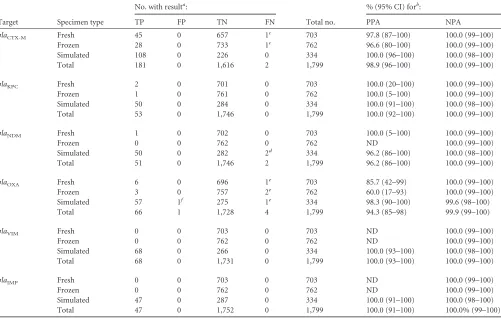
Identification of resistance markers in monomicrobial cul-tures.The prevalence of the 6 genetic resistance determinants detected by the BC-GN assay was low (0.0% to 0.6%) among 1,465 prospectively or retrospectively collected fresh and frozen speci-mens, with the exception ofblaCTX-M(5.0%). Therefore, to ade-quately assess the clinical performance of the BC-GN assay for the
identification of these targets, we relied on the use of simulated broths (see Materials and Methods).
The PPA of the BC-GN assay was 100.0% for the identification ofblaKPC(53/53),blaVIM(68/68), andblaIMP(47/47) in prospec-tive, retrospecprospec-tive, and simulated blood culture broths (Table 3). The majority of theblaKPC-positive results (46/53 [87%]) were called in broths containingK. pneumoniae, but there were also blaKPC-positive results in broths containingK. oxytoca(n⫽2),E. coli(2),Citrobactersp. (n⫽1),Enterobactersp. (n⫽1), andP. aeruginosa(n⫽1). Of interest, 8 specimens called positive for blaKPC were positive for a second resistance determinant (5 blaCTX-Mand 3blaVIM), andblaKPCwas correctly identified in two cultures in which the BC-GN assay failed to detect the organism recovered by reference culture (one each ofK. pneumoniaeandK. oxytoca). Eight differentblaIMPtypes (as determined by the refer-ence methods) were detected by the BC-GN assay in the 47 broths with positive results. Among these,blaIMP-4was most common (14/47 [29.8%]), followed byblaIMP-1 (9/47 [19.1%]),blaIMP-2 (8/47 [17.0%]),blaIMP-5andblaIMP-7(4/47 [8.5% each]),blaIMP-18 (3/47 [6.4%]),blaIMP-13andblaIMP-27 (2/47 [4.3% each]), and blaIMP-16(1/47 [2.1%]). Detection ofblaIMPwas most common in broths containing P. aeruginosa (15/47 [31.9%]); however, blaIMPwas detected in at least one broth containing each of the 8 bacteria identified by the BC-GN assay. ThreeblaVIMtypes were identified by reference methods in 68 broths positive for this marker. These included 53 broths containingblaVIM-1, 14 contain-ingblaVIM-2, and 1 containingblaVIM-17. The majority of these (28/68 [41.2%]) were identified in broths containingK. pneu-moniae; however,blaVIMwas detected in at least one broth con-taining each of the 8 bacteria identified by the BC-GN assay. Eight broths positive forblaVIMwere positive for a second genetic resis-tance marker, including 4blaCTX-M, 3blaKPC, and 1blaOXA-48.
The PPA of the BC-GN assay for identifying blaCTX-M was 98.9% (181/183) overall. Both false-negative results were in broths containingE. coliandblaCTX-M-1type enzymes by reference meth-ods. Among the positive results,blaCTX-M-1 type was the most prevalent (145/181 [80.1%]), followed byblaCTX-M-9type (26/181 [14.4%]). Among the 108 simulated cultures containingblaCTX-M, 88 (81.5%) contained a second genetic marker of resistance, again demonstrating the ability of the BC-GN assay to correctly identify multiple resistance markers in a single broth culture. TheblaCTX-M resistance determinant was detected primarily in cultures con-tainingE. coli; however, the marker was also prevalent in broths containingKlebsiellaspp. andEnterobacterspecies. TheblaCTX-M marker was not detected in any broth containingP. aeruginosa, Citrobacterspp., orAcinetobacterspp., so detection of this resis-tance determinant in these genera was not established. Of note, blaCTX-Mwas correctly identified in two broths containing Mor-ganellaspp., which is not a target identified by the BC-GN assay. While potentially useful, the detection of resistance determinants in the absence of a genus or species identification by the BC-GN assay is not a part of the FDA-clearedin vitrodiagnostic (IVD) assay.
The BC-GN assay correctly identifiedblaNDMin 51/53 broths containingblaNDMby reference methods, for a PPA of 96.2%. The two false-negative results were in broths containingE. coliandK. pneumoniae strains, both of which also harbored blaCTX-M-1. Among the 51 positive results, 26 (51.0%) were found in cultures containingK. pneumoniae, 13 (25.5%) in broths containingE. coli, 2 (3.9%) each in broths containingCitrobacterspp. and
on May 16, 2020 by guest
http://jcm.asm.org/
ellaspp., and 1 (2.0%) in a broth containingAcinetobacterspecies. The majority (35/51 [68.6%]) of the broths positive forblaNDM were also positive forblaCTX-M. There were no positiveblaNDM results in broths containingProteusspp.,K,oxytoca,P. aeruginosa, orProteusspecies.
Among the 8 genetic resistance determinants identified by the BC-GN assay, the detection ofblaOXAwas the least sensitive, at 94.3% (66/70). The 4 false-negative results were distributed across fresh (n⫽1), frozen (n⫽2), and simulated (n⫽1) specimen types. All were confirmed to beblaOXA-23type by discrepant se-quence analysis. Among the positive results,blaOXA-48type was most common (46/66 [69.7%]), with the remaining positive broths containing theblaOXA-23,blaOXA-40, orblaOXA-58types. A second resistance determinant was correctly identified in 60.6% (40/66) of the broths positive for blaOXA. Broths positive for blaOXAcontainedAcinetobacterspp. (n⫽20),Citrobactersp. (n⫽ 1),Klebsiellaspp. (n⫽23),Enterobacterspp. (n⫽3),Shewanella sp. (n⫽1),Serratiasp. (n⫽1), orMorganellaspecies (n⫽2).
Taken together, these data demonstrate the ability of the BC-GN assay to correctly identify six genetic resistance determi-nants, irrespective of the bacterium being present in the specimen or the presence of multiple resistance determinants in a single specimen. It is important to note that while the BC-GN assay is
capable of detecting several alleles of theblaCTX-M,blaVIM,blaIMP, blaOXA,blaKPC, andblaNDMtypes, it does not differentiate among the various subtypes.
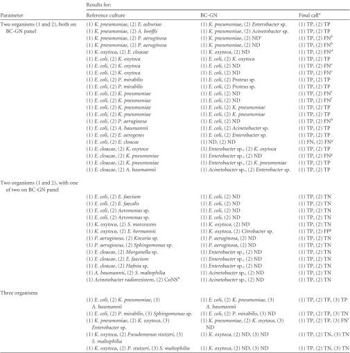
Polymicrobial cultures.A total of 48 broths analyzed by the BC-GN assay contained multiple organisms by reference culture. This included 26/729 (3.6%) fresh specimens, 19/781 (2.4%) fro-zen specimens, and 3/337 (0.9%) simulated cultures. Eight of these cultures contained Gram-negative organisms that are not part of the BC-GN panel, includingS. maltophilia,Serratiaspp., Achromobacterspp.,Sphingomonasspp.,Delftiaspp., andPantoea species. The BC-GN result was correctly reported as not detected for all targets in these cultures. Of the remaining 40 broths, 22 contained two organisms that were both targets of the BC-GN assay, 13 contained two organisms, of which only one was a target of the BC-GN assay, and 5 contained three organisms (Table 4).
The BC-GN assay correctly identified at least one organism in 21/22 (95.4%) of broths and correctly identified all organisms in 12/22 (54.5%) broths. Among broths with false-negative results, P. aeruginosawas not detected in 2 broths containingK. pneu-moniaeandP. aeruginosa. The BC-GN assay failed to detectK. oxytocain 2/3 broths containingE. coliandK. oxytocaand failed to detectK. pneumoniaein 2/4 broths containingE. coliandK. pneu-moniae(Table 4). The BC-GN assay also failed to detectP.
aerugi-TABLE 3Identification of genetic markers of resistance in monomicrobial cultures using BC-GN assay
Target Specimen type
No. with resulta:
Total no.
% (95% CI) forb:
TP FP TN FN PPA NPA
blaCTX-M Fresh 45 0 657 1
c 703 97.8 (87–100) 100.0 (99–100)
Frozen 28 0 733 1c 762 96.6 (80–100) 100.0 (99–100)
Simulated 108 0 226 0 334 100.0 (96–100) 100.0 (98–100)
Total 181 0 1,616 2 1,799 98.9 (96–100) 100.0 (99–100)
blaKPC Fresh 2 0 701 0 703 100.0 (20–100) 100.0 (99–100)
Frozen 1 0 761 0 762 100.0 (5–100) 100.0 (99–100)
Simulated 50 0 284 0 334 100.0 (91–100) 100.0 (98–100)
Total 53 0 1,746 0 1,799 100.0 (92–100) 100.0 (99–100)
blaNDM Fresh 1 0 702 0 703 100.0 (5–100) 100.0 (99–100)
Frozen 0 0 762 0 762 ND 100.0 (99–100)
Simulated 50 0 282 2d 334 96.2 (86–100) 100.0 (98–100)
Total 51 0 1,746 2 1,799 96.2 (86–100) 100.0 (99–100)
blaOXA Fresh 6 0 696 1e 703 85.7 (42–99) 100.0 (99–100)
Frozen 3 0 757 2e 762 60.0 (17–93) 100.0 (99–100)
Simulated 57 1f 275 1e 334 98.3 (90–100) 99.6 (98–100)
Total 66 1 1,728 4 1,799 94.3 (85–98) 99.9 (99–100)
blaVIM Fresh 0 0 703 0 703 ND 100.0 (99–100)
Frozen 0 0 762 0 762 ND 100.0 (99–100)
Simulated 68 0 266 0 334 100.0 (93–100) 100.0 (98–100)
Total 68 0 1,731 0 1,799 100.0 (93–100) 100.0 (99–100)
blaIMP Fresh 0 0 703 0 703 ND 100.0 (99–100)
Frozen 0 0 762 0 762 ND 100.0 (99–100)
Simulated 47 0 287 0 334 100.0 (91–100) 100.0 (98–100)
Total 47 0 1,752 0 1,799 100.0 (91–100) 100.0% (99–100)
a
TP, true positive, FP, false positive, TN, true negative, FN, false negative.
bPPA, positive percent agreement; NPA, negative percent agreement; 95% CI, 95% confidence interval; ND, not detected.
c
Confirmed to beblaCTX-M-1by sequence analysis.
dOne of two confirmed to bebla
NDMby sequence analysis. One was not available for discrepant analysis.
e
Confirmed to beblaOXA-23by sequence analysis.
fNegative forbla
OXAby sequence analysis.
on May 16, 2020 by guest
http://jcm.asm.org/
[image:7.585.43.544.78.396.2]TABLE 4Identification of Gram-negative organisms in polymicrobial cultures using BC-GN assay
Parameter
Results for:
Reference culture BC-GN Final calla
Two organisms (1 and 2), both on BC-GN panel
(1)K. pneumoniae, (2)E. asburiae (1)K. pneumoniae, (2)Enterobactersp. (1) TP, (2) TP (1)K. pneumoniae, (2)A. lwoffii (1)K. pneumoniae, (2)Acinetobactersp. (1) TP, (2) TP (1)K. pneumoniae, (2)P. aeruginosa (1)K. pneumoniae, (2) NDc (1) TP, (2) FNb (1)K. pneumoniae, (2)P. aeruginosa (1)K. pneumoniae, (2) ND (1) TP, (2) FNb (1)K. oxytoca, (2)E. cloacae (1)K. oxytoca, (2) ND (1) TP, (2) FNd (1)E. coli, (2)K. oxytoca (1)E. coli, (2)K. oxytoca (1) TP, (2) TP (1)E. coli, (2)K. oxytoca (1)E. coli, (2) ND (1) TP, (2) FNe (1)E. coli, (2)K. oxytoca (1)E. coli, (2) ND (1) TP, (2) FNe (1)E. coli, (2)P. mirabilis (1)E. coli, (2)Proteussp. (1) TP, (2) TP (1)E. coli, (2)P. mirabilis (1)E. coli, (2)Proteussp. (1) TP, (2) TP (1)E. coli, (2)K. pneumoniae (1)E. coli, (2) ND (1) TP, (2) FNf (1)E. coli, (2)K. pneumoniae (1)E. coli, (2) ND (1) TP, (2) FNf (1)E. coli, (2)K. pneumoniae (1)E. coli, (2)K. pneumoniae (1) TP, (2) TP (1)E. coli, (2)K. pneumoniae (1)E. coli, (2)K. pneumoniae (1) TP, (2) TP (1)E. coli, (2)P. aeruginosa (1)E. coli, (2) ND (1) TP, (2) FNb (1)E. coli, (2)A. baumannii (1)E. coli, (2)Acinetobactersp. (1) TP, (2) TP (1)E. coli, (2)E. aerogenes (1)E. coli, (2)Enterobactersp. (1) TP, (2) TP (1)E. coli, (2)E. cloacae (1) ND, (2) ND (1) FN, (2) FNg (1)E. cloacae, (2)K. oxytoca (1)Enterobactersp., (2)K. oxytoca (1) TP, (2) TP (1)E. cloacae, (2)K. pneumoniae (1)Enterobactersp., (2) ND (1) TP, (2) FNg (1)E. cloacae, (2)K. pneumoniae (1)Enterobactersp., (2)K. pneumoniae (1) TP, (2) TP (1)E. cloacae, (2)A. baumannii (1)Acinetobactersp., (2)Enterobactersp. (1) TP, (2) TP
Two organisms (1 and 2), with one of two on BC-GN panel
(1)E. coli, (2)E. faecium (1)E. coli, (2) ND (1) TP, (2) TN (1)E. coli, (2)E. faecalis (1)E. coli, (2) ND (1) TP, (2) TN (1)E. coli, (2)Aeromonassp. (1)E. coli, (2) ND (1) TP, (2) TN (1)E. coli, (2)Aeromonassp. (1)E. coli, (2) ND (1) TP, (2) TN (1)K. oxytoca, (2)S. marcescens (1)K. oxytoca, (2) ND (1) TP, (2) TN (1)K. oxytoca, (2)E. hermannii (1)K. oxytoca, (2)Citrobactersp. (1) TP, (2) FPg (1)P. aeruginosa, (2)Kocuriasp. (1)P. aeruginosa, (2) ND (1) TP, (2) TN (1)P. aeruginosa, (2)Sphingomonassp. (1)P. aeruginosa, (2) ND (1) TP, (2) TN (1)E. cloacae, (2)Morganellasp. (1)Enterobactersp., (2) ND (1) TP, (2) TN (1)E. cloacae, (2)E. faecium (1)Enterobactersp., (2) ND (1) TP, (2) TN (1)E. cloacae, (2)Hafniasp. (1)Enterobactersp., (2) ND (1) TP, (2) TN (1)A. baumannii, (2)S. maltophilia (1)Acinetobactersp., (2) ND (1) TP, (2) TN (1)Acinetobacter radioresistens, (2) CoNSh (1)Acinetobactersp., (2) ND (1) TP, (2) TN
Three organisms
(1)E. coli, (2)K. pneumoniae, (3) A. baumannii
(1)E. coli, (2)K. pneumoniae, (3) A. baumannii
(1) TP, (2) TP, (3) TP
(1)E. coli, (2)P. mirabilis, (3)Sphingomonassp. (1)E. coli, (2)P. mirabilis, (3) ND (1) TP, (2) TP, (3) TN (1)K. pneumoniae, (2)K. oxytoca, (3)
Enterobactersp.
(1)K. pneumoniae, (2)K. oxytoca, (3) ND
(1) TP, (2) TP, (3) FNi
(1)K. oxytoca, (2)Pseudomonas stutzeri, (3) S. maltophilia
(1)K. oxytoca, (2) ND, (3) ND (1) TP, (2) TN, (3) TN
(1)K. oxytoca, (2)P. stutzeri, (3)S. maltophilia (1)K. oxytoca, (2) ND, (3) ND (1) TP, (2) TN, (3) TN
aFinal call versus reference culture result. TP, true positive; TN, true negative; FP, false positive; FN, false negative.
b
P. aeruginosaconfirmed by sequence analysis. cND, not detected.
d
Identification of the organisms in this specimen could not be resolved by sequence analysis. eK. oxytocaconfirmed by sequence analysis.
f
K. pneumoniaeconfirmed by sequence analysis.
gSpecimens (15131, 17155, and 17111) were not available for sequence analysis.
h
CoNS, coagulase-negativeStaphylococcusspecies.
iSequence analysis failed to identifyK. oxytocain the specimen.
on May 16, 2020 by guest
http://jcm.asm.org/
nosa,Enterobacter cloacae, andK. pneumoniaein additional cul-tures positive for another target. A single culture was falsely negative for both targets recovered by reference culture (E. coli andE. cloacae).
In all 13 cultures in which one of two organisms was a target of the BC-GN assay, the target organism was correctly identified, for a PPA of 100% (Table 4). In one culture containingK. oxytocaand Escherichia hermannii, the BC-GN assay reportedK. oxytocaand Citrobacterspecies. In one of two broths containing 3 organisms present on the BC-GN panel, all three targets were correctly iden-tified (E. coli,K. pneumoniae, andA. baumannii).
DISCUSSION
The ability to rapidly identify Gram-negative organisms from sub-culture or directly from positive blood sub-culture broths has proven beneficial to patient outcomes, antimicrobial stewardship, and overall cost of care for patients with these infections (15,19,20). For example, the use of MALDI-TOF MS to rapidly identify Gram-negative organisms from subculture contributed to a 51-h reduction in the time to optimal therapy and a 17% reduction in 30-day mortality in patients with Gram-negative BSI (15). Impor-tantly, 61.4% of the antibiotic modifications were based on organ-ism identification alone. In another study, direct analysis of posi-tive blood culture broths using MALDI-TOF MS combined with a rapid susceptibility testing method reduced the length of hospital stay by 2.6 days and reduced the total cost of care by $19,000 (19). An early evaluation of the BC-GN assay estimated that the detec-tion ofblaKPCdirectly from positive blood cultures could have reduced the time to most appropriate therapy by approximately 14 h in 56% of patients infected withK. pneumoniae carbapen-emase (KPC)-producingK. pneumoniae(20). When combined with an active antimicrobial stewardship intervention, these out-come studies demonstrate the potential value of rapid identifica-tion of organisms and genetic resistance markers among blood cultures containing Gram-negative bacteria.
A number of studies have already evaluated the BC-GN assay and demonstrated a sensitivity and specificity of 81.0% to 100% and 98.0% to 100%, respectively, for identifying individual bacte-rial and resistance markers (20,27–30). However, these studies were limited by a small sample size (31 to 125 cultures) (20,26,
29), a large proportion of simulated specimens (63% to 74% of evaluated cultures) (29,30), or a potential lack of strain diversity due to the single center-based design of the study (20,27,30). The present study describes the most comprehensive evaluation of the BC-GN assay to date. In addition to the large total number of specimens evaluated (n ⫽1,847), seven types of blood culture media representing the three primary manufacturers of blood cul-ture systems were included. The NPA for specimens tested using all blood culture medium types included in this study was ⬎99.8%, and no statistical difference in NPA was found between any of the blood culture types included in this study (P⫽0.736, Fisher’s exact test). The difference in PPA between specimens tested using the various blood culture media ranged from 94.3% (Bactec Standard/10 Aerobic/F) to 100% (BacT/Alert SA). This difference was statically significant (P⫽0.030, Fisher’s exact test); however, it is important to note that BacT/Alert SA accounted for only 20 (1.1%) of the blood cultures tested, while Bactec Stan-dard/10 Aerobic/F accounted for 292 (15.8%) of the cultures. The low number of total cultures tested using BacT/Alert SA might have impacted the calculation of statistical significance between
these two medium types. The sample size, diversity of culture me-dia, and inclusion of 12 geographically distinct clinical centers within the United States are specific strengths of this study.
The overall PPA of the BC-GN assay for eight bacterial identi-fication targets was 97.9% (1,590/1,623). The PPA of the BC-GN assay was highest in fresh prospectively tested cultures, including 100% PPA for 6/8 genus or species identification targets on the panel. The majority of the false-negative results (6/7, excludingK. pneumoniae/K. variicola, as discussed below) were obtained when testing cultures that had been frozen prior to analysis. The insta-bility of the nucleic acid targets resulting from freezing and thaw-ing of these samples may have contributed to the false-negative results, although this hypothesis was not tested.
The BC-GN assay contains capture probe targets that enabled an identification of the organism present in approximately 90% of the prospectively collected blood cultures containing Gram-neg-ative bacilli. Among targets not present on the BC-GN assay, the most commonly encountered wasK. variicola(12/703 [1.7%] of prospective cultures).K. variicolais phenotypically similar toK. pneumoniaeand is easily misidentified as K. pneumoniaewhen using automated phenotypic identification methods (31) and MALDI-TOF MS (our unpublished data). This may lead to per-ceived false-negative BC-GN results in cultures containingK. va-riicolathat are reported as not detected by the BC-GN assay. Among prospectively collected cultures phenotypically identified asK. pneumoniae, 10.4% (range, 0% to 25% among clinical cen-ters in this study) were subsequently identified asK. variicolaby sequence analysis. Although initially associated with environmen-tal sources and plant material (32),K. variicolamay comprise up to 10% of allKlebsiellaspecies isolates obtained from clinical spec-imens (31,33,34). One report details a fatal case of sepsis, which was initially attributed toK. pneumoniaebut was later resolved as K. variicola(31). Literature exploring the relative virulence or sus-ceptibility patterns ofK. variicolais sparse, but a larger proportion of isolates ofK. variicolamay be susceptible to piperacillin and cephalosporins thanK. pneumoniae(34). Given these data, labo-ratories that identifyK. pneumoniaein blood cultures resulted as not detected by the BC-GN assay may be advised to confirm the identity with additional sequence analysis or definitive biochem-ical tests prior to reporting.
In addition toK. variicola, a potential weakness of the BC-GN assay is the omission of some other relatively common Gram-negative pathogens from the panel, including Serratia spp., S. maltophilia, and non-aeruginosa Pseudomonas, which combined made up 4.4% (31/703) of the positive cultures. The BC-GN assay also lacks targets for detectingNeisseria meningitidisand Haemo-philus influenzae. While the prevalence of these organisms is rela-tively rare (neither was isolated in 703 prospecrela-tively collected cul-tures in this study), their presence in blood culture is indicative of serious life-threatening or invasive infection.
A specific advantage of the BC-GN assay versus other rapid identification methods (e.g., MALDI-TOF MS and peptide nu-cleic acid-FISH [PNA-FISH]) is the ability to identify 6 genetic markers associated with resistance to various classes of-lactam antibiotics. Gram-negative bacteria harboring extended-spec-trum-lactamases (ESBLs) or carbapenemases are of concern be-cause of the limited therapeutic options available to effectively treat infections with these organisms (35). Specifically, bacteria harboring carbapenemases are resistant to many, if not all, classes of-lactams and are often resistant to several other classes of
on May 16, 2020 by guest
http://jcm.asm.org/
antibiotics. This contributes to an increase in morbidity and mor-tality in patients infected with these organisms (6). While the prevalence of carbapenemases in prospectively collected cultures was low in this study (KPC, 2/703 [0.3%]; NDM, 1/703 [0.1%]), larger surveillance efforts have indicated a steady rise in the prev-alence of KPC both in the United States and abroad since its initial characterization in 1996 (36–38). Carbapenem-resistant Entero-bacteriaceae(CRE) currently account for⬎9,000 serious infec-tions per year in the United States and have been reported in 44 states, including New York, Pennsylvania, and New Jersey, where isolates producingblaKPCare considered endemic (36,37,39). The increasing prevalence and a lack of effective therapeutic op-tions to treat infecop-tions caused by CRE have made early detection and implementation of infection control measures a key aspect in the management of these patients (6,40).
Current phenotypic methods for detecting CRE, including the modified Hodge test (MHT), have a reported sensitivity of 93% to 100% for class A (e.g.,blaKPC) and class D (e.g.,blaOXA) carbap-enemases; however, subjective interpretation of the test and low-level carbapenemase activity of other-lactamases result in spec-ificity of only 39% to 76% (41,42). Further, the sensitivity of the MHT may be as little as 27% to 50% for detecting class BblaNDM enzymes (42,43). The Carba NP assay is a newer phenotypic test based on detection of in vitro hydrolysis of imipenem. This method appears to be as sensitive as the MHT for detecting strains harboringblaKPCand also demonstrates 94% to 100% sensitivity for detectingblaNDMwhile maintaining⬎99% specificity (44,45). Unfortunately, the Carba NP assay demonstrated reduced sensi-tivity for other carbapenemases, including only 60.6% sensisensi-tivity for detecting class DblaOXA-48(44,46). In addition, both MHT and the Carba NP assay require the isolation of organisms from positive blood culture broths prior to phenotypic testing. Com-bined with assay setup and incubation, the total time to result for these methods can be 48 to 72 h. The molecular detection of blaKPC, blaNDM, andblaOXAusing the BC-GN assay is advanta-geous in that the PPA (94.3% to 100%) and NPA (99.9% to 100%) for each of these targets was high, and results are not based on a subjective interpretation of phenotypic characteristics. Addition-ally, the ability to detect these resistance markers directly from positive blood cultures within 2 h greatly reduces the time to re-porting of positive results compared with that with phenotypic methods. This has the potential to positively impact both the se-lection of appropriate antimicrobial therapy and implementation of infection control measures for patients testing positive for one of these targets (20).
In addition to identifying genes encoding carbapenemase en-zymes, the BC-GN assay also identifiesblaCTX-M, the most preva-lent ESBL worldwide (47). ESBL enzymes, including blaSHV, blaTEM, and blaCTX-M, are broad-spectrum enzymes capable of hydrolyzing penicillins, cephalosporins, and aztreonam but re-main susceptible to carbapenems. ESBL activity in blaSHVand blaTEMarises from point mutations in narrow spectrum “parent” enzymes, which makes the differentiation of narrow- versus ex-panded-spectrum -lactamases difficult without full-sequence analysis (48,49). In contrast, allblaCTX-Menzymes have native ESBL activity (49), which allows comparatively easy molecular identification of these ESBLs using nucleic acid probes. Accurate identification of isolates harboring ESBLs is of clinical importance because of the poor correlation betweenin vitrosusceptibility and
the clinical efficacy of cephalosporins for the treatment of infec-tions caused by ESBL-producing bacteria (50,51).
Phenotypic screening methods to identify strains carrying ESBLs were recommended by the Clinical and Laboratory Standards Institute (CLSI); however, the presence of AmpC enzymes in members of theEnterobacteriaceaefamily can lead to false-positive screen results or obscure the activity of ESBL enzymes during confirmatory disk diffusion tests (52–54). In response to these limitations, the CLSI elected to lower the susceptibility break-points for several cephalosporins in an attempt to eliminate the need for phenotypic ESBL tests (55). Despite the lowered break-points, strains harboringblaCTX-MESBLs may still test as suscep-tible to ceftazidime, cefepime, and aztreonam (56). Specifically, 14% to 45% ofE. coliisolates and 85% to 96% ofP. mirabilis isolates harboringblaCTX-Mtested as susceptible to these antibiot-ics based on the lower breakpoints (56). This potential undercall-ing of resistance was especially pronounced in isolates harborundercall-ing CTX-M-9 enzymes (56). Limitations in ESBL detection exist for both phenotypic and genetic approaches, and no one-size-fits-all approach has emerged. The rapid detection of key prevalent resis-tance factors that are detected using the BC-GN system provide a front-line approach in the early identification of emerging resis-tance, which is likely to positively impact patient care. The BC-GN assay demonstrated 98.9% PPA and 100% NPA for identification ofblaCTX-Mand is inclusive of⬎100 individual types in the sub-groups M-1, M-2, M-8, M-9, and CTX-M-25 (see package insert). Combined, the rapid time to result and accuracy of detectingblaCTX-Mhas the potential to aid in earlier modification of empirical therapy and reduce the time to imple-mentation of infection control practices.
Despite the potential benefits of molecular detection of cepha-losporinases and carbapenemases, it is important to recognize that resistance may be mediated by alternative mechanisms, including other classes of-lactamases, active drug efflux pumps, alterna-tive or modified drug targets, and point mutations that reduce membrane permeability. These mechanisms are especially com-mon in some species ofEnterobacteriaceae(Serratiaspp.,P. vul-garis,Citrobacterspp., andEnterobacterspp.) and genera outside the familyEnterobacteriaceae, includingP. aeruginosaand Acin-etobacterspecies. These factors resulted in a negative predictive value of only 78.6% for carbapenem resistance inP. aeruginosain one evaluation of the BC-GN assay (20). Therefore, the absence of detection of any of the specific genetic markers by the BC-GN assay cannot be interpreted as an indication of a susceptible iso-late. Laboratories, in conjunction with pharmacy and antimicro-bial stewardship teams, may decide to tailor reporting structures within their individual institutions. This may include the addition of interpretive statements with each positive report that aid in guiding antimicrobial therapy choices. For example, a positive report of CTX-M may include a statement indicating the detec-tion of an ESBL and advise against the use of penicillin, cephalo-sporin, or aztreonam antibiotics until full phenotypic susceptibil-ity results are available. All cultures should include a statement acknowledging that a result of not detected for antimicrobial re-sistance markers does not indicate susceptibility.
Polymicrobial cultures compose 6 to 12% of all positive blood cultures and present a particular challenge for any technology uti-lized for direct analysis of these specimens (18,22,24,57). The BC-GN assay correctly identified at least one organism present in ⬎95% of the polymicrobial cultures tested; however, a correct