Bmp4-directed nuclear cyan fluorescent protein provides
a tool for live imaging and reveals cellular resolution
of Bmp4 expression patterns during embryogenesis
CHUAN-WEI JANG
1, LIANG GAO
2,4, MARY E. DICKINSON
1,2,4and RICHARD R. BEHRINGER*
,1,3 1Program in Developmental Biology, 2Department of Molecular Physiology and Biophysics, Baylor Collegeof Medicine, 3Department of Genetics, University of Texas M.D. Anderson Cancer Center and 4Department of Bioengineering, Rice University, Houston, Texas, USA
ABSTRACT The genesis of hair follicles in mammals involves multiple inductive and suppressive signaling interactions between epithelial cells and the underlying mesenchymal tissue. Bmp4 (Bone Morphogenetic Protein 4) is expressed in the mesenchymal tissue surrounding developing hair follicles. The BMP signaling pathway is suggested to play a suppressive role in hair follicle induction. In addition, it is also found to be important for specification of different cell types in mature hair follicles. Knowledge of the precise expression pattern of Bmp4 during hair follicle differentiation should provide insights into how these suppressive and differentiative roles regulate hair follicle development. However, in situ hybridization studies do not provide sufficient cellular resolution, and three-dimensional reconstructions of serial sections are tedious. We have targeted a nuclear-localized cyan fluorescent protein (CFP) reporter into the endogenous Bmp4 locus. Nuclear CFP expression was detected in embryonic tissues in a Bmp4-specific pattern, including lateral mesoderm, limb bud, dorsal optic cup, lung bud epithelium, heart outflow tract, otocyst, branchial arches, nasal pits, mammary buds, vibrassa and hair follicles. In developing hair follicles, the nuclear CFP reporter provided precise cellular resolution of Bmp4 expression patterns. These mice provide a novel tool for visualizing Bmp4 expression patterns in live and fixed tissues with cellular resolution. In addition, these studies reveal a novel view of the arrangement and dynamic changes of Bmp4-expressing cells of different stage hair follicles.
KEY WORDS: BMP4, fluorescent protein, hair follicle, live imaging
Introduction
Bone morphogenetic proteins (BMPs) are a group of secreted signaling molecules belonging to the transforming growth factor-beta superfamily (Hogan, 1996). BMPs signal through binding of receptor kinase proteins on the cell membrane and trigger tran-scriptional regulation of downstream effector genes (Derynck and Zhang, 2003). BMP4 is one of the most broadly expressed BMPs and has been implicated to play important roles in an array of biological processes and the genesis of many organ systems (Bitgood and McMahon, 1995, Zhao, 2003). The generation of novel fluorescent protein variants that localize to various subcel-lular locations provides new opportunities to examine
develop-doi: 10.1387/ijdb.092911cj
BIOLOGY
www.intjdevbiol.com*Address correspondence to: Richard R. Behringer. Department of Genetics, 1515 Holcombe Blvd. Unit 1010, Houston, TX 77030, USA. e-mail: [email protected]
Supplementary Material for this paper (three videos) is available at: http://dx.doi.org/10.1387/ijdb.092911cj
Accepted: 2 June 2009. Final author corrected PDF published online: 8 January 2010.
ISSN: Online 1696-3547, Print 0214-6282
© 2010 UBC Press Printed in Spain
Abbreviations used in this paper: BMP, bone morphogenetic protein; CFP, cyan fluorescent protein.
mental expression patterns (Hadjantonakis and Papaioannou, 2004). Here we report the generation of a new Bmp4 knock-in allele in the mouse that labels the nuclei of Bmp4-expressing cells with a CFP. The CFP expression pattern in Bmp4CFP mice was
1 kb
Primer set A Primer set B
600
reiterative changes in the pattern of Bmp4-expressing cells in developing hair follicles suggest complex roles for BMP4 at different stages of hair follicle development.
Results
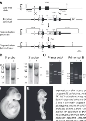
Generation of a nuclear cyan fluorescent protein (CFP) knock-in allele of Bmp4 in the mouse
We originally designed the Bmp4 knock-in allele to simulta-neously mark the cell nuclei of Bmp4-expressing cells and the secreted BMP4 ligand with different fluorescent proteins and observe the structure of BMP4 morphogen gradients in vivo. Therefore, we targeted the endogenous Bmp4 allele with a gene construct comprising two different fluorescent proteins in mouse embryonic stem (ES) cells (Fig. 1A). In the targeted allele, a human histone H2b-fused CFP gene is inserted at the endogenous translational start codon. The expressed histone H2b-fused fluorescent protein (FP) will be incorporated into chromatin and serves as a nuclear and mitotic chromosome marker (Hadjantonakis and Papaioannou, 2004). A yellow fluorescent protein (YFP) is inserted in frame into the coding sequence of BMP4 after the furin cleavage site, so that the
mature BMP4 peptide will have an N-terminal YFP tag. A similar fluorescent protein tagging strategy was used for the Droso-phila BMP4 orthologue Dpp to observe morphogen gradients in the wing maginal disc by overexpression of the chimeric protein (Teleman and Cohen, 2000). Between the H2b-CFP and BMP4-YFP coding sequences, a self-cleavage viral 2A peptide se-quence was incorporated to mediate co-expression and post-translational separation of the two gene products (Szymczak and Vignali, 2005). Thus, Bmp4-expressing cells should be marked in their nuclei with CFP and the BMP4 ligand tagged with YFP should be secreted and function as a wild-type allele. We successfully targeted the construct into the endogenous
Parental Genotypes Number of Live Born Pups of Specific Genotypes
Bmp4CFP/+ X Bmp4LacZ/+
GENETIC TEST OF BMP4CFP ALLELE FUNCTION
(a) Total n = 40 from 7 litters. (b) Total n = 41 from 6 litters.
locus in ES cells and generated mice bearing the allele named Bmp4CFP (Fig. 1 B and C). The neomycin
resistance (neo) expression cassette was then re-moved by crossing to the Sox2-Cre mouse line (Hayashi et al., 2003), and all experiments were conducted on mice with the neo cassette removed. Unfortunately, Bmp4CFP proved to be a functionally
null allele. Bmp4LacZ is a null allele that has part of the Bmp4 coding sequence replaced by a beta-galactosi-dase reporter gene (Lawson et al., 1999). We were unable to recover any live born animals with a Bmp4CFP/ LacZ genotype in crosses between Bmp4CFP/+ and
Bmp4LacZ/+ animals (Table 1). In addition, we could
not recover any live born Bmp4CFP/CFP animals in
intercrosses of Bmp4CFP/+ mice (Table 1). Bmp4CFP/ CFP embryos show growth retardation at E9.5
(embry-onic day 9.5) (Fig. 1E), resembling the homozygous Bmp4 null phenotype (Winnier et al., 1995). Although CFP expression was readily observed in Bmp4CFP/+
mice (see below), no YFP fluorescence was detected, suggesting that undetectable levels of BMP4 are pro-duced from the targeted allele. However, Bmp4CFP/+
expression in the mouse germ line. A 5’ probe and a 3’ probe are used to identify correctly targeted ES cell clones. Hcfp: histone H2b-CFP; Neo: Pgk-Neo-bpA positive selection cassette; TK: MC1-thimidine kinase negative selection cassette; B: BamHI sites. (B) Southern analysis of
BamHI-digested genomic DNA isolated from ES cells. Lanes 1 and 3, wild-type controls, lanes 2 and 4 correctly targeted clone (clone 2C9) used to generate germline chimeras. (C) PCR genotyping results of tail DNA. Primer set A allows for differentiation between wild-type, CFP
and LacZ alleles. Lanes 1 and 2, Bmp4CFP/+ and Bmp4LacZ/+ animals, respectively. Primer set B
allows for detection of the deletion of Neo cassette in the targeted allele. Lanes 3 and 4, heterozygous animals carrying one CFP allele before and after Cre-mediated deletion of the Neo selection cassette, respectively. (D) E9.5 wild-type embryo. (E) E9.5 Bmp4CFP/CFP embryo,
showing growth retardation. Scale bar, 1 mm.
Fig. 1. Generation of Bmp4CFP knock-in allele and
heterozygous animals appear normal and are fertile. Hence, we focused our study on the nuclear CFP signal patterns in the heterozygous animals.
CFP expression patterns in Bmp4CFP/+ are consistent with
Bmp4 expression
Because H2b-CFP has been inserted into the Bmp4 locus, expression should be under the control of the endogenous Bmp4 regulatory elements. Thus, the fluorescent signal in
Bmp4CFP/+ animals should reflect the endogenous Bmp4
ex-pression pattern and agree with exex-pression studies observed by other methods, i.e. in situ hybridization or X-gal staining with a lacZ knock-in allele (Lawson et al., 1999). We examined whole embryos or specific fetal organs at different stages and observed CFP signals in the known Bmp4-specific expression patterns.
Prior to E9.5, CFP signals in Bmp4CFP/+ embryos were either
undetectable or very weak. In the E9.5 Bmp4CFP/+ embryo, the
CFP signal is strong in the lateral mesoderm and the developing heart (Fig. 2A), in agreement with a previous report (Jones et al., 1991). In the early stage limb bud, Bmp4 is expressed in both the mesoderm and the apical ectodermal ridge (AER), and, together with other BMPs, plays key roles in skeletal genesis and patterning in the limb (Bandyopadhyay et al., 2006, Maatouk et al., 2009, Selever et al., 2004). The CFP signal in the E10.5~11.5 Bmp4CFP/+ embryo shows strong expression in
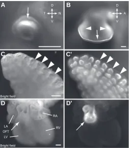
the AER and the broader expression domains in the limb mesoderm (Fig. 2B and 3B). Bmp4 was also shown to be essential for patterning and development of the eye (Behesti et al., 2006, Furuta and Hogan, 1998). In the E10.5 Bmp4CFP/+
Fig. 2. CFP signal observed in known Bmp4-expressing tissues. Different stage Bmp4CFP/+ heterozygous embryos
imaged under fluorescent stereo micro-scope with CFP filters. The embryos are at E9.5 (A), E10.5 (B), E11.5 (C), E12.5
(D), E13.5 (E), E14.5 (F) stages. Arrows point to the CFP signal positive tissues/ organs that are known to express Bmp4. BA, branchial arches; E, eye; H: heart; HF, hair follicle; L, limb; LM, lateral me-soderm; MG, mammary gland; NP, na-sal pit; OV, otic vesicle; VF, vibrissa follicle. Scale bars, 1 mm. Insert in (D) shows a higher magnification view of mammary gland; scale bar, 100 μm.
Fig. 3. CFP signal shows specific Bmp4 expression patterns in developing organs. (A) E10.5 eye. CFP is detected in the dorsal optic cup (arrow). (B) E11.5 forelimb bud, CFP is expressed in the AER (arrow) and the mesodermal regions (arrowheads). (C) Brightfield image of E13.5 lung. (C’) CFP view of (C). CFP is specifically elevated in the epithelium cells at the tip of lung buds (arrow heads). (D) Brightfield image of E13.5 heart. (D’) CFP view of (D). CFP is expressed in the outflow tract arrow. LA, left atrium; LV, left ventricle; RA, right atrium; RV, right ventricle. Orientation: D, dorsal; Vm ventral; N, nasal; T, temporal; A, anterior; P, posterior. Scale bars, 200 μm.
H
LM
L
OV
E
NP
MG
VF
HF
BA
E9.5
E13.5
E10.5
E11.5
E12.5
E14.5
B
C
D
E
F
A
V D
T N
V D
P A
RA
RV
LV LA
OFT Bright field
Bright field
B
C
D
C'
D'
A
embryo, the CFP signal is present in the eye and is restricted to the dorsal domain of the optic cup, reconfirming the known Bmp4 expression pattern (Fig. 2B and 3A). In the developing lung, Bmp4 is expressed in the epithelium at the tip of lung buds and plays a key role in branching morphogenesis (Bellusci et al., 1996, Que et al., 2006). The CFP signal in the E13.5
Bmp4CFP/+ embryo recapitulates the specific Bmp4 expression
pattern in the embryonic lung (Fig. 3C’). In the developing heart, Bmp4 is expressed in the outflow tract myocardium and en-docardial cushion of outflow tract ridges in E12~14 stage embryos and plays an essential role in heart formation (Abdelwahid et al., 2001, McCulley et al., 2008). The CFP signal in the E13.5 Bmp4CFP/+ embryo highlights the outflow
patterning or differentiation of hair follicles. These signaling molecules and some of their antagonists need to be expressed in proper domains and at correct time points for normal hair follicle formation. (Schmidt-Ullrich and Paus, 2005).
Taking advantage of the nuclear localized CFP signal in the Bmp4CFP/+ mice that provides precise information of
Bmp4-ex-pressing cells, we performed a detailed analysis of the Bmp4 expression pattern at different stages of hair follicle development in the embryo. The CFP signal in the ectoderm is initiated between E13.5 and E14.5, forming focal spots in E14.5 embryos (Fig. 2F, 4A and 4D). This timing correlates with the onset of primary hair placode formation (Schmidt-Ullrich and Paus, 2005). On the skin of E15.5 embryos, the CFP signal outlines many oval-shaped structures with an accompanying brighter spot asymmetrically on one side of each oval ring, and the rings are evenly spaced and surrounded by small focal spots (Fig. 4B and 4E). These ovals with their associated asymmetric spots are coordinately oriented in the same direction. In E16.5 embryos, the CFP signal continues to form more rings with spots, while some bigger rings have their accompanying spot apparently moving further away from the ring (Fig. 4C and 4F). In later stage embryos, the CFP signal basically repeats similar patterns as that in E16.5 but with more foldings of the skin as the epithelium thickens (data not shown). With confo-cal microscopy, we observed the nuclear CFP pattern at
sub-Fig. 4. Bmp4 expression pattern in the skin changes through different developmental stages. E14.5 (A,D), E15.5 (B,E) and E16.5 (C,F) skin. At E14.5, CFP signal is first observed on the skin as evenly distributed focal spots (arrowheads in D). At E15.5, the ring and spot structures form (arrow in E). At E16.5, the spot (arrowhead in F) associated with a ring is further away from the ring in more mature follicles (arrow in F). Scale bars, 200 μm. (A-C) are on the same scale; (D-F) are on the same scale.
in Bmp4CFP/+ embryos also reflects
several Bmp4 expression domains that have been reported, including the oto-cyst (Fig. 2B) (Grotewold et al., 2001), branchial arches (Fig. 2B) (Grotewold et al., 2001), nasal pit (Fig. 2C) (Jones et al., 1991), mammary buds (Fig. 2D) (Phippard et al., 1996), vibrassa fol-licles (Fig. 2E) (Jones et al., 1991, Ozeki et al., 2004), and hair follicles (Fig. 2F) (Botchkarev et al., 1999).
Cellular resolution Bmp4 expression patterns in developing hair follicles
The development of hair follicles in-volves complex signaling mechanisms between adjacent tissues, including Wnts (Andl et al., 2002, Reddy et al., 2001), Shh (St-Jacques et al., 1998), Edar (Headon and Overbeek, 1999, Mikkola et al., 1999) and BMPs (Botchkarev and Sharov, 2004), that have been implicated in the initiation,
Fig. 5. Bmp4 expressing cells form different structures on the skin at different developmental stages. Three-dimensionally reconstructed confocal images of CFP signals provide cellular resolution on Bmp4
expression pattern at different stages. (A) At E14.5, Bmp4-expressing cells form focal spots (red circle) with cells in the center fluorescing more strongly. (B,C) At E15.5, the Bmp4-expressing cells form the ring (yellow circle) and spot (red circle) structures. (D) In more mature hair follicle of E16.5 skin, the bright spot (red circle) appears to move further away from the ring (yellow circle). (A,B,C,D) Views onto the surface of the skin, whereas (C’) is a tilted view of (C), and (D’) is a 90 degree rotated view of (D). The spot is actually located beneath the ring in the ring and spot structures, as shown in (C’,D’). Ruler numbers indicate length in μm.
P A
D V
P A
D V
B
C
D
E
F
A
B
C
C'
D
D'
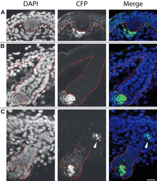
cellular resolution of in live fetal skin and recon-structed three dimensional images. In E14.5 skin, the expression is concentrated in an aggregate of cells, with cells having stronger fluorescence lo-cated at the center of every focal spot (Fig. 5A). In E15.5 skin, a ring structure forms with a central region of non-expressing cells surrounded by ex-pressing cells and a group of strongly fluorescing cells residing on one side of the ring (Fig. 5B and 5C). The group of cells with strong signal is actu-ally located beneath the ring of structure, revealed by rotating the reconstructed three-dimensional images ~45 degrees (Fig. 5C’). In E16.5 skin, the bright spot is farther away from the ring (Fig. 5D and 5D’). We also generated videos rotating the reconstructed images for a better understanding of the structures formed by Bmp4-expressing cells (Supplementary Materials Online). To know the positional relationship of these Bmp4-expressing cells to the developing hair follicles, we made frozen sections of fixed skin tissue. In early stage hair placodes, the Bmp4-expressing cells are in the mesenchymal layer subjacent to the hair pla-code (Fig. 6A). In more mature hair follicles, Bmp4 is expressed in the dermal papilla of involuted hair follicles (Fig. 6B). The Bmp4-expressing dermal papilla cells eventually become surrounded by non-expressing cells in the bulb structure in late stage hair follicles (Fig. 6C).
Discussion
We have generated a novel Bmp4 allele that expresses a nuclear-localized CFP reporter for live imaging and cellular resolution of Bmp4 ex-pression patterns. The observations of CFP sig-nals in various tissues compared with the known pattern of Bmp4 expression show that the CFP signal faithfully reflects the expression pattern of Bmp4. Thus, the Bmp4CFP/+ mice can be used as
an imaging tool to visualize Bmp4 expression
Fig. 6. Bmp4 expressing cells associate with developing hair follicles. Frozen sections of E15.5 (A), E16.5 (B) and E18.5 (C) skin show the respective localization of Bmp4 -expressing cells by CFP signals in developing hair follicles. The epithelial layer and hair follicles are outlined by red dashed lines. (A) Bmp4 is first expressed by mesenchymal cells (arrow) underneath the hair placode. (B,C) At later stages, the dermal papillae express Bmp4
(arrows). Weaker CFP positive cells are seen near the base of hair follicles (arrowhead in C).
DAPI CFP Merge
B
C
A
patterns in live tissues. In addition, Bmp4CFP/+ mice provide a
novel resource for the isolation of Bmp4-expressing cells in live tissues using gross dissection or flow cytometry for the analysis of gene expression profiles or biochemical studies. However, the fluorescent signal is very weak at E8.5 and almost undetectable in E7.5 and earlier stage embryos, although Bmp4 expression is detected at these time points by in situ hybridization or beta-galactosidase detection using the lacZ knock-in allele (Lawson et al., 1999, Winnier et al., 1995). However, both in situ and beta-galactosidase staining techniques detect enzymatically amplified signals and are very sensitive, whereas with the Bmp4CFP allele,
the signal strength largely depends on absolute level of fluores-cent protein produced and is especially prone to photobleaching at low levels.
We were surprised that the Bmp4CFP allele is functionally a null
or very severe hypomorphic allele because in vitro studies of the BMP4-YFP fusion protein in transient tissue culture assays or Xenopus embryos suggested that the fusion protein was active
(Jang, Ueno, and Behringer, unpublished observations). In our targeting strategy, no endogenous Bmp4 sequence was deleted in the targeted allele, so that all of the cis regulatory sequences are present. Integration of exogenous sequences into an endogenous locus with exons and introns is different from cDNA expression from plasmids and expression of synthetic mRNA. We speculate that our generation of a null allele may have to do with expression and/or mRNA processing from a genomic locus. It is possible that the YFP-tagged BMP4 protein is destabilized in vivo, and this stability issue was masked by overexpression in cell culture and Xenopus embryos. In addition, western blot analysis of protein extracts from tissues of Bmp4CFP/+ embryos shows a very weak or
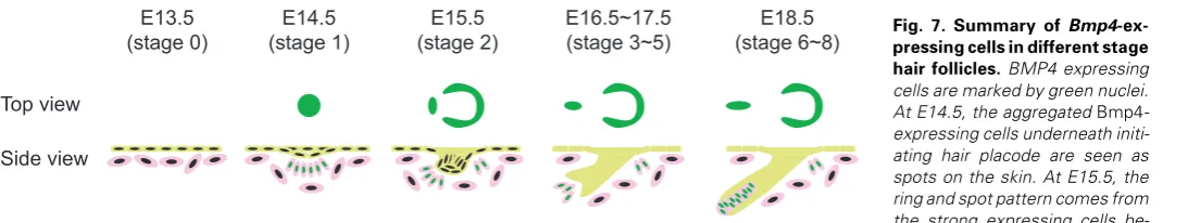
undetectable YFP-BMP4 fusion protein band (data not shown). We summarize our findings of Bmp4 expression in the embry-onic skin using the Bmp4CFP allele in Fig. 7. In E13.5 skin, prior to
epithelial cells in primary hair follicles start to invaginate, cells with stronger Bmp4 expression form a cap structure at the tip of the invaginated epithelium structure with a ring of Bmp4-expressing cells surrounding the invagination at the base. At E16.5, Bmp4 is expressed in the dermal papilla adjacent to the tip of hair follicles that are extending further into dermis. At E18.5, Bmp4 expression is maintained in the dermal papilla cells surrounded in the bulb structure in late stage hair follicles.
The suppressive activity of BMPs on hair placode formation has been implicated in different experimental systems (Botchkarev et al., 1999, Jung et al., 1998, Noramly and Morgan, 1998). In addition, the importance of the BMP signaling pathway in speci-fication of different cell types in the hair follicle has been well documented (Botchkarev and Sharov, 2004). The ring-shaped expression pattern of developing hair follicles is quite unique, among other signaling molecules that are expressed in the hair follicle and required for proper development (Laurikkala et al., 2002, Mou et al., 2006). This may reflect a unique role BMP4 plays or the advantage of using fluorescent protein labeling technique since many gene expression patterns have only been examined on sections on which the three dimensional structure is not easily noticed. It will be interesting to determine how the dynamic Bmp4-expressing cell rings and focal aggregates that we have discov-ered relate to these inductive and differentiation roles of BMP4 during hair follicle development.
Here we have shown the feasibility of visualizing Bmp4 expres-sion in live tissues with our knock-in allele, and analyzed Bmp4 expressing domains associated with developing hair follicles. This allele provides a new experimental tool for studying BMP4 signaling mechanisms in live tissues. In addition, BMP reporter transgenic mouse lines that express EGFP in BMP signal-receiv-ing cells have been created (Blank et al., 2008, Monteiro et al., 2008). EGFP and CFP are spectrally distinguishable (Dickinson et al., 2002, Lansford et al., 2001, Zimmermann, 2005). The possibility of combining our Bmp4CFP allele with the BMP
signal-ing GFP reporter lines may further advance the study of BMP signaling by live tissue imaging techniques.
Materials and Methods
Generation of Bmp4CFP knock-in allele in the mouse
The targeting construct was generated by subcloning a 2.1-kb 5’
homologous region upstream from the endogenous start site of Bmp4 to
fuse with a human histone H2b-Cerulean (an improved CFP) coding
sequence (Hadjantonakis and Papaioannou, 2004, Rizzo et al., 2004),
followed by a self-cleavage 2A sequence from the virus Thosea asigna
(Szymczak and Vignali, 2005), followed by the endogenous coding sequence for BMP4 as an in-frame fusion. A floxed Pgk-neo-bpA cassette
Top view
[from pL452 (Liu et al., 2003)] was inserted into the Eco RI site into the
preserved third intron in reverse orientation. An improved YFP, Citrine
(Griesbeck et al., 2001), coding sequence was inserted into the fourth
exon as an in-frame fusion after the furin cleavage site that generates the mature peptide. An additional homologous region of 3.3 kb was included in the targeting construct after the Citrine coding sequence. In our targeting strategy, no endogenous sequence was deleted. An MC1-TK cassette was placed next to the 3’ homologous region for negative
selection. G4 ES cells (George et al., 2007) were electroporated with
linearized targeting construct DNA and subjected to G418 and FIAU double drug selection. Surviving clones were screened by Southern
blotting with BamHI enzyme digestion of genomic DNA. A 1-kb 5’ probe
and a 1.4-kb 3’ probe outside of the homologous region on the targeting construct were used to identify correctly targeted clones. One of these clones was expanded and injected into C57Bl/6J blastocysts to generate chimeras. The floxed Neo cassette was removed through crossing with Sox2-Cre transgenic animals (Hayashi et al., 2003). Heterozygous
ani-mals carrying the CFP knock-in Bmp4 allele without Neo are maintained
on an outbred SWISS Webster (Taconic, NY) genetic background and used for analysis in this report.
Primers for PCR-genotyping animals:
PCR genotyping with Primer Set A with 60.5 oC annealing temperature
generates a 550-bp product from the wild-type Bmp4 allele, a 700-bp
product from the null lacZ knock-in allele (Lawson et al., 1999), or an
850-bp product from the CFP knock-in allele. PCR genotyping with Primer Set
B with 55 oC annealing temperature generates a 320-bp product for the
wild-type allele, a 500-bp product for the floxed Neo cassette in the CFP
allele, or a 400-bp product for the footprint loxP site after Neo is removed
by Cre recombinase.
Imaging of CFP expression patterns in live and fixed tissues For live imaging of whole embryos, embryos of desired stages were isolated from the uterus of pregnant females from timed matings. Em-bryos were immersed in phosphate buffered saline (PBS) and imaged at low magnification with a Carl Zeiss Lumar fluorescent stereo microscope system (http://www.zeiss.de/). Lungs and heart were dissected from E13.5 embryos for imaging with the same settings. For higher resolution CFP confocal images of the skin, live embryos were placed on a glass bottom dish with PBS and imaged with a Zeiss LSM 510 META laser confocal microscopy system, using a C-Apochromat 40X/W, NA=1.2 Corr objective (Carl Zeiss Inc.). Three-dimensional images were reconstructed using the image processing software Imaris 5.0.3 (Bitplane, St. Paul, MN, USA) (http://www.bitplane.com/).
For frozen sections of skin, whole embryos were fixed in 4% paraform-aldehyde in phosphate buffered saline for one hour and washed through
10%, 20% and 30% sucrose solutions, and then embedded in O.C.T. compound [Tissue-Tek (http://www.tedpella.com/)]. Sections were mounted with DAPI containing medium and then imaged with the same laser confocal microscopy system and objective as that for live skin imaging.
Acknowledgements
We thank Jim Martin for providing Bmp4 genomic DNA plasmids, Andras Nagy for the G4 mouse ES cells, Jian Min Deng for tissue culture assistance, and Henry Adams for advice on microscopy. Supported by National Institutes of Health (NIH) grants NIH HL077187 and EB005173 to M.E.D and NIH HD30284 and the Ben F. Love Endowment to R.R.B. DNA sequencing and veterinary resources were supported by the NIH Cancer Center Support Grant CA16672.
References
ABDELWAHID, E., RICE, D., PELLINIEMI, L.J. and JOKINEN, E. (2001). Overlap-ping and differential localization of Bmp-2, Bmp-4, Msx-2 and apoptosis in the endocardial cushion and adjacent tissues of the developing mouse heart. Cell Tissue Res 305: 67-78.
ANDL, T., REDDY, S.T., GADDAPARA, T. and MILLAR, S.E. (2002). WNT signals are required for the initiation of hair follicle development. Dev Cell 2: 643-53.
BANDYOPADHYAY, A., TSUJI, K., COX, K., HARFE, B.D., ROSEN, V. and TABIN, C.J. (2006). Genetic analysis of the roles of BMP2, BMP4, and BMP7 in limb patterning and skeletogenesis. PLoS Genet 2: e216.
BEHESTI, H., HOLT, J.K. and SOWDEN, J.C. (2006). The level of BMP4 signaling is critical for the regulation of distinct T-box gene expression domains and growth along the dorso-ventral axis of the optic cup. BMC Dev Biol 6: 62.
BELLUSCI, S., HENDERSON, R., WINNIER, G., OIKAWA, T. and HOGAN, B.L. (1996). Evidence from normal expression and targeted misexpression that bone morphogenetic protein (Bmp-4) plays a role in mouse embryonic lung morpho-genesis. Development 122: 1693-702.
BITGOOD, M.J. and MCMAHON, A.P. (1995). Hedgehog and Bmp genes are coexpressed at many diverse sites of cell-cell interaction in the mouse embryo. Dev Biol 172: 126-38.
BLANK, U., SETO, M.L., ADAMS, D.C., WOJCHOWSKI, D.M., KAROLAK, M.J. and OXBURGH, L. (2008). An in vivo reporter of BMP signaling in organogen-esis reveals targets in the developing kidney. BMC Dev Biol 8: 86.
BOTCHKAREV, V.A., BOTCHKAREVA, N.V., ROTH, W., NAKAMURA, M., CHEN, L.H., HERZOG, W., LINDNER, G., MCMAHON, J.A., PETERS, C., LAUSTER, R. et al. (1999). Noggin is a mesenchymally derived stimulator of hair-follicle induction. Nat Cell Biol 1: 158-64.
BOTCHKAREV, V.A. and SHAROV, A.A. (2004). BMP signaling in the control of skin development and hair follicle growth. Differentiation 72: 512-26.
DERYNCK, R. and ZHANG, Y.E. (2003). Smad-dependent and Smad-independent pathways in TGF-beta family signalling. Nature 425: 577-84.
DICKINSON, M.E., WATERS, C.W., WOLLESCHENSKY, R., BEARMAN, G., TILLE, S. and FRASER, S.E. (2002). Sensitive imaging of spectrally overlap-ping fluorochromes using the LSM 510 META. In Multiphoton Microscopy in the Biomedical Sciences, vol. 4620 (ed. PERIASAMY, A. and SO, P. T.), pp. 123-136: Proceedings of the International Society for Optical Engineering (SPIE).
FURUTA, Y. and HOGAN, B.L. (1998). BMP4 is essential for lens induction in the mouse embryo. Genes Dev 12: 3764-75.
GEORGE, S.H., GERTSENSTEIN, M., VINTERSTEN, K., KORETS-SMITH, E., MURPHY, J., STEVENS, M.E., HAIGH, J.J. and NAGY, A. (2007). Develop-mental and adult phenotyping directly from mutant embryonic stem cells. Proc Natl Acad Sci USA 104: 4455-60.
GRIESBECK, O., BAIRD, G.S., CAMPBELL, R.E., ZACHARIAS, D.A. and TSIEN, R.Y. (2001). Reducing the environmental sensitivity of yellow fluorescent protein. Mechanism and applications. J Biol Chem 276: 29188-94.
GROTEWOLD, L., PLUM, M., DILDROP, R., PETERS, T. and RUTHER, U. (2001). Bambi is coexpressed with Bmp-4 during mouse embryogenesis. Mech Dev 100: 327-30.
HADJANTONAKIS, A.K. and PAPAIOANNOU, V.E. (2004). Dynamic in vivo
imag-ing and cell trackimag-ing usimag-ing a histone fluorescent protein fusion in mice. BMC Biotechnol 4: 33.
HAYASHI, S., TENZEN, T. and MCMAHON, A.P. (2003). Maternal inheritance of Cre activity in a Sox2Cre deleter strain. Genesis 37: 51-3.
HEADON, D.J. and OVERBEEK, P.A. (1999). Involvement of a novel Tnf receptor homologue in hair follicle induction. Nat Genet 22: 370-4.
HOGAN, B.L. (1996). Bone morphogenetic proteins: multifunctional regulators of vertebrate development. Genes Dev 10: 1580-94.
JONES, C.M., LYONS, K.M. and HOGAN, B.L. (1991). Involvement of Bone Morphogenetic Protein-4 (BMP-4) and Vgr-1 in morphogenesis and neurogenesis in the mouse. Development 111: 531-42.
JUNG, H.S., FRANCIS-WEST, P.H., WIDELITZ, R.B., JIANG, T.X., TING-BERRETH, S., TICKLE, C., WOLPERT, L. and CHUONG, C.M. (1998). Local inhibitory action of BMPs and their relationships with activators in feather formation: implications for periodic patterning. Dev Biol 196: 11-23.
LANSFORD, R., BEARMAN, G. and FRASER, S.E. (2001). Resolution of multiple green fluorescent protein color variants and dyes using two-photon microscopy and imaging spectroscopy. J Biomed Opt 6: 311-8.
LAURIKKALA, J., PISPA, J., JUNG, H.S., NIEMINEN, P., MIKKOLA, M., WANG, X., SAARIALHO-KERE, U., GALCERAN, J., GROSSCHEDL, R. and THESLEFF, I. (2002). Regulation of hair follicle development by the TNF signal ectodysplasin and its receptor Edar. Development 129: 2541-53.
LAWSON, K.A., DUNN, N.R., ROELEN, B.A., ZEINSTRA, L.M., DAVIS, A.M., WRIGHT, C.V., KORVING, J.P. and HOGAN, B.L. (1999). Bmp4 is required for the generation of primordial germ cells in the mouse embryo. Genes Dev 13: 424-36.
LIU, P., JENKINS, N.A. and COPELAND, N.G. (2003). A highly efficient recombineering-based method for generating conditional knockout mutations. Genome Res 13: 476-84.
MAATOUK, D.M., CHOI, K.S., BOULDIN, C.M. and HARFE, B.D. (2009). In the limb AER Bmp2 and Bmp4 are required for dorsal-ventral patterning and interdigital cell death but not limb outgrowth. Dev Biol.
MCCULLEY, D.J., KANG, J.O., MARTIN, J.F. and BLACK, B.L. (2008). BMP4 is required in the anterior heart field and its derivatives for endocardial cushion remodeling, outflow tract septation, and semilunar valve development. Dev Dyn 237: 3200-9.
MIKKOLA, M.L., PISPA, J., PEKKANEN, M., PAULIN, L., NIEMINEN, P., KERE, J. and THESLEFF, I. (1999). Ectodysplasin, a protein required for epithelial morphogenesis, is a novel TNF homologue and promotes cell-matrix adhesion. Mech Dev 88: 133-46.
MONTEIRO, R.M., DE SOUSA LOPES, S.M., BIALECKA, M., DE BOER, S., ZWIJSEN, A. and MUMMERY, C.L. (2008). Real time monitoring of BMP Smads transcriptional activity during mouse development. Genesis 46: 335-46.
MOU, C., JACKSON, B., SCHNEIDER, P., OVERBEEK, P.A. and HEADON, D.J. (2006). Generation of the primary hair follicle pattern. Proc Natl Acad Sci USA 103: 9075-80.
NORAMLY, S. and MORGAN, B.A. (1998). BMPs mediate lateral inhibition at successive stages in feather tract development. Development 125: 3775-87.
OZEKI, H., KURIHARA, Y., TONAMI, K., WATATANI, S. and KURIHARA, H. (2004). Endothelin-1 regulates the dorsoventral branchial arch patterning in mice. Mech Dev 121: 387-95.
PHIPPARD, D.J., WEBER-HALL, S.J., SHARPE, P.T., NAYLOR, M.S., JAYATALAKE, H., MAAS, R., WOO, I., ROBERTS-CLARK, D., FRANCIS-WEST, P.H., LIU, Y.H. et al. (1996). Regulation of Msx-1, Msx-2, Bmp-2 and Bmp-4 during foetal and postnatal mammary gland development. Development 122: 2729-37.
QUE, J., CHOI, M., ZIEL, J.W., KLINGENSMITH, J. and HOGAN, B.L. (2006). Morphogenesis of the trachea and esophagus: current players and new roles for noggin and Bmps. Differentiation 74: 422-37.
REDDY, S., ANDL, T., BAGASRA, A., LU, M.M., EPSTEIN, D.J., MORRISEY, E.E. and MILLAR, S.E. (2001). Characterization of Wnt gene expression in develop-ing and postnatal hair follicles and identification of Wnt5a as a target of Sonic hedgehog in hair follicle morphogenesis. Mech Dev 107: 69-82.
SCHMIDT-ULLRICH, R. and PAUS, R. (2005). Molecular principles of hair follicle induction and morphogenesis. Bioessays 27: 247-61.
SELEVER, J., LIU, W., LU, M.F., BEHRINGER, R.R. and MARTIN, J.F. (2004). Bmp4 in limb bud mesoderm regulates digit pattern by controlling AER devel-opment. Dev Biol 276: 268-79.
ST-JACQUES, B., DASSULE, H.R., KARAVANOVA, I., BOTCHKAREV, V.A., LI, J., DANIELIAN, P.S., MCMAHON, J.A., LEWIS, P.M., PAUS, R. and MCMAHON, A.P. (1998). Sonic hedgehog signaling is essential for hair development. Curr Biol 8: 1058-68.
SZYMCZAK, A.L. and VIGNALI, D.A. (2005). Development of 2A peptide-based
Further Related Reading, published previously in the Int. J. Dev. Biol.
See Special Issue Pattern Formation edited by Michael K. Richardson and Cheng-Ming Chuong at: http://www.ijdb.ehu.es/web/contents.php?vol=53&issue=5-6
Analyses of regenerative wave patterns in adult hair follicle populations reveal macro-environmental regulation of stem cell activity
Maksim V. Plikus, Randall B. Widelitz, Rob Maxson and Cheng-Ming Chuong Int. J. Dev. Biol. (2009) 53: 857-868 (doi: 10.1387/ijdb.072564mp)
Frontiers in fluorescence microscopy
José Rino, José Braga, Ricardo Henriques and Maria Carmo-Fonseca Int. J. Dev. Biol. (2009) 53: 1569-1579 (doi: 10.1387/ijdb.072351jr)
Transdifferentiation of corneal epithelium: evidence for a linkage between the segregation of epidermal stem cells and the induction of hair follicles during embryogenesis
David J Pearton, Corinne Ferraris and Danielle Dhouailly Int. J. Dev. Biol. (2004) 48: 197-201
Hair follicle differentiation and regulation. George E Rogers
Int. J. Dev. Biol. (2004) 48: 163-170
5 yr ISI Impact Factor (2008) = 3.271
strategies in the design of multicistronic vectors. Expert Opin Biol Ther 5: 627-38.
TELEMAN, A.A. and COHEN, S.M. (2000). Dpp gradient formation in the Droso-phila wing imaginal disc. Cell 103: 971-80.
WINNIER, G., BLESSING, M., LABOSKY, P.A. and HOGAN, B.L. (1995). Bone morphogenetic protein-4 is required for mesoderm formation and patterning in the mouse. Genes Dev 9: 2105-16.
ZHAO, G.Q. (2003). Consequences of knocking out BMP signaling in the mouse. Genesis 35: 43-56.