Original Research Article
Abnormal uterine bleeding in perimenopausal women: sonographic and
histopathological correlation and evaluation of uterine endometrium
Rupali Modak
1, Amitrajit Pal
2, Amitava Pal
3*, Kausik Bose
4INTRODUCTION
In 2001, stages of reproductive aging workshop (STRAW) defined perimanopause as the period beginning with menopausal transition and ending 12
months after the last menstrual period.1,2 The
premenopausal condition is often characterized by menstrual cycle irregularities in frequency, duration and
volume due to fluctuation of estrogen hormone levels. Although irregular bleeding pattern are a normal and expected part of perimenopusal period the incidence of uterine pathology and associated medical condition also increase in this age group.3 AUB occurs in 50% of
perimenopausal women and significantly impacting quality of life and imposing financial burden.4,5 This is
mainly due to long anovulatory periods with unopposed
ABSTRACT
Background: The objectives of the study was to establish the role of histopathological diagnosis of uterine endometrial lesions in patients of AUB at perimenopausal age and to correlate the transvaginal sonographic (TVS) finding with histopathological examination.
Methods: This prospective observational study was carried out over 1 and 1/2 years in the two apex level teaching hospitals in eastern India. A total of 197 women in the age group of 40-49 years and ≥50 years (up to 55 years) who presented with abnormal uterine bleeding were included in the study. After selecting the patient with eligibility criteria in the OPD, detailed clinical history, systemic and gynecological examinations and investigations were done as per proforma. TVS study of endometrial pattern and thickness was measured followed by dilatation and curettage (D and C) and HPE of the endometrial curetting was done.
Results: Menorrhagia (44.67%) was the most common clinical finding. Mean endometrial thickness measured by TVS was 7.04±2.11 mm in proliferative phase and 10.25±1.27 mm in the secretory phase. Proliferative endometrium (37.06%) was the most frequent finding in HPE followed by secretory endometrium (20.3%). Hyperplasia of endometrium was noted in 27 cases (100%) at 12-15 mm of endometrial thickness on TVS whereas endometrial hyperplasia with and without atypia and endometrial carcinoma was noted in 25 cases (92.59%) at the same thickness of 12-15 mm of uterine endometrium on HPE. Endometrial hyperplasia and polyp both had sensitivity of 84.21% and 71.43% respectively on TVS as compared with histopathology.
Conclusions: Increased endometrial thickness and echo pattern by TVS correlated well with abnormal endometrial tissue histopathology in perimenopausal women with AUB.
Keywords: Abnormal uterine bleeding, Endometrial thickness, Dilatation and curettage, Menorrhagia, Transvaginal sonography
DOI: http://dx.doi.org/10.18203/2320-1770.ijrcog20201788
1Department of Obstetrics and Gynecology, R. G. Kar Medical College, Kolkata, West Bengal, India
2Department of Pharmacology, Grant Medical College, Mumbai, Maharashtra, India
3Department of Obstetrics and Gynecology, Burdwan Medical College, Purba Barddhaman, West Bengal, India
4Department of Pathology, Burdwan Medical College, Purba Barddhaman, West Bengal, India
Received: 16 February 2020
Accepted: 20 March 2020
*Correspondence:
Dr.Amitava Pal,
E-mail: [email protected]
estrogen stimulation result in endometrial hyperplasia thus increasing the risk of endometrial cancer.6 Heavy
menstrual bleeding may be associated with fibromyoma, adenomyosis or endometrial polyp. Polymenorrhagia and metrorrhagia represent other types of AUB; their underlying cause is other type of endometrial alterations
The International Federation of gynecology and obstetrics working group on menstrual disorder was proposed a classification system (PALM-COEIN) for causes of AUB
in women.7 PALM-COEIN represents polyp,
adenomyosis, leiomyoma, malignancy and hyperplasia, coagulopathy, ovulatory dysfunction, endometrial and not yet classified. Using a non-invasive and a convenient diagnostic technique such as TVS is preferable at the first instance for studying the endometrial patterns and its thickness accurately and at the same time to exclude organic uterine pathology in AUB followed by the invasive technique of dilatation and curettage (D and C).
Dilatation and curettage remain the standard diagnostic procedure for assessment of AUB and for early detection of atypical or typical endometrial hyperplasia, but it has a drawback of being a blind procedure with a chance of missing of a small or focal lesion.8 TVS and D and C
procedures still remain a very cost effective, practical and dependable approach for investigating AUB.
Objectives of the present study were to determine the efficacy of TVS in depicting the pattern of endometrium and to correlate transvaginal endometrial thickness with
histopathology of endometrium in AUB at 2019. A total of 197 patients with the complaints of AUB in perimenopausal age group of 40-55 years were invited to participate in the study. After ethical clearance, all the candidates who fulfill the following inclusion and exclusion criteria and who gave informed consent were enrolled in the study.
Inclusion criteria
• Perimenopausal women (40-55 years)
• Having AUB
• Uterus <12 weeks size.
Exclusion criteria
• Active pelvic infection
• Uterus >12 weeks size
• Vaginal or cervical bleeding
• Pregnancy complications
•
Detailed menstrual, contraceptive, obstetric, medical and surgical history of the eligible candidates were taken along with H/O presenting complains and general, physical, systemic and gynecological examination was done as per proforma. Every patient was subjected to following laboratory investigations including CBC, blood group, RBG, coagulation profile, liver and kidney function tests, urine routine and microscopy and UPT.
All the eligible candidates were subjected to trans vaginal sonography (TVS). TVS was done by 7.5 MHz trans vaginal probe and various sonographic parameters such as endometrial thickness, uterine pathology, adnexal and any other pelvic pathology was noted. Endometrial thickness was measured at 1 cm from the fundus and described in longitudinal section. Endometrium was considered thickened or hyperplasic when endometrial thickness was ≥12 mm in premenopausal women and ≥ 5 mm in postmenopausal women and considered atrophic when thin, homogenous, echogenic and pencil line endometrium with thickness of less than 4-5 mm noted. This was followed by D and C procedure under short general anesthesia in OT. The endometrial tissue sample for histopathological examination was fixed in 10% formalin and sent to the college pathology department for analysis. After tabulating the finding of TVS, it was compared with histopathology and the sensitivity, specificity, PPV and NPV of TVS was calculated.
Statistical analysis
Categorical variables were expressed as number of patients and percentage of patients and compared across the groups using Pearson‘s Chi Square test for independence of attributes. Continuous variables were expressed as mean, median and standard deviation (SD) and compared across the groups using unpaired students’ t-test. Sensitivity, specificity, PPV, NPV and diagnostic accuracy was calculated. The statistical software SPSS version 20 was used for the analysis. An alpha level of 5% has been taken, i.e. p value of < 0.05, was considered as significant.
RESULTS
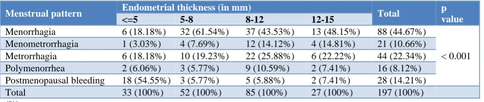
Table 1: Correlation between endometrial thickness and abnormal menstrual pattern.
Menstrual pattern Endometrial thickness (in mm) Total p
value
Table 2: Findings of histopathology.
Histopathology Number Percentage
Proliferative endometrium 73 37.06%
Secretory endometrium 40 20.3%
Atrophic endometrium 16 8.12%
Endometritis 12 6.09%
Disordered proliferative phase 11 5.58%
Endometrial polyp 7 3.55%
Endometrial hyperplasia without atypia 24 12.18%
Endometrial hyperplasia with atypia 12 6.09%
Endometrial carcinoma 2 1.02%
Total 197 100.0%
Table 3: Correlation between TVS finding in mm and HPE findings at different endometrial thickness.
ET
PE, proliferative endometrium; SE, secretory endometrium; AE, atrophic endometrium; EP, endometrium polyp; HE, hyperplastic endometrium;; E, endometritis; DP, disordered proliferative phase; EH, endometrial hyperplasia; WOA, without atypia; WA, with atypia; EC, endometrial carcinoma. Mean endometrial thickness (mean±SD, 8.70±3.30 mm); Proliferative endometrial thickness (mean ± SD, 7.04±2.11mm); Secretory endometrial thickness (mean±SD,10.25± 1.27mm)
Table 4: Sensitivity, specificity, NPV of TVS findings in comparison to histopathology.
Proliferative endometrium (PE) having thickness of 5-8 mm was present in 49 cases and majority (n=47) of secretory endometrium had thickness of 8-12 mm on TVS examination but on histopathology secretory endometrium was confirmed by 38 cases at the same
endometrial thickness. Normal endometrial
histopathology (PE+SE) with the thickness of 5-12 mm by TVS was present in 113 patients. Hyperplastic endometrium was present in 27 cases at endometrial thickness of 12-15 mm by TVS examination, but histopathology confirmed fourteen cases of endometrial hyperplasia without atypia and 9 cases with atypia at ET of 12-15 mm (Table 3).
Sensitivity, specificity, PPV, NPV of TVS for proliferative phase was 95.89%, 89.52%, 84.34% and 97.37%; for secretory phase was 95%, 93.63%, 79.17% and 98.66% and for hyper plastic phase 84.21%, 98.11%, 91.43% and 96.30% respectively (Table 4).
Figure 1: Endometrial thickness (mm) in relation to uterine pathology: group 1 (leiomyoma), group 2 (adenomyosis), group 3 (endometrial polyp), group 4
(thickened endometrium).
The box-plot diagram shows relationship of ET with uterine pathology. The endometrial thickness in ‘mm’ was assessed by TVS in patients with leiomyoma (n=50), adenomyosis (n=37), endometrial polyp (n=13) and thickened endometrium (n=36). There was statistically significant difference of endometrial thickness in patients with leiomyoma vs. thickened endometrium, and those with endometrial polyp versus thickened endometrium (p <0.001) (Figure 1)First quartile(Q1), third quartile(Q3), median value , maximum and minimum values of each group were plotted in each box- plot diagram as per data summary shown in the Figure 1.
DISCUSSION
Abnormal uterine bleeding is the most common
gynecological complaints among women in
perimenopausal women.
In the present study menorrhagia was the most common clinical presentation in 44.67 % (n=88) of cases followed by menometrorrhagia (10.66%) which is very similar to the study by Jetley et al and Pillai.6,9 In this prospective
study histopathology report revealed that most of the
women had proliferative endometrium (37.06%),
followed by secretory endometrium (20.3%) and hyperplastic endometrium (18.27%) and 3.55% of cases had endometrial polyp. In Thulasi et al, study of histopathological reports showed that proliferative endometrium, secretory endometrium and endometrial hyperplasia were present in 33.33%, 20% and 40% of cases respectively.10 Proliferative endometrium was the
most common histopathological finding like other studies.6,11 The present study showed that the mean
endometrial thickness was 7.04±2.11 mm in proliferative phase and 10.25±1.27 mm in the secretory phase. These followed by proliferative endometrium.9,12 In this study
secretory endometrium was present only in 20.3% of cases on HPE.
Transvaginal sonography to measure endometrial thickness is easier and better. In the present study endometrial hyperplasia on TVS was found in 35 cases. On comparison with histopathology report, 32 cases were truly diagnosed having hyperplastic endometrium and 3 cases misdiagnosed, out of which two were endometritis and one had normal secretory endometrium.
In the present study the cut-off value of endometrial thickness (ET) was 12 mm in premenopausal age group. Six patients had endometrial hyperplasia when ET was of 11-12 mm and other endometrial abnormalities on HPE at the above-mentioned thickness was present in 26 cases only. No major endometrial abnormality was detected in perimenopausal age group when ET was <11 mm. Study finding corresponds well with the finding of Singh et al, but Gazala and Pillai did not found any major endometrial pathology when ET was <14 mm and <14.9 mm, respectively.6,13,14 The present study showed no case
of endometrial atypia or malignancy when ET was <5 mm. The same result was also noted in different other studies.15-18 All patients with thickened endometrium
diagnosed on TVS must have a through endometrial evaluation by D and C in perimenopausal age groups.
hyperplasia and cancer.19-22 The likelihood ratio (LR)
reflects the degree in which a practical method may increase or lower the probability of diagnosing the certain diseases. Thus, an increased positive LR (44.632) associated with low negative LR (0.161) for TVS in this study reflects the high acceptance of this method for the diagnosis of uterine endometrial hyperplasia. Dasgupta et al found a low positive LR (2.8-3.8) for TVS and easy and relatively inexpensive method of first choice to find out various etiologies of AUB. In perimenopausal women evaluation of endometrial echo pattern and measurement of ET by TVS is a useful tool to differentiate normal and abnormal uterine endometrium in relation to the clinical findings. Histopathology plays a major role in the definitive diagnosis and for accurate evaluation of uterine endometrium which correlates well with the TVS finding. The method of measuring the endometrium, the experience and skill of radiologist influences the TVS result, whereas histopathology of hyperplastic uterine endometrium is the gold standard for accurate diagnosis of typical and atypical pattern with endometrial carcinoma.
Funding: No funding sources Conflict of interest: None declared
Ethical approval: The study was approved by the Institutional Ethics Committee
REFERENCES
1. Soules MR, Sherman S, Parrott E. Stages of
reproductive aging workshop (STRAW). J Womens Health Gender Based Med. 2001;10:843-8.
2. Awwad T, Toth TL, Schiff I. Abnormal uterine
bleeding in the perimenopause. Int. J Fertil Menopausal Stud. 1993;38(5):261-9.
3. Speroff L, Fritz MA, Menopause and the
perimenopausal transition, clinical endocrinology. In: Speroff L, Fritz MA, eds. Clinical gynecologic endocrinology and infertility.7th ed. Philadelphia,
London: Lippincott Williams and Wilkins; 2005:628. 4. Oriel KA, Schrager S. Abnormal uterine bleeding.
Am Fam Phys Discus. 1999;60(5):1371-80.
5. Fraser IS, Langham S, Uhi-Hochgraeber K. Health related quality of life and economic burden of abnormal uterine bleeding. Expert Rev Obstet Gynecol. 2009;4(2):179-89.
6. Pillai SS. Sonographic and histological correlation and evaluation of endometrium in perimenopausal
women with abnormal uterine bleeding. Int J Reprod Contracept Obstet Gynecol. 2014;3:113-7.
7. Munro MG, Critchley HO, Fraser IS, FIGO
menstrual disorder working group. The FIGO classification of causes of abnormal uterine bleeding
in the reproductive years. Fertil Steril.
2011;95(7):2204-8.
8. Albert JR, Hull SK, Wesley RM. Abnormal uterine bleeding. Am Fam Phys. 2004;69:1915-26.
9. Jetley S, Rama S, Jairajpuri ZS. Morphological
spectrum of endometrial pathology in middle aged women with atypical uterine bleeding- a study of 219 cases. Midlife Health. 2013;4:216-20.
10. Thulasi P, Balakrishnan R, Shanthi M. Correlation of endometrial thickness by transvaginal sonography and histopathology in women with abnormal peri- menopausal and postmenopausal bleeding - A prospective study. Indian J Obstet Gynecol Res. 2018;5:44-8.
11. Najeeb R, Awan AS, Bakhtiar U, Akhtar S. Role of
transvaginal sonography in assessment of abnormal uterine bleeding in perimenopausal agegroup. J Ayub Med Coll Abbottabad. 2010;22(1):87-90
12. Acharya V, Mehta S, Rander A. Evaluation of
dysfunctional uterine bleeding by TVS, hysteroscopy and histopathology. J Obstet Gynecol India. 2003;53:170-7.
13. Gazala AA. Role of TVS in case of abnormal uterine
bleeding. Prof Med J. 2009;1:127-34.
14. Singh M, Sachan R, Yadav A. Significance of
endometrial thickness on transvaginal sonography in heavy menstrual bleeding. J Curr Sci Med. 2019;5:28-32.
15. Sharma J, Dhiman B, Sud N, Kaushik A. Synergistic
approach in diagnosing endometrial disease in women with postmenopausal bleeding. Int J Reprod Contracept Obstet Gynecol. 2017;6:4081-89.
16. Epstein E, Ramirez A, Skoog L, Valentin L.
Dilatation and curettage fails to detect most focal lesions in the uterine cavity in women with postmenopausal bleeding. Acta Obstet Gynecol Scand. 2001;80:1131-6.
17. Gull B, Karlsson B, Milsom I, Granberg S. Can ultrasound replace dilation and curettage? A longitudinal evaluation of postmenopausal bleeding and transvaginal sonographic measurement of the endometrium as predictors of endometrial cancer. Am J Obstet Gynecol. 2003;188:401-8.
18. Bruchim I, Biron-Shental T, Altaras MM, Fishman A, Beyth Y, Tepper R, et al. Combination of endometrial thickness and time since menopause in predicting endometrial cancer in women with postmenopausal bleeding. J Clin Ultrasound. 2004;32(5):219-24.
19. Jain M, Chakroborty S. Evaluation of abnormal
uterine bleeding with trans vaginal sonography. Int J Reprod Contracept Obstet Gynecol. 2017;6:2794-9.
20. Deshmukh V, Yelikar K, Devile M. Clinical study of
by transvaginal sonography and its histopathological correlation. J Evol Med Dent Sci. 2013;2:2440-5. 21. Choudhary J, Acharya V, Jain M. Evaluation of
abnormal uterine bleeding with trans vaginal sonography and hysteroscopy in perimenopausal women, Int J Reprod Contracept Obstet Gynecol. 2017;6:3607-13.
22. Mukhopadhyay S, Bhattacharyya SK, Ganguly RP,
Patra KK, Bhattacharya N, Barman SC. Comparative evaluation of perimenopausal abnormal uterine bleeding by transvaginal sonography, hysteroscopy and endometrial biopsy. J Indian Med Assoc. 2007;105:624-8.
23. Dasgupta S, Chakraborty B, Karim R, Aich RK,
Mitra PK, Ghosh TK. Abnormal uterine bleeding in
peri-menopausal age: diagnostic options and
accuracy. J Obstet Gynaecol India. 2011;61(2):189-94.
Cite this article as: Modak R, Pal A, Pal A, Bose K. Abnormal uterine bleeding in perimenopausal women: sonographic and histopathological