Hepatic Medicine: Evidence and Research
Dove
press
O R I G I N A L R E S E A R C H
open access to scientific and medical research
Open Access Full Text Article
Presence and consequence of tooth periapical
radiolucency in patients with cirrhosis
Lea Ladegaard Grønkjær1
Palle Holmstrup2
Søren Schou3
Kristoffer Schwartz4
Johanne Kongstad2
Peter Jepsen1,5
Hendrik Vilstrup1
1Department of Hepatology and
Gastroenterology, Aarhus University Hospital, Aarhus, 2Section of
Periodontology, Microbiology, and Community Dentistry, Department of Odontology, Faculty of Health and Medical Sciences, 3Section for Oral
and Maxillofacial Surgery, Department of Odontology, Faculty of Health and Medical Sciences, University of Copenhagen, Copenhagen,
4Department of Oral and Maxillofacial
Surgery, 5Department of Clinical
Epidemiology, Aarhus University Hospital, Aarhus, Denmark
Background: Periapical radiolucency is the radiographic sign of inflammatory bone lesions around the apex of the tooth. We determined the prevalence and predictors of periapical radio-lucency in patients with cirrhosis and the association with systemic inflammation status and cirrhosis-related complications.
Methods: A total of 110 cirrhosis patients were consecutively enrolled. Periapical radiolucency was defined as the presence of radiolucency or widening of the periapical periodontal ligament space to more than twice the normal width. Predictors of periapical radiolucency and the asso-ciation with systemic inflammation markers and cirrhosis-related complications were explored by univariable and multivariable logistic regression analyses.
Results: Periapical radiolucency was present in one or more teeth in 46% of the patients. Strong predictors were gross caries (odds ratio [OR] 3.12, 95% confidence interval [CI] 1.43–6.79) and severe periodontitis (OR 3.98, 95% CI 1.04–15.20). Also old age (OR 1.10, 95% CI 1.01–1.19) and smoking (OR 3.24, 95% CI 1.02–17.62) were predictors. However, cirrhosis etiology (alcoholic vs nonalcoholic) or severity (Model of End-Stage Liver Disease score) were not predictors. The patients with periapical radiolucency had higher C-reactive protein (15.8 mg/L vs 8.1 mg/L, P=0.02) and lower albumin contents (25 g/L vs 28 g/L, P=0.04) than those without. Furthermore, the patients with periapical radiolucency had a higher prevalence of cirrhosis-related complications such as ascites, hepatic encephalopathy, and/or variceal bleeding (46% vs 27%, P=0.05).
Conclusion: Periapical radiolucency is often present as an element of poor oral health status and likely has an adverse clinical significance, which should motivate diagnostic and clinical attention to the findings.
Keywords: apical periodontitis, cirrhosis, C-reactive protein, inflammation status, MELD score, periapical radiolucency
Introduction
Periapical radiolucency is the descriptive term for radiographic changes which are most often due to apical periodontitis and radicular cysts, that is, inflammatory bone lesions around the apex of the tooth which develop if bacteria are spread from the oral cavity through a caries-affected tooth with necrotic dental pulp.1,2 Clinical signs
and symptoms such as pain, tenderness, and swelling may occur in varying degrees, depending on the diagnosis.3
Although recent studies of cirrhosis patients have provided evidence for poor oral health and increased prevalence of periodontal diseases,4–7 which may
acceler-ate the progression of the liver disease,8 the prevalence of periapical radiolucency
Correspondence: Lea Ladegaard Grønkjær
Department of Hepatology and Gastroenterology, Aarhus University Hospital, Nørrebrogade 44, 8000 Aarhus C, Denmark
Tel +45 26 66 81 84 Fax +45 78 46 28 53 Email [email protected]
Journal name: Hepatic Medicine: Evidence and Research Article Designation: ORIGINAL RESEARCH
Year: 2016 Volume: 8
Running head verso: Grønkjær et al
Running head recto: Periapical radiolucency in cirrhosis DOI: http://dx.doi.org/10.2147/HMER.S113485
Hepatic Medicine: Evidence and Research downloaded from https://www.dovepress.com/ by 118.70.13.36 on 22-Aug-2020
For personal use only.
This article was published in the following Dove Press journal: Hepatic Medicine: Evidence and Research
13 September 2016
Dovepress
Grønkjær et al
in these patients has so far been subjected to only a few studies.5,9,10 It is thus unknown whether alcoholic cirrhosis
and decompensation of the cirrhosis disease are associated with periapical radiolucency as some of these patients are generally more likely to have poor oral care habits and dental caries.7
Infection as a complication of cirrhosis is a frequent cause of increased morbidity and mortality.11,12 Periapical
radiolucency due to apical periodontitis may contribute to this problem as it has been reported that apical periodontitis can precipitate a systemic inflammation activation in both healthy persons and patients with a variety of diseases.13,14
However, it remains unknown whether this is also true for cirrhosis patients.
Thus, the aim of this study was to determine the preva-lence and identify the predictors of periapical radiolucency and to examine the association between periapical radiolu-cency and systemic inflammation status and cirrhosis-related complications in a cohort of patients with cirrhosis.
Methods
The study was conducted at Aarhus University Hospital, Denmark, between April 2013 and December 2015. The par-ticipating cirrhosis patients were consecutively recruited at the Department of Hepatology and Gastroenterology, which has a large local catchment population and also receives referred patients. The patient cohort was partly the same as previously presented using other data.15 Regardless of
etiology and severity, all adult patients with an established diagnosis of cirrhosis who were able to give consent and to cooperate in a clinical and radiographic dental examination, and who had two or more teeth, were invited to participate. The diagnosis of cirrhosis was based on either biopsy and/ or clinical, biochemical, and ultrasonic findings. The exami-nation was carried out with the understanding and written consent of each patient and according to the Declaration of Helsinki. The study was approved by the Central Denmark Region Committees on Health Research Ethics (number 1-10-72-128-12).
Radiographic examination
The periapical status was assessed using digital panoramic radiography. Three trained and experienced dental hygien-ists performed the panoramic radiography using a Planmeca ProMax 3D. The method of viewing the radiographies was standard. The images were examined in a room with con-trollable ambient lighting, using a computer with Planmeca Romexis software. The number of teeth present and the
location and number of teeth having identifiable periapical lesions were recorded for each patient.
The periapical status of each tooth was assessed at 0 for normal: the periapical periodontal ligament space and the surrounding bone showed no alteration – and 1 for periapical radiolucency: the presence of radiolucency or widening of the periapical periodontal ligament space to more than twice the normal width.16
A senior resident in oral and maxillofacial surgery (author KS) with extensive clinical experience examined the radiographs after thorough training and calibration at the Department of Odontology, Aarhus University.
In connection with the radiographic examination, a clinical examination was performed by one of three dental hygienists at the Department of Hepatology and Gastro-enterology, Aarhus University Hospital. Number of teeth, gross caries, that is, a form of caries with advanced dental decay that is easily detected clinically,17 plaque score,
clini-cal probing depth, cliniclini-cal attachment level, and bleeding on probing were registered to determine the dental status and the prevalence of severe periodontitis in the patients. Severe periodontitis was defined by having ≥2 interproximal sites with clinical attachment loss ≥6 mm (not of the same tooth) and ≥1 interproximal site with pocket depth ≥5 mm.18 A
detailed description of the examination is shown elsewhere.15
Data collection
Questions were asked or data were retrieved from the medical charts with respect to patients’ age, sex, smok-ing habits, alcohol consumption, burden of comorbidity (Charlson Comorbidity Index),19 cirrhosis etiology,
cir-rhosis severity, current circir-rhosis-related complications (ie, episodes of ascites, hepatic encephalopathy, and/or variceal bleeding), and oral care habits (ie, frequency of tooth brushing, frequency of dental visits, and feeling of dry mouth). Smoking status was classified as no smoking (if the patient had never smoked or was a former smoker) or as currently smoking. The patients’ Model of End-Stage Liver Disease (MELD) score, C-reactive protein (CRP), and plasma albumin (P-albumin) were obtained from routine blood samples, and taken on the same day as the oral and radiographic examinations.
Statistical analyses
Univariable and multivariable logistic regression analyses were used to determine predictors associated with periapi-cal radiolucency and to evaluate the association between the presence of periapical radiolucency and systemic
Hepatic Medicine: Evidence and Research downloaded from https://www.dovepress.com/ by 118.70.13.36 on 22-Aug-2020
Dovepress Periapical radiolucency in cirrhosis
inflammation status and cirrhosis-related complications. The candidate predictor variables were as follows: age, male sex (yes/no), smoking (yes/no), comorbidity, visit dentist annually (yes/no), gross caries, severe periodontitis (yes/no), alcoholic cirrhosis (yes/no), MELD score, current cirrhosis-related complications (yes/no), CRP, and P-albumin. Beside the cirrhosis and systemic inflammation measures, the predictor variables were selected from those reported to be associated with periapical lesions and apical periodontitis, as described in previous studies.20 Continuous variables
were entered into the analyses in untransformed form. Two multivariable, logistic regression analyses were performed. In the first, all the predictor variables were entered into one multivariable, logistic regression analysis and assessed simultaneously. In the second, a backward, stepwise logistic regression with P>0.221 was used to identify a small set of
strong predictors and evaluate the association between sys-temic inflammation status/cirrhosis-related complications and periapical radiolucency. In all tests, a P-value of 0.05 or less was considered to be statistically significant. The
data were analyzed using Stata version 12.0 (StataCorp LP, College Station, TX, USA).
Results
A total of 110 patients participated in the study. Their mean age was 59 years (range 39–82 years), and 76% were men. Their demographic and clinical characteristics in relation to the pres-ence of periapical radiolucency status are presented in Table 1. The overall prevalence of periapical radiolucency in all the examined teeth was 4%, and one or more teeth with periapical radiolucency (range 1–9) were found in 46% of the patients. A majority of the patients had only one (59%) or two (32%) teeth with periapical radiolucency.
There were no difference in age, sex, smoking status, alcohol use, comorbidity, cirrhosis etiology or severity, number of teeth, brushing frequency, and feeling of dry mouth in patients with periapical radiolucency compared with patients without. However, patients with one or more periapical radiolucencies attended the dentist less often than patients without periapical radiolucency (Table 1).
Table 1 Characteristics of the patient cohort
Characteristics Patients with periapical radiolucency Patients without periapical radiolucency
Number of patients 51 59
Age (years) 60 (53–64) 59 (50–63)
Female/male (%) 24/76 45/55
Smoking status (%)
Yes/no 51/49 31/69
Alcohol use (%)
Yes/no 44/56 25/75
Charlson Comorbidity Index (%)
3+ 2 1 0
4 7 27 63
0 13 24 63
Oral care habits (%)
Brushing teeth twice daily Visiting dentist annually
Dry mouth (nearly always or always)
32 24 25
49 46 24
Oral measures
Number of teeth Gross caries (%) Severe periodontitis (%)
22 (17–26) 46 66
26 (16–28) 10 39
Cirrhosis etiology (%)
Alcohol Cryptogenic
Autoimmune or cholestatic
76 17 7
65 8 27
Cirrhosis severity
MELD score
Patients with current cirrhosis-related complications (%)
11 (8–15) 46
11 (7–15) 27
Inflammation status
C-reactive protein (mg/L) P-albumin (g/L)
15.8 (6.9–40.3) 25 (22–31)
8.1 (5.6–24.0) 28 (25–35)
Note: Data are presented as number, percentage, or median and interquartile range.
Abbreviation: MELD, Model of End-Stage Liver Disease.
Hepatic Medicine: Evidence and Research downloaded from https://www.dovepress.com/ by 118.70.13.36 on 22-Aug-2020
Dovepress
Grønkjær et al
Oral health markers in the form of gross caries (odds ratio [OR] 3.12, 95% confidence interval [CI] 1.43–6.79) and severe periodontitis (OR 3.98, 95% CI 1.04–15.20) were predictors of periapical radiolucency. The same was found for old age (OR 1.10, 95% CI 1.01–1.19) and smoking (OR 3.24, 95% CI 1.02–17.62). Alcoholic versus nonalcoholic cirrhosis and MELD score were not associated with periapical radiolucency (Table 2).
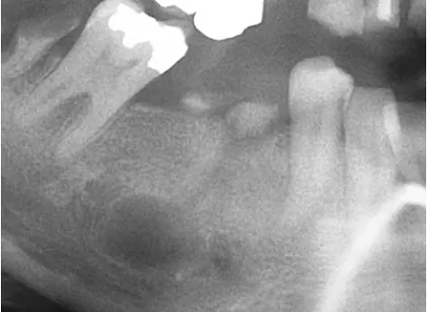
The median CRP concentration was 15.8 mg/L in the group of patients with one or more teeth with periapical radiolucency compared with 8.1 mg/L in the patients without periapical radiolucency (P=0.02). Likewise, P-albumin was slightly lower in the patients with periapical radiolucency than in those without (25 vs 28 g/L, P=0.04). Furthermore, the patients with periapical radiolucency had a higher prevalence of current cirrhosis-related complications such as ascites, episodes of hepatic encephalopathy, and/or variceal bleeding (46% vs 27%, P=0.05) (Table 2). Figure 1 shows a panoramic radiography of periapical radiolucency.
Discussion
In this study, nearly half of the patients with cirrhosis had periapical radiolucency, independently of alcoholic versus nonalcoholic etiology and cirrhosis severity. Gross caries, periodontitis, age, and smoking emerged as predictors. The presence of periapical radiolucency was associated with signs of systemic inflammation activation and with more frequent cirrhosis-related complications.
Periapical radiolucency may be caused by a variety of diseases, with apical periodontitis and radicular cysts being the most common and cementoblastoma and periapi-cal cemental dysplasia being less frequent. Still, it is not
possible to translate the radiographic findings to a specific diagnosis.22,23 In the Scandinavian countries, the reported
prevalence of apical periodontitis ranges from 1.5% to 3.4% at tooth level and 30%–50% at individual level.20,24,25 Lins
et al9 have found radiographic signs of periapical lesions in
48% of their cirrhosis cohort, which is in line with our find-ings, but below the 79% found in a study of liver transplant candidates.10 This might suggest that the frequency of
peri-apical radiolucency that we report in our cirrhosis patients is not higher than in the background population. However, it should be taken into account that the criteria for periapical radiolucency vary markedly among the available studies. In our study, periapical radiolucency was diagnosed from digital panoramic radiographs using preestablished criteria16 rather
than the “periapical index”,10,26 which is used in population
studies and which highly overestimates the prevalence of the lesions.27
Table 2 Univariable and multivariable logistic regression analyses of the association between the predictor variables and the outcome variables in periapical radiolucency
Variables Univariable Multivariable Stepwise multivariable
Odds ratio 95% CI Odds ratio 95% CI Odds ratio 95% CI
Age, per year increase Male sex (yes/no) Smoking (yes/no) Comorbidity
Visiting dentist annually (yes/no)
1.03
2.53
2.31 1.04
0.36
0.99–1.07
1.02–6.26
0.97–5.48 0.60–1.79
0.15–0.90
1.09
1.71
3.31
1.13 0.87
1.00–1.20
0.50–7.21
1.09–16.63
0.51–2.34 0.23–3.24
1.10
3.24
1.01–1.19
1.02–17.62
Gross caries
Severe periodontitis (yes/no)
2.63 3.05
1.37–5.04 1.28–7.23
2.99 3.39
1.45–6.17 1.00–16.70
3.12 3.98
1.43–6.79 1.04–15.20
Alcoholic cirrhosis (yes/no) MELD score
Current cirrhosis compilations (yes/no)
1.55 1.01
2.39
0.61–3.94 0.95–1.08
1.58–5.78
1.05 1.00
2.34
0.31–1.29 0.90–1.11
1.54–34.99 2.90 1.91–32.68
CRP, per mg/L increase P-albumin, per g/L increase
1.02 0.94
1.01–1.04 0.89–0.99
1.02 0.96
1.01–1.06 0.89–0.99
1.02 0.96
1.01–1.06 0.89–0.99
Note: Statistically significant associations are shown in bold.
Abbreviations: CI, confidence interval; CRP, C-reactive protein; MELD, Model of End-Stage Liver Disease.
Figure 1 Radiograph of right mandibular second premolar showing gross caries and periapical radiolucency.
Hepatic Medicine: Evidence and Research downloaded from https://www.dovepress.com/ by 118.70.13.36 on 22-Aug-2020
Dovepress Periapical radiolucency in cirrhosis
The registration of periapical radiolucency was collected from panoramic radiographs because of their suitability for the clinical setting, although this method may be considered as a less sensitive diagnostic tool compared with full-mouth radiographs. However, the authors have argued that the use of panoramic radiography in epidemiological studies is acceptable, and they found a high specificity and sensitivity to the detection of periapical pathology compared with full-mouth radiographs.28 The risk of underestimating the score
of periapical lesions on panoramic radiographs is thus most likely low, and the overall validity of evaluating the periapical conditions is therefore reliable.29
No association emerged between periapical radiolucency and cirrhosis etiology. This is unexpected as alcoholic cir-rhosis is often linked to a lifestyle of self-neglect, leading to a large number of carious teeth and extensive periodontal disease,7 and as autoimmune liver disorders such as primary
biliary cirrhosis are related to Sjögren’s syndrome, which may cause rapid tooth decay.4 However, no difference was
found in the feeling of dry mouth between the patients with or without periapical radiolucency.
Likewise, in this study, the MELD score was not a predictor for periapical radiolucency, although a recent study has reported a correlation between oral infections and accelerated progression of liver disease measured by the MELD score.8 However, our findings indicate that it is
the cirrhosis and the associated poor oral health status that predispose to periapical radiolucency rather than the etiol-ogy or severity of cirrhosis.
Older age and smoking were predicatively associated with periapical radiolucency in our cirrhosis patients. The importance of these factors in the general population is well documented.20,30 Likewise, oral care habits have been
asso-ciated with periapical lesions.20 This is consistent with our
study where an association was found between annual visits to the dentist and periapical radiolucency in the univariable analysis.
The predictive value of gross caries and severe peri-odontitis is mechanistically relevant as both lesions are main gateways for bacteria entering into the surrounding tissue and thereby providing access to the bloodstream.31,32
Previous studies report that apical periodontitis may lead to systemic inflammation activation.14,33,34 In accordance
with this finding and the assumption that most of our cases of periapical radiolucency were due to apical periodontitis, it is noteworthy that the cirrhosis patients with radiolucent lesions had a double level of CRP and a slightly lower level of P-albumin than those without. This has not earlier been
shown in patients with cirrhosis, but it may evidently have untoward consequences in the clinical course of the cirrhosis disease which is often fraught with infectious episodes as major precipitating factors for the well-known cirrhosis complications, thus contributing to the high morbidity and mortality of the disease.12 The level of systemic
inflamma-tion activainflamma-tion as judged from CRP and P-albumin was not very marked in the patients with periapical radiolucency. However, similar to intestinal translocation, the dental infec-tions may well be a whole-time, latent “internal” source of bacterial contamination of the bloodstream, resulting in low-grade inflammation, which is known to be a source of systemic complications.35 The present findings of more
cirrhosis-related complications among patients with periapi-cal radiolucency are in line with this view, and it may imply that more clinical attention should be directed toward oral examination and clinical care.
There are limitations to this study. First, the cross-sectional study design provides information only on asso-ciations among the study variables and not on causality. Second, the observed associations could to some extent be due to confounding variables, such as unmeasured biologi-cal variables, or socioeconomic conditions, although other studies indicate that these do not have obvious implica-tions for periapical disease.36 Finally, no intra-examiner
reliability testing was carried out. It was important that the participating patients were not disturbed more than absolutely necessary, and as the radiographs were taken by a different department it hampered the recall of the patients for intra-examiner testing. This is problematic and impairs the reliability of the examinations, even though it has been argued that the benefits of intra-examiner testing are limited.37 However, a high reliability can be expected
due to the fact that the examiner is a senior resident with extensive clinical experience. Furthermore, the examiner related his findings to written criteria throughout the whole evaluation.
In conclusion, the present results contribute new infor-mation to the sparse knowledge of periapical disease. It was observed that almost half of the patients presented periapical radiolucency. Neither cirrhosis etiology nor severity was predictors. However, periapical radiolucency was associated with systemic inflammation activation and clinical cirrhosis complications. Further studies are needed to confirm and expand these findings and to evaluate whether improved clinical care of oral health may improve the disease burden related to inflammation and infection in patients with cirrhosis.
Hepatic Medicine: Evidence and Research downloaded from https://www.dovepress.com/ by 118.70.13.36 on 22-Aug-2020
Dovepress
Grønkjær et al
Acknowledgments
The authors would like to thank dental hygienists Nanna Jensen, Natasja Nielsen, and Susanne Hedegaard for performing clinical examinations and the Department of Oral and Maxillofacial Surgery, Aarhus University Hos-pital, Aarhus, Denmark, for performing the radiographic examinations.
This work is supported by grants from Aarhus University Hospital, Aase and Ejnar Danielsen’s Foundation, A.P. Møller Foundation, Central Denmark Region Foundation for Health Research, and Novo Nordisk Foundation.
Disclosure
The authors report no conflicts of interest in this work.
References
1. Ricucci D, Mannocci F, Ford TR. A study of periapical lesions correlating the presence of a radiopaque lamina with histologi-cal findings. Oral Surg Oral Med Oral Pathol Oral Radiol Endod. 2006;101(3):289–394.
2. Stashenko P. Aetiology and pathogenesis of pulpitis and apical periodon-titis. In: Ørstavik D, Ford P, editors. Essential Endodontology. Oxford: Blackwell Science; 1998:42–67.
3. Bukmir RP, Grgic MJ, Brumini G, Spalj S, Pezelj-Ribaric S, Prso IB. Influence of tobacco smoking on dental periapical condition in a sample of Croatian adults. Wien Klin Wochenschr. 2016;128(7–8):260–265. 4. Grønkjær LL, Vilstrup H. Oral health in patients with liver cirrhosis.
Eur J Gastroenterol Hepatol. 2015;27(7):834–839.
5. Helenius-Hietala J, Meurman JH, Höckerstedt K, Lindqvist C, Isoniemi H. Effect of the aetiology and severity of liver disease on oral health and dental treatment prior to transplantation. Transpl Int. 2012;25(2):158–165.
6. Oettinger-Barak O, Barak S, Machtei EE, Ardekian L, Baruch Y, Peled M. Periodontal changes in liver cirrhosis and post-transplantation patients. I: clinical findings. J Periodontol. 2001;72(9):1236–1240. 7. Novacek G, Plachetzky U, Pötzi R, Lentner S, Slavicek R, Gangl A,
Ferenci P. Dental and periodontal disease in patients with cirrhosis – role of aetiology of liver disease. J Hepatol. 1995;22(5):576–582. 8. Åberg F, Helenius-Hietala J, Meurman J, Isoniemi H. Association
between dental infections and the clinical course and chronic liver disease. Hepatol Res. 2014;44(3):349–353.
9. Lins L, Bittencourt PL, Evangelista MA, et al. Oral health profile of cir-rhotic patients awaiting liver transplantation in the Brazilian Northeast.
Transplant Proc. 2011;43(4):1319–1321.
10. Castellanos-Cosano L, Machuca-Portillo G, Segura-Sampedro JJ, Torres-Lagares D, López-López J, Velasco-Ortega E, Segura-Egea JJ. Prevalence of apical periodontitis and frequency of root canal treat-ments in liver transplant candidates. Med Oral Patol Oral Cir Bucal. 2013;18(5):773–779.
11. Preda S, Trifan A, Girleanu I, Stanciu C, Cojocariu C. Infectious com-plications in patients with liver cirrhosis. Rev Med Chir Soc Med Nat
Iasi. 2014;118(3):590–597.
12. Jalan R, Fernandez J, Wiest R, et al. Bacterial infections in cirrhosis: a position statement based on the EASL Special Conference 2013.
J Hepatol. 2014;60(6):1310–1324.
13. Vidal F, Fontes TV, Marques TVF, Goncalves LS. Association between apical periodontitis lesions and plasmatic levels of C-reactive protein, interleukin 6 and fibrinogen in hypertensive patients. Int Endod J. 2015;24:1–9.
14. Gomes MS, Blattner TC, Santana Filho M. Can apical periodontitis modify systemic levels of inflammatory markers? A systematic review and meta-analysis. J Endod. 2013;39(10):1205–1217.
15. Grønkjær LL, Holmstrup P, Schou S, Kongstad J, Jepsen P, Vilstrup H. Periodontitis in cirrhosis. World J Gastroenterol. Epub 2016 July 13. 16. De Moor RJ, Hommez GM, De Boever JG, Delme KI, Mertens GE.
Periapical health related to the quality of root canal treatment in a Belgian population. Int Endod J. 2000;33(2):113–120.
17. Hillson S. Teeth. 2nd ed. UK: Cambridge University Press; 2005. 18. Page RC, Eke PI. Case definitions for use in population-based
surveil-lance of periodontitis. J Periodontol. 2007;78(7 Suppl):1387–1399. 19. Quan H, Li B, Couris CM, et al. Updating and validating the
Charl-son comorbidity using data from 6 countries. Am J Epidemiol. 2011; 173(6):676–682.
20. Kirkevang LL, Wenzel A. Risk indicators for apical periodontitis.
Com-munity Dent Oral Epidemiol. 2003;31(1):58–67.
21. Budtz-Jørgensen E, Keiding N, Grandjean P, Weihe P. Confounder selection in environmental epidemiology: assessment of health effects of prenatal mercury exposure. Ann Epidemiol. 2007;17(1):27–35. 22. Razavi SM, Kiani S, Khalesi S. Periapical lesions: a review of
clini-cal, radiographic, and histopathologic features. Avicenna J Dent Res. 2014;6:e19435.
23. Peters E, Lau M. Histopathologic examination to confirm diagnosis of periapical lesions: a review. J Can Dent Assoc. 2003;69(9):598–600. 24. Odesjo B, Hellden L, Salonen L, Langeland K. Prevalence of previous
endodontic treatment, technical standard and occurrence of periapical lesions in a randomly selected adult, general population. Endod Dent
Traumatol. 1990;6(6):265–272.
25. Eriksen HM, Bjertness E, Orstavik D. Prevalence and quality of end-odontic treatment in an adult urban population in Norway. Endod Dent
Traumatol. 1988;4(3):122–126.
26. Ørstavik D, Kerekes K, Eriksen HM. The periapical index: a scoring system for radiographic assessment of apical periodontitis. Endod Dent
Traumatol. 1986;2(1):20–34.
27. Gomes MS, Hugo FN, Hilgert JB, et al. Apical periodontitis and incident cardiovascular events in the Baltimore Longitudinal Study of Ageing.
Int Endod J. 2016;49(4):334–342.
28. Ahlqwist M, Halling A, Hollender L. Rational panoramic radiography in epidemiological studies of dental health. Comparison between panoramic radiographs and intraoral full mouth surveys. Swed Dent J. 1986;10(1–2):73–84.
29. Molander B, Ahlqwist M, Gröndahl HG. Panoramic and restrictive intraoral radiography in comprehensive oral radiographic diagnosis.
Eur J Oral Sci. 1995;103(4):191–198.
30. Walter C, Rodriguez FR, Taner B, Hecker H, Weiger R. Association of tobacco use and periapical pathosis – a systemic review. Int Endod J. 2012;45(12):1065–1073.
31. Kirkevang LL, Ørstavik D, Bahrami G, Wenzel A, Vaeth M. Prediction of periapical status and tooth extraction. Int Endod J. 2015;1:1–10. 32. Jansson L, Ehnevid H, Lindskog S, Blomlöf L. Relationship between
periapical and periodontal status: A clinical retrospective study. J Clin
Periodontol. 1993;20(2):117–123.
33. Van der Waal SV, Lappin DF, Crielaard W. Does apical periodontitis have systemic consequences? The need for well-planned and carefully conducted studies. Br Dent J. 2015;218(9):513–516.
34. Cotti E, Dessi C, Piras A, Mercuro G. Can a chronic dental infection be considered a cause of cardiovascular disease? a review of the literature.
Int J Cardiol. 2011;148(1):4–10.
35. Belstrøm D, Damgaard C, Nielsen CH, Holmstrup P. Does a causal rela-tion between cardiovascular disease and periodontitis exist? Microbes
Infect. 2012;14(5):411–418.
36. Frisk F, Hakeberg M. Socio-economic risk indicators for apical peri-odontitis. Acta Odontol Scand. 2006;64(2):123–128.
37. Reit C. The influence of observer calibration on radiographic periapical diagnosis. Int Endod J. 1987;20(2):75–81.
Hepatic Medicine: Evidence and Research downloaded from https://www.dovepress.com/ by 118.70.13.36 on 22-Aug-2020
Dovepress
Hepatic Medicine: Evidence and Research
Publish your work in this journal
Submit your manuscript here: https://www.dovepress.com/hepatic-medicine-evidence-and-research-journal Hepatic Medicine: Evidence and Research is an international,
peer-reviewed, open access journal covering all aspects of adult and pediatric hepatology in the clinic and laboratory including the following topics: Pathology, pathophysiology of hepatic disease; Investigation and treat-ment of hepatic disease; Pharmacology of drugs used for the treattreat-ment
of hepatic disease. Issues of patient safety and quality of care will also be considered. The manuscript management system is completely online and includes a very quick and fair peer-review system, which is all easy to use. Visit http://www.dovepress.com/testimonials.php to read real quotes from published authors.
Dove
press
Periapical radiolucency in cirrhosis
Hepatic Medicine: Evidence and Research downloaded from https://www.dovepress.com/ by 118.70.13.36 on 22-Aug-2020