Article
Efficient Single Base Editing in Mouse Using Cytosine Base Editor 4
Salah Adlat1, Ping Yang1, Yang Chen1, Rajiv Kumar Sah1, Zin Mar Oo1, May Zun Zaw Myint1, Farooq Hayel1, Noor Bahadar1, Mahmoud Al-Azab3, Fatoumata Binta Bah1, Luqing Zhang4*, Yaowu Zheng*1,2 and Xuechao Feng*1,2
1 Transgenic Research Center, School of Life Sciences, Northeast Normal University, Changchun, Jilin 130024, China, [email protected] (S.A.); [email protected] (P.Y.); [email protected] (Y.C.);
[email protected] (R.K.S.); [email protected] (Z.M.O.); [email protected] (M.Z.Z.M.); [email protected] (F.H.); [email protected] (N.B.); [email protected] (M.A.); [email protected] (F.B.B.)
2 Key Laboratory of Molecular Epigenetics of Ministry of Education, School of Life Sciences, Northeast Normal University, Changchun, Jilin 130024, China.
3 Immunology Laboratory, Guangzhou Institute of Pediatrics, Guangzhou Women and Children Medical Centre, Guangzhou, China, [email protected] (M.A.)
4 Cardiovascular Research Institute, University of California, San Francisco, CA; [email protected] (L.Z.) *Corresponding author: [email protected] (Y.W.Z.); [email protected] (X.C.F.); Tel.:+86-0431-85098583 (Y.W.Z., and X.C.F)
Abstract: Most human genetic disease arises from point mutations. These genetic diseases can theoretically be corrected by gene therapy but clinic practice is still far from mature. Nearly half of the pathogenic single-nucleotide polymorphisms (SNPs) are caused by G:C>A:T or T:A>C:G base changes. The best current methods to repair these changes are by base editing without footprint using recently developed CRISPR-Cas9 technology by David Liu’s lab. These base editing methods have been confirmed with precision and efficiency in cultured mammalian cells, but it is barely confirmed and the efficiency is still very low. Animal models are important in dissecting pathogenic mechanism for human genetic diseases and efficacy testing of base correction in vivo. Cytidine base editor BE4 is a newly developed version of cytidine base editing system that converts cytidine (C) to uridine (U) in cultured mammalian cells but has not been proven in vivo. In this study, we have tested this system in cells to inactivate GFP gene and in mice by introducing single-base substitution that leads to a stop codon in tyrosinase gene. High percentage albino colored mice were obtained from black coat-colored donor zygotes after pronuclei microinjection. Sequencing results showed that expected base changes were obtained with high precision and efficiency (56.25%). There are no off-targeting events identified in predicted off-target sites. Results confirm BE4 system can work in vivo with high precision and efficacy, and has great potentials in clinic to repair human genetic mutations.
Keywords: BE4, CRISPR-Cas9; Tyr; Cytosine base editing; Mouse model
1. Introduction
Point mutations are the most common events in human genetic diseases and nearly 50% of disease-associated mutations are C>T and G>A substitutions [1]. Animal modeling of human genetic diseases are valuable in study of pathogenic mechanism, testing of drug efficacy and proof of gene therapy reliability. CRISPR Cas9 system is an adaptive immune system in bacteria that protects its
genome from invading viruses [2,3]. CRISPR Cas9 system has been successfully applied to genetic engineering in variety of cells and organisms. Targeted insertion and mutation usually requires homologous recombination that is accomplished through cultured embryonic stem cells (ES cells), selection for positive clones and ES cell/blastocyst injection. This process is time consuming and costly. CRISPR-Cas9 system has been applied to targeted gene engineering but efficiency in vivo has been very low. Homology-directed repair (HDR) with CRISPR-Cas9 system requires DNA double-strand breaks (DSBs) at the target and a DNA template with homologous arms [4-7]. Cells respond to DSBs often with nonhomologous end joining (NHEJ) that may introduce insertions or deletions (indels) and lead to disruption of corresponding genes and reduce the expected targeting events [8,9]. In addition, HDR often leaves footprint that is not doable for base changes. David Liu’s lab has developed many versions of CRISPR-based base editing systems with different accuracy and efficacy that eventually permit precise and efficient base change in cultured cells.
CRISPR/Cas9-based cytidine base editors (CBEs) have recently been developed to generate precise base changes from cytidine to thymidine with high efficiency [1; 10; 11; 12]. CBEs system consists of a CRISPR-Cas9-derived DNA-binding module and a cytidine deaminase, and is able to introduce nucleotide substitutions of C>T [13-15] and G>A [16] without DSBs. It has been demonstrated successful in various cells and some organisms [15].
Base editing systems have gone through various stage of improvement to broaden their applicability and utility in editing of single nucleotide and have been widely applied to cell lines, various animals and plants. The fourth generation of base editor 4 (BE4) is the most advanced system in base editing with high precision and efficiency in cultured mammalian cells. BE4 has a cytidine deaminase (rAPOBEC1) with two copies of uracil glycosylase inhibitor (UGI) that are directly fused to C terminus of Cas9n, a Cas9 mutant with a D10A amino acid substitution, through a 32 amino acid linker (Fig. 1A). BE4 enables direct conversion of cytidine (C) to uridine (U) in chosen bases of DNA sequence [9] . However, feasibility and efficacy of this system has not been assessed in vivo. In the current study, we have tested and confirmed that BE4 system is able to perform a multiplexed base editing with high precision and efficiency in mice. BE4 system shows great potentials in modeling human genetic diseases and validation for gene therapy.
2. Results
2.1 Screening for efficient sgRNAs in HEK293 cells
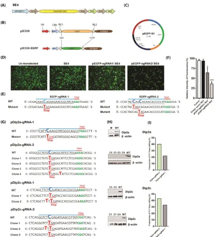
transfected with pEGFP-sgRNA1 still express relatively high levels of EGFP (64%), while cells transfected with pEGFP-sgRNA2 exhibited weak signal with an intensity of 36%, indicating EGFP was knocked down more efficiently (Fig. 1D, F). Genomic DNA was extracted for PCR and sequencing to confirm successful base editing.
Figure 1. Screening of base editing in cells. (A) Architecture of cytosine base editor 4 (BE4). (B) Replacement of
2.2 Knockout of Dip2a and Dip2c genes in tumor cells using BE4
Next, BE4 system was analyzed in murine tumor cell B16-F10. Dip2a and Dip2c genes were each targeted with two sgRNAs (Fig. 1G, Supplementary Table 1). Base substitution was screened by PCR amplification, sequencing and western blotting. pDip2a-sgRNA-1 transfection results showed Q54Z mutation with an efficiency of 22% while pDip2c-sgRNA-1 showed mutations S72F and R73Z with a total efficiency of 33%. Knockout of Dip2a and Dip2c proteins using sgRNAs-1 are shown in Fig. 1G, H, I, Supplementary Fig. 2S, and 3S. Similarly, pDip2a-sgRNA-2 and pDip2c-sgRNA-2 were transfected together with BE4 plasmid. Both pDip2a-sgRNA-2 and pDip2c-sgRNA-2 appeared to work more efficiently and induced 40% and 43% mutations at targeted sites respectively (Fig. 1G, H, I, Supplementary Fig. 4S, 5S). Expression of Dip2a and Dip2c genes from WT and mutated clones were shown in Supplementary Fig. 6S.
2.3 BE4 can induce C>T substitution in mice
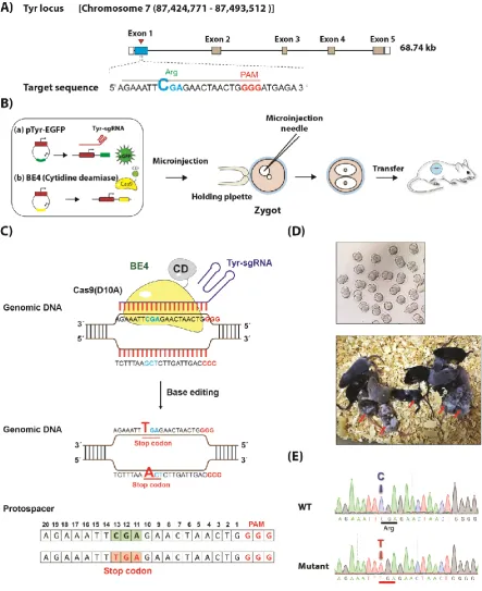
Figure 2. BE4 mediated C>T base editing in mice. (A) Schematic of sgRNA design at Tyr locus. (B) Working
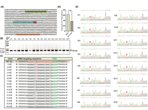
Figure 3. Screening of mutations in mice by genomic PCR and sequencing. (A) Sequences of Tyr gene target
Figure 4. Chromatogram sequencing analysis of potential off-target sites (POTs) for sgRNAs predicted according
to the online platform (https://benchling.com/)
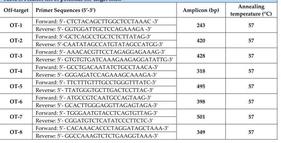
Table 3. Primers list of potential off-target sites.
Off-target Primer Sequences (5’-3’) Amplicon (bp) Annealing
temperature (°C)
OT-1 Forward: 5’- CTCTACAGCTTGGCTCCTAAAC -3’ 243 57
Reverse: 5’- GGTGGATTGCTCCAGAAAGA -3’
OT-2 Forward: 5’-GCTCAGCCTGCTCTCTTATAG-3’ 420 57
Reverse: 5’-CAATATAGCCATGTATAGCCATGG-3’
OT-3 Forward: 5’- AAACACGTTCCTAGAGGAGAAAG-3’ 428 57
Reverse: 5’- GTGTGTGATCAAAGAAGAGGATATTG-3’
OT-4 Forward: 5’- GCCTGACAATATCTGCCTAACA-3’ 318 57
Reverse: 5’- GGGAGATCCAGAAAGCAAAGA-3’
OT-5 Forward: 5’- TTCTTTGTTTGCCTGGGTTTATC-3’ 495 57
Reverse: 5’- TTATGGGTGCTTGACTCCTTAC-3’
OT-6 Forward: 5’- ATGCCGTCAATGCCAGTAAG-3’ 398 57
Reverse: 5’- GCACTTGGGAGGTTAGAGTAGA-3’
OT-7 Forward: 5’- TGGGAATGTACCTCAGTGTTAG-3’ 501 57
Reverse: 5’- CGGATGTCTCATATCCCTTCTC-3’
OT-8 Forward: 5’- CACAAACACCCTAGGATAGCTAAA-3’ 349 57
Reverse: 5’- GGCCAAAGTCTCTGAAGGTAAA-3’
5. Discussion
Majority of human genetic diseases arise from point mutations. G:C>A:T or T:A>C:G point mutations represent nearly half of all pathogenic single-nucleotide polymorphisms (SNPs) [1,17]. While most animal models gene targeting are generated by HDR using embryonic stem cells, which Table 2. List of potential off-target sites
Off-target Sequence (5’-3’) PAM Score Mismatch
is time-consuming and costly, CRISPR/Cas9 system has revolutionized the way of generating mouse models. However, CRISPR/Cas9 system can give unpredictable deletion, insertion and off target mutation. Generation of point mutation mouse model is most time consuming with low success rate [17,18]. The point mutation mouse models are the best human disease models that can precisely mimic human pathology. Gene therapy involves mostly correction of point mutations. Previous reports have demonstrated that cytosine base editing (CBE) systems are versatile and potentially applicable in cultured cells, different animal models and plants [17-21]. Moreover, CBE is a secure system that can modify genomic DNA without DSBs with very low off-target effects [10,15]. Yet, applications of base substitutions in animal models are still limited. David Liu has developed a variety of versions of base editing systems but the newest system BE4 has not been tested in vivo.
In this study, we have tested BE4 plasmid along with sgRNA expression plasmid in cell culture and applied to animal model by transgenic microinjection. We designed a precise base editing method which knockout tyrosinase gene and results in loss-of-pigmentation (Albinism). High percentage of albino offspring was seen. We achieved successful C>T transition with high efficiency. C>T conversions have occurred exclusively within the approximate editing window of protospacers (positions~4–8). BE-mediated STOP-codon generation disrupts gene function by converting C to T in coding sequences (CAG, CAA, CGA). The study demonstrates that BE4 system is highly efficient and most precise in base editing. In addition, the stop codon generation provides a secure approach to generate knockout animal models with minimum change of genome structure, mimicking most of the genetic diseases [21-23]. Our results highlighted that BE4 system can introduce site-specific and single-base substitution with high precision and efficiency in mouse embryos with no off-target mutation. This study demonstrate the great values of BE4 editing in human disease modeling and future gene therapy.
4. Materials and Methods 4.1 Animals
All mice and experimental protocols used in this project has been approved by Institutional Animal Care and Use Committee for Animal Experimental Ethics Committee of Northeast Normal University (NENU/IACUC, AP2018011) and carried out in accordance with recommendations in Guide for Care and Use of Laboratory Animals of National Institutes of Health as well. Mice were bred and maintained under specific pathogen-free condition in animal facility with controlled temperature at 21±1°C, 30%-60% humidity, 12:12 light/dark cycles and free access to food and water.
4.2 Reagents
4.3 Plasmid construction and sgRNA design
Cas9 coding region of pX330 plasmid (Gifted from Dr. Feng Zhang, Addgene accession no. 42230) was replaced with EGFP cDNA (Fig. 1B). EGFP sequence was PCR amplified from pEGFP-N1 (Clontech cat# 6059-1) (Fig. 1C) using following primers: EGFP-F:
5’-GGCCACCGGTGATCCACCGGTCGCCACCAT-3’ (20bp) and EGFP-R: 5’-
GGCCGAATTCTTACTTGTACAGCTCGTCCATG-3’ (22bp) with AgeI site at 5’-end and EcoRI site at 3’-end (AgeI and EcoRI are shown by underline). PCR was performed at 94°C for 4min, 24 cycles of 94°C for 30s, 56°C for 30s, 72°C for 1min and 72°C for 10min. EGFP PCR products were digested with AgeI and EcoRI (NEB) and inserted into AgeI and EcoRI sites of pX330. Resultant pX330-EGFP plasmid (Fig. 1B) was confirmed by sequencing. Oligos coding for sgRNA targets were synthesized by Genewiz (Beijing, China), annealed at 95°C for 5min and ramped down to 25°C (-5°C/min) and then subcloned into BbsI sites of pX330-EGFP. BE4 plasmid (Fig. 1A) was gifted from David Liu lab (Addgene access no. 100802). The sgRNAs were designed using online platform https://benchling.com/ and all sgRNAs oligos are listed in Supplementary Table 1.
4.4 Cells culture and EGFP stable expression
Human embryonic kidney (HEK293) cells were from American Type Culture Collection (ATCC CRL-1573, Manassas, USA) and cultured in Dulbecco’s modified Eagle’s Medium (DMEM, Sigma) supplemented with fetal bovine serum (10%) and penicillin/streptomycin (Gibco, Life Technologies). Cells were maintained at 37oC and 5% CO2 in a humidified incubator. To stably express EGFP in
HEK293, cells were seeded in 12-well plates with 1ml of DMEM. When cells reached 60–80% confluency, medium was replaced with Opti-MEM (Gibco, Life Technologies). Then cells were transfected using Lipofectamine 2000 (Invitrogen) according to the manufacturer’s protocol. One μg of pEGFP-N1 was transfected with 2μL Lipofectamine 2000. Medium was replaced with fresh DMEM medium with serum 6hrs after transfection. 48hrs later, cells were treated with G418 (500 μg/mL, Sigma) for 15 days with medium changed every 3 days. Colonies were picked into 96 wells and expanded into 6-well plates before genomic DNA extraction, PCR amplification, and sequencing.
4.5 Plasmid transfection
SgRNA oligos (Supplementary Table 1) were annealed and cloned into pX330-EGFP plasmid. HEK293 and B160F10 cells were transfected according to the manufacturer’s protocols (Invitrogen, Cat. No. 11668-027). In brief, HEK293 and murine B16-F10 cells were seeded on 12-well plates in 1ml of DMEM. When cells reached 60–80% confluency, medium was changed to Opti-MEM. Cells were then transfected with Lipofectamine 2000 (Invitrogen) according to manufacturer’s protocol. One μg pX330-sgRNAs and 2μg BE4 plasmids were mixed with 2μL Lipofectamine 2000. Six hours later, medium was replaced with fresh DMEM. Cells were then subjected to G418 treatment as described above.
4.6 Oocyte/DNA microinjection and oviduct transfer
CO., Ltd, Ningbo, Zhengjiang, China) and followed by 5IU of human chorionic gonadotropin (Ningbo Hormone Products CO., Ltd, Ningbo, Zhengjiang, China) 48hrs later. Superovulated B6D2F1 females were crossed with B6D2F1 males. Fertilized eggs at pronucleus stage were collected in M2 medium. Mixtures of pTyr-gRNAs (2.35ng/ul) and BE4 plasmids (2.64ng/ul) were injected into nucleus in a droplet of M2 medium using inverted microscope equipped with a pair of micromanipulators (Olympus, Tokyo, Japan). Then the injected embryos were incubated in M16 culture medium at 37 °C, 6% CO2 overnight, followed by transfer into the oviduct of a recipient
mother at two-cell stage.
4.7 Genomic DNA extraction and genotyping
Genomic DNA was extracted from mouse tail tips using G-NTK lysis buffer [24] and proteinase K (1mg/ml) (Beijing Solarbio Science & Technology Co., Ltd., Beijing, China) at 55oC overnight. Proteinase K was deactivated at 95°C for 15min and PCR was performed in 25μl reaction volume with diluted tail DNA and genotyping primers (supplementary table 2). PCR master mix was as follow: 1.2μl of each primer (10μM), 16.4μl of ddH2O, 1.5μl of 25mM MgCl2, 2.5μl of 10X PCR buffer,
0.5μl of 10mM dNTP Mix and 0.25μl of Taq DNA Polymerase. The PCR conditions were as follows: 95ºC for 5 min, 32 cycles of 95ºC 30sec, 58ºC 30sec and 72ºC 30sec, and 72ºC 10min using PCR machine by Bio-Rad, Hercules, CA, USA.
4.8 RNA extraction
One ml RNAiso plus reagent (Takara, Dalian, China) was added to cells on 100mm Petri dish. Cell lysates were collected and incubated at room temperature for 5min. Cells were then centrifuged at 13500 ×g for 5min at 4°C. A 200μL of CHCl3 was added, followed by 30sec mixing
and 5min incubation at RT, samples were centrifuged at 13500 ×g for 15min at 4°C to separate RNA into aqueous phase. Aqueous phase (about 600ul) was transferred to a new tube and RNA was precipitated with 750 μL of absolute isopropanol at RT for 10min and then centrifuged at 13500 ×g for 10min at 4°C. Precipitate was washed with 1mL 70% ethanol, followed by centrifugation at 13500 ×g for 5min at 4°C. RNA pellet was resuspended in 50μL of DEPC -treated water. RNA concentrations were determined using NanoDrop 2000 (Thermo Fisher Scientific, USA). RNA integrity was checked on 0.8% agarose gel.
4.9 RT-qPCR
One μg of total RNA was reverse-transcribed into first-strand complementary DNA (cDNA) with Prime Script RT Reagent Kit (Perfect Real Time, TaKaRa, Dalian, China) according to the manufacturer’s instructions. Real-time PCR was performed with 50ng of cDNA using One-Step SYBR PrimeScriptTM RT-PCR kit (Takara, Dalian, China). All reactions were performed in triplicate. All primers were initially evaluated for efficiency using relative standard curve and electrophoresis on gel. Primer sequences are listed in Supplementary Table 3.
Purified PCR products were extracted using gel extraction kit (Qiagen, Germany) and cloned into pMD18-T plasmid (TaKaRa, Dalian, China). Positive clones were sequenced in two directions utilizing M13 forward and reverse primers. Mutations were identified by alignment to wild-type sequences.
4.11 Western blot
Total proteins were extracted using RIPA buffer (0.5% Nonidet P-40, 0.1% sodium deoxycholate, 150mM NaCl, 50mM Tris-Cl, pH7.5 and 1x protease inhibitor cocktail). Cell lysates were subjected to high-speed centrifugation at 12000xg for 15min at 4°C. Protein concentrations were measured using Coomassie (Bradford) protein assay kit. Total soluble proteins were then separated on 10% SDS-PAGE and transferred into polyvinylidene difluoride membrane (Millipore, Billerica, MA). Membrane was blocked with 5% nonfat dry milk for 1h followed by incubation with diluted the primary antibodies (β-actin, 1:2000, Signalway antibody; Dip2a, 1:500, Novus; Dip2c, 1:1000, Abcam) for overnight at 4°C. Then the membrane was washed in TBST for three times, 5 min each and then incubated with secondary antibody (anti-rabbit horseradish peroxidase conjugate, 1:5,000; anti-mouse horseradish peroxidase conjugate, 1:5,000; Transgene) for 30min, followed by washing three times with TBST. Signals were detected using enhanced chemiluminescence Amersham™ ECL™ (GE Healthcare, USA) reagents. β-actin protein served as a loading control.
4.12 Off-target detection
Eight potential off-target sites (POTs) were identified according to an online design tool (https://benchling.com/). Selected POTs (Table 2) were amplified by PCR and sequenced. Sequences were compared with wild type. All primers used for off-target assay were listed in Table 3.
4.13 Statistical Analysis
Statistical analyses and graphics were performed with GraphPad Prism 5.01 (GraphPad Software Inc.) and SPSS software version 25.0 (IBM Inc., New York, USA). Parametric unpaired Student’s t test was used to assess difference between the groups. P-values were two-sided; a P-value < 0.05 was considered statistically significant
Supplementary Materials: Supplementary materials can be found online.
Author Contributions: “Conceptualization, S.A., L.Z., Y.Z. and X.F.; methodology S.A., P.Y., Y.C. R.K.S, and
M.A.; software S.A., Z.O., M.Z.Z.M, R.K.S., M.A. and F.B.B.; validation L.Z., Y.Z., and X.F.; formal analysis, S.A., F.H, N.B. and M.A.; investigation, S.A., P.Y., Y.C.; resources, Y.Z. and X.F.; writing—original draft preparation, S.A., L.Z., Y.Z. and X.F.; writing—review and editing, S.A., L.Z., Y.Z. and X.F.; visualization, S.A.; supervision, L.Z., Y.Z. and X.F.; project administration, L.Z., Y.Z. and X.F.; funding acquisition, L.Z., Y.Z. and X.F. All authors have read and agreed to the published version of the manuscript.”
Funding: This work was funded supported in part or in whole by National Natural Science Foundation of China
(81270953), the Natural Science Foundation of Jilin Province (20160101344JC) and Science and Technology Project of Jilin Provincial Education Department (JJKH20180023KJ). The funders had no roles in this study, including study design, data collection, data analysis or decision to publish or preparation of the manuscript.
Acknowledgments: We are grateful to Ms. Huiyan Wu for microinjection and managing mouse colony.
Abbreviations
CRISPR Clustered Regularly Interspaced Short Palindromic Repeats Cas9 CRISPR-associated protein 9
SNPs Single-Nucleotide Polymorphisms BE4 Base Editor4
DSBs DNA double-strand breaks HDR Homology-directed repair NHEJ Nonhomologous end joining CBEs Cytosine Base Editors SgRNA Single guided RNA
EGFP Enhanced Green Fluorescence Protein POTs Potential Off-Target sites
UGI Uracil Glycosylase Inhibitor HEK293 Human Embryonic Kidney 293 Dip2a Disco Interacting Protein 2 Homolog a Dip2c Disco Interacting Protein 2 Homolog c
References
1. Gaudelli, N.M.; Komor, A.C.; Rees, H.A.; Packer, M.S.; Badran, A.H.; Bryson, D.I.; Liu, D.R. Programmable base editing of A• T to G• C in genomic DNA without DNA cleavage. Nature 2017, 551, 464.
2. Rath, D.; Amlinger, L.; Rath, A.; Lundgren, M. The CRISPR-Cas immune system: biology, mechanisms and applications. Biochimie 2015, 117, 119-128.
3. Sontheimer, E.J.; Barrangou, R. The bacterial origins of the CRISPR genome-editing revolution. Hum Gene Ther 2015, 26, 413-424.
4. Wu, H.; Liu, Q.; Shi, H.; Xie, J.; Zhang, Q.; Ouyang, Z.; Li, N.; Yang, Y.; Liu, Z.; Zhao, Y. Engineering CRISPR/Cpf1 with tRNA promotes genome editing capability in mammalian systems. Cell Mol Life Sci
2018, 75, 3593-3607.
5. Yang, Y.; Wang, K.; Wu, H.; Jin, Q.; Ruan, D.; Ouyang, Z.; Zhao, B.; Liu, Z.; Zhao, Y.; Zhang, Q. Genetically humanized pigs exclusively expressing human insulin are generated through custom endonuclease-mediated seamless engineering. J Mol Cell Biol 2016, 8, 174-177.
6. Yang, L.; Guell, M.; Byrne, S.; Yang, J.L.; De Los Angeles, A.; Mali, P.; Aach, J.; Kim-Kiselak, C.; Briggs, A.W.; Rios, X. Optimization of scarless human stem cell genome editing. Nucleic Acids Res 2013, 41, 9049-9061.
7. Yin, H.; Xue, W.; Chen, S.; Bogorad, R.L.; Benedetti, E.; Grompe, M.; Koteliansky, V.; Sharp, P.A.; Jacks, T.; Anderson, D.G. Genome editing with Cas9 in adult mice corrects a disease mutation and phenotype. Nat Biotechnol 2014, 32, 551.
8. Davis, A.J.; Chen, D.J. DNA double strand break repair via non-homologous end-joining. Translational cancer research 2013, 2, 130.
9. Komor, A.C.; Zhao, K.T.; Packer, M.S.; Gaudelli, N.M.; Waterbury, A.L.; Koblan, L.W.; Kim, Y.B.; Badran, A.H.; Liu, D.R. Improved base excision repair inhibition and bacteriophage Mu Gam protein yields C: G-to-T: A base editors with higher efficiency and product purity. Sci Adv 2017, 3, eaao4774. 10. Komor, A.C.; Kim, Y.B.; Packer, M.S.; Zuris, J.A.; Liu, D.R. Programmable editing of a target base in
11. Nishida, K.; Arazoe, T.; Yachie, N.; Banno, S.; Kakimoto, M.; Tabata, M.; Mochizuki, M.; Miyabe, A.; Araki, M.; Hara, K.Y. Targeted nucleotide editing using hybrid prokaryotic and vertebrate adaptive immune systems. Science 2016, 353, aaf8729.
12. Ma, Y.; Zhang, J.; Yin, W.; Zhang, Z.; Song, Y.; Chang, X. Targeted AID-mediated mutagenesis (TAM) enables efficient genomic diversification in mammalian cells. Nat Methods 2016, 13, 1029.
13. Xie, J.; Ge, W.; Li, N.; Liu, Q.; Chen, F.; Yang, X.; Huang, X.; Ouyang, Z.; Zhang, Q.; Zhao, Y. Efficient base editing for multiple genes and loci in pigs using base editors. Nat Commun 2019, 10, 2852. 14. Kim, K.; Ryu, S.-M.; Kim, S.-T.; Baek, G.; Kim, D.; Lim, K.; Chung, E.; Kim, S.; Kim, J.-S. Highly efficient
RNA-guided base editing in mouse embryos. Nat Biotechnol 2017, 35, 435.
15. Kim, D.; Lim, K.; Kim, S.-T.; Yoon, S.-h.; Kim, K.; Ryu, S.-M.; Kim, J.-S. Genome-wide target specificities of CRISPR RNA-guided programmable deaminases. Nat Biotechnol 2017, 35, 475.
16. Liu, Z.; Lu, Z.; Yang, G.; Huang, S.; Li, G.; Feng, S.; Liu, Y.; Li, J.; Yu, W.; Zhang, Y. Efficient generation of mouse models of human diseases via ABE-and BE-mediated base editing. Nat Commun 2018, 9, 2338. 17. Liu, Z.; Chen, M.; Chen, S.; Deng, J.; Song, Y.; Lai, L.; Li, Z. Highly efficient RNA-guided base editing
in rabbit. Nat Commun 2018, 9, 2717.
18. Zhang, Y.; Qin, W.; Lu, X.; Xu, J.; Huang, H.; Bai, H.; Li, S.; Lin, S. Programmable base editing of zebrafish genome using a modified CRISPR-Cas9 system. Nat Commun 2017, 8, 118.
19. Zong, Y.; Wang, Y.; Li, C.; Zhang, R.; Chen, K.; Ran, Y.; Qiu, J.-L.; Wang, D.; Gao, C. Precise base editing in rice, wheat and maize with a Cas9-cytidine deaminase fusion. Nat Biotechnol 2017, 35, 438.
20. Li, G.; Liu, Y.; Zeng, Y.; Li, J.; Wang, L.; Yang, G.; Chen, D.; Shang, X.; Chen, J.; Huang, X. Highly efficient and precise base editing in discarded human tripronuclear embryos. Protein Cell 2017, 8, 776-779.
21. Yang, G.; Zhu, T.; Lu, Z.; Li, G.; Zhang, H.; Feng, S.; Liu, Y.; Li, J.; Zhang, Y.; Chen, J. Generation of isogenic single and multiplex gene knockout mice by base editing-induced STOP. Sci Bull 2018, 63, 1101-1107.
22. Kuscu, C.; Parlak, M.; Tufan, T.; Yang, J.; Szlachta, K.; Wei, X.; Mammadov, R.; Adli, M. CRISPR-STOP: gene silencing through base-editing-induced nonsense mutations. Nat Methods 2017, 14, 710.
23. Billon, P.; Bryant, E.E.; Joseph, S.A.; Nambiar, T.S.; Hayward, S.B.; Rothstein, R.; Ciccia, A. CRISPR-mediated base editing enables efficient disruption of eukaryotic genes through induction of STOP codons. Mol Cell 2017, 67, 1068-1079. e1064.