Article
Susceptibility Analysis of COVID-19 in Smokers
Based on ACE2
Jin Wang 1, Qiulin Luo1, Rui Chen 2, Tao Chen 1 and Jian-xiang Li 1*
1 Department of Toxicology, School of Public Health, Medicine College, Soochow University, Suzhou,
215123, China.
2 Department of Respiratory Medicine, The Second Affiliated Hospital of Soochow University, Suzhou,
215004, China;
* Correspondence: [email protected]; Tel.: +86-0512-65881038
Abstract: Background: Cigarette smoking (CS) is a global public health problem and a high-risk factor for various diseases. In December 2019, a novel coronavirus (COVID-19) was identified in Wuhan, China. Because ACE2 has been identified as a receptor for COVID-19, we hypothesize that CS affects the expression pattern of ACE2 in respiratory tract, causing differences in susceptibility to the virus. Methods: Three datasets (GSE994, GSE17913, and GSE18344), were downloaded from the Gene Expression Omnibus (GEO) database. Correlation and enrichment analysis were used to evaluate the function of ACE2. Also, the different expression of ACE2 in different groups of three datasets were analyzed. Results: Genes associated with ACE2 were enriched in important biological processes such as viral processes and immune response. Elevated ACE2 were found in intrapulmonary airways (GSE994) and oral epithelial cells (GSE17913) of smokers but not those of non-smokers or former smokers. Significant dose- and time-dependent relationships between CS and ACE2 expression were observed in mouse lung tissues, and long periods without smoking were found to significantly reduce ACE2 expression. Conclusions: Both human and rat data confirmed that CS could induce increased ACE2 in the respiratory tract, indicating that smokers have a higher susceptibility to COVID-19.
Keywords: Cigarette smoke; ACE2; COVID-19; susceptibility
1. Introduction
Cigarette smoking is a global public health problem. There are about 1.3 billion smokers in the world, and about one third of them live in China, which directly threatens the health of smokers and passive smokers [1]. The health problems caused by smoking are a hot issue of public concern, but it is still difficult to solve because it involves personal living habits. Cigarette smoking can cause lung cancer and cardiovascular and reproductive system problems [2]. Studies have demonstrated that smokers are susceptible to Middle East Respiratory Syndrome Coronavirus (MERS-CoV) [3].
Coronaviruses can infect humans, various birds, and mammals worldwide. In February and March 2003 there were major outbreaks of Severe Acute Respiratory Syndrome (SARS) in many countries. As of July 31, 2003, 8098 probable cases were reported, with a death toll of 774 (9.6%)[4]. Between March 2012 and November 2015, MERS broke out in several countries, including Saudi Arabia (1255 cases and 539 deaths), South Korea (185 cases and 36 deaths), and the United Arab Emirates (81 cases and 11 deaths) [5]. In December of 2019, a novel coronavirus (2019-nCov, COVID-19) was identified in Wuhan, Hubei Province, China. As of February 23, 2020, COVID-19 has infected 77,262 people and caused 2595 deaths in China, with rapidly growing numbers of cases reported internationally. People of all ages can be infected by the new coronavirus (COVID-19). Elderly people and people with pre-existing diseases, such as asthma, diabetes, and heart disease seem to be more susceptible to the virus or have more severe symptoms [6]. There was also a significant gender
difference in incidence with 0.31 (male) vs. 0.27 (female) patients per 100,000 people [7]. The difference might be caused by smoking behavior.
The COVID-19 genome is approximately 80% identical to SARS-CoV and approximately 96% identical to bat coronavirus [8]. Structural analyses have established atomic-level interactions between SARS-CoV spike glycoprotein (S protein) receptor-binding domain (RBD) and its receptor, angiotensin-converting enzyme 2 (ACE2), which contribute to the cross-species and human-to-human transmissions of SARS-CoV [9,10]. It has been shown that the S protein of COVID-19, which is the same as that of SARS-CoV, may exploit ACE2 for host infection [11-13]. One recent study suggests that the affinity between ACE2 and the RBD of COVID-19 is 10–20 times greater than that of SARS-CoV [13]. The level of expression of ACE2 may reflect the susceptibility to COVID-19.
In this study, we analyzed the patterns of expression of ACE2 in the respiratory tract tissues of humans and mice with different smoking states based on three Gene Expression Omnibus (GEO) datasets. We aimed to determine whether cigarette smoking is a susceptibility factor for COVID-19.
2. Materials and Methods 2.1 Data source
Three GEO datasets, GSE994, GSE17913, and GSE18344, were obtained from the GEO database (http://www.ncbi.nlm.nih.gov/geo). The samples in GSE994 were obtained from intrapulmonary airways from normal smoking and non-smoking volunteers (including 34 current smokers, 23 never smokers, and 18 former smokers). The overall design of GSE17913 involved oral biopsy from 40 current smokers and 40 age- and gender-matched never smokers. We also extracted 55 samples from 14 different groups in the GSE18344 dataset, including a sham group (sham) and exposure group. The mice in the exposure group were continuously exposed to cigarette smoke (CS, 750 μg total particulate matter/L) for 2, 3, or 4 h/day (our low, medium, and high dose groups, respectively). Exposure time included 1 day, 2 months, and 5 months, as well as 5 months + 1 day recovery and 5 months + 13 days recovery (medium dose only). There were four replicates for each group in GSE18344.
2.2 Correlation and enrichment analysis
R software was used to identify and import the list of genes correlated with ACE2 in the GSE994 and GSE17913 datasets. These related genes were then imported into the DAVID online tool for Gene Oncology (GO) enrichment analysis.
2.3 Analysis of ACE2 expression in patients with different smoking histories
All samples in the GSE994 and GSE17913 datasets were grouped according to smoking history. GSE994 was divided into three groups, including (1) never smokers (never), (2) current smokers (current), and (3) former smokers (former); GSE17913 was divided into two groups including (1) never smokers (never) and (2) current smokers (current). Changes in ACE2 expression were evaluated between groups in the datasets.
2.4 Statistical analyses and plots
In the correlation analysis, genes with a |correlation coefficient| > 0.3 and P < 0.05 were considered significantly related genes. The t-test in GraphPad Prism 7 software was used to analyze the correlation between smoking history and ACE2 gene expression in the GSE994 and GSE17913 datasets and the differences in gene expression between different treatment groups in the GSE18344 dataset. P <0.05 is considered statistically significant
3.1. Functional enrichment of ACE2-related genes
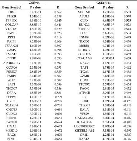
A total of 1370 positively and 1871 negatively ACE2-related genes were identified in the GSE994 intrapulmonary airway dataset, and 544 positively and 182 negatively related genes were found in the GSE17913 oral epithelial cell dataset (Table 1, 2). The genes in GSE994 were significantly enriched in essential biological processes including cell–cell adhesion, viral processes, viral transcription, intracellular transport of viruses, and the TGF-β receptor signaling pathway (Figure 1A). In GSE17913, the genes were significantly enriched in biological processes including immune response, viral processes, T cell immunity, and apoptosis (Figure 1B).
Table 1 Summary of genes related to ACE2 expression in two datasets
Datasets Positive Negative
GSE994 1370 1871
GSE17913 544 182
Table 2 Top 20 genes related to ACE2 expression in two datasets
GSE994 GSE17913
Gene Symbol P value R Gene Symbol P value R
CBX3 6.45E-11 0.667 SECTM1 1.71E-08 0.583
PHKB 1.34E-10 0.659 APOL1 4.28E-08 0.570
PPP1CC 4.16E-10 0.645 CLPX 6.65E-07 0.525
GOLGA7 5.83E-10 0.641 RUNX3 7.65E-07 0.523
CDC123 1.50E-09 0.629 BTN3A3 1.32E-06 0.513
RAP1B 2.32E-09 0.623 EDC3 2.16E-06 0.504
PPT1 4.17E-09 0.616 PSMB9 8.02E-06 0.479
PAIP1 8.23E-09 0.606 TLCD2 9.34E-06 0.476
TSPAN31 1.60E-08 0.597 MSRB1 9.74E-06 0.475
CASP7 1.63E-08 0.596 S100A12 1.02E-05 0.474
TM9SF3 2.05E-08 0.593 CORO1A 1.31E-05 0.469
COPB1 2.09E-08 0.593 CEACAM7 0.000014 0.468
APOBEC3G 2.15E-08 0.592 NKG7 1.62E-05 0.464
CCDC6 2.33E-08 0.591 TAP1 1.78E-05 0.462
PSMD7 2.81E-08 0.589 ITGAL 2.17E-05 0.458
PARP1 3.14E-08 0.587 GZMB 2.18E-05 0.458
ADO 3.21E-08 0.587 CUX1 2.21E-05 0.458
SAT1 3.35E-08 0.586 TTC39C 2.59E-05 0.454
THOC7 3.39E-08 0.586 PAOX 2.91E-05 0.452
DERA 4.53E-08 0.581 ATP10B 3.29E-05 0.449
SEPW1 1.10E-12 -0.709 C7 7.17E-05 -0.432
CRIP1 1.66E-12 -0.705 BUB1 1.02E-04 -0.423
SCAMP4 2.59E-12 -0.701 CHRM5 1.38E-04 -0.416
CNOT3 6.50E-12 -0.691 RALA 1.51E-04 -0.414
CLDN3 7.30E-12 -0.690 VPS29 1.56E-04 -0.413
STRN4 1.78E-11 -0.681 CADM3-AS1 2.00E-04 -0.407
CABIN1 3.49E-11 -0.674 KIAA1456 2.55E-04 -0.400
ITGA3 3.61E-11 -0.673 LOC101929002 2.70E-04 -0.399
MFSD10 4.01E-11 -0.672 KIRREL3-AS2 3.13E-04 -0.395
BAG6 4.89E-11 -0.670 OR1I1 4.28E-04 -0.387
NPIPA1 9.88E-11 -0.662 LOC200830 4.40E-04 -0.386
ABHD11 1.89E-10 -0.655 GRK5 4.57E-04 -0.385
CST3 2.20E-10 -0.653 RP11-214K3.19 4.85E-04 -0.384
MED12 2.59E-10 -0.651 ANXA2 5.36E-04 -0.381
RNH1 2.95E-10 -0.649 RP11-354I10.1 6.03E-04 -0.378
MORN1 3.09E-10 -0.649 AGAP11 6.27E-04 -0.376
TJP3 4.07E-10 -0.645 ZNF582-AS1 6.54E-04 -0.375
PYY 4.92E-10 -0.643 FKSG49 6.63E-04 -0.375
GLB1L2 6.76E-10 -0.639 PEG3 6.75E-04 -0.374
Figure 1. Histogram of Gene Ontology enrichment analysis of genes associated with ACE2 expression in the (A) GSE994 and (B) GSE17913 datasets.
3.2. ACE2 expression and correlation with smoking history
In the GSE994 intrapulmonary airway dataset, the level of expression of ACE2 in current smokers is significantly higher than that in never smokers (t = 2.295, P = 0.026) (Figure 2A). There was no significant difference between never smokers and reformed smokers. In addition, the ACE2 expression level was much lower in reformed smokers than in current smokers (t = 2.709, P = 0.001) (Figure 2A). In the GSE17913 oral epithelial cell dataset, the level of expression of ACE2 in current smokers was significantly higher than in never smokers (t = 3.674, P < 0.001) (Figure 2B).
Figure 2. ACE2 expression levels in volunteers with different smoking history in the (A) GSE994 and (B) GSE17913 datasets.
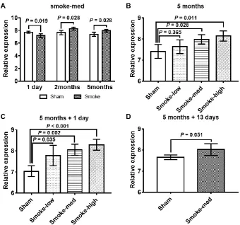
After 1 day of CS treatment, the level of expression of ACE2 in the medium CS-exposed group was significantly downregulated relative to the control group (Figure 3A) However, ACE2 expression level was significantly higher in the medium CS-exposed group after 2 and 5 months of treatment (Figure 3A). After 5 months of CS exposure, ACE2 expression levels in the medium- and high-dose groups were upregulated in a dose-dependent manner (Figure 3B). A significant dose-dependent increase in ACE2 expression was also observed in the lungs of mice exposed to 5 months CS plus 1 day recovery (Figure 3C). In addition, the ACE2 expression level was higher in the medium-dose group after 5 months CS treatment plus 13 days of recovery (Figure 3D).
Figure 3. Data analysis of smoking-exposed mouse model. (A) ACE2 expression levels measured at different times after medium smoking (smoke-med) exposure; (B) ACE2 expression levels in different dose groups after 5 months of smoking exposure; (C) ACE2 expression levels in different dose groups after 5 months of smoking exposure and 1 day of recovery; (D) ACE2 expression level in the smoke-med group after 5 months of smoking exposure and 13 days of recovery.
4. Discussion
Studies have established that ACE2 is the receptor for the SARS-CoV [9,10] and Wuhan new coronavirus (COVID-19) [11-13]. Based on correlation and enrichment analysis of two human datasets (GSE994 and GSE17913), we found smoke-induced changes in ACE2 expression to be correlated with essential biological processes including viral processes and immune response, indicating that ACE2 is involved in virus infection and immune response.
It is well known that CS is a high risk factor to such diseases as cardiovascular disease, chronic obstructive pulmonary disease, and cancer [17]. Studies have confirmed the relationship between CS and the susceptibility of influenza infections [18]. In addition, CS affects platelet-activating factor metabolism and may contribute to the elevated incidence of bacterial superinfection in people who develop influenza [19]. Moreover, influenza antibodies decreased more rapidly in smokers than in nonsmokers [20]. Another study found that CS can damage the host’s antiviral response, contributing to the increased rate of influenza infection and the incidence of lower respiratory tract disease in smokers [21]. In this study, elevated ACE2 expression was found in intrapulmonary airways and oral epithelial cells in smokers compared with non-smokers, indicating that smokers are susceptible to 2019-CoV. Importantly, the ACE2 expression level was lower in reformed smokers, suggesting that quitting smoking can reduce susceptibility to 2019-CoV. Animal experiments have also shown significant dose- and time-dependent relationships between CS exposure and ACE2 expression in the lung tissues of mice. Quitting smoking for a long time but not short time could reverse the overexpression of ACE2 in the lungs of mice.
5. Conclusions
In conclusion, our results indicated that CS could induce elevated ACE2 expression in the respiratory tract, indicating that smokers have a higher susceptibility to COVID-19 than non-smokers. Since CS-induced changes in ACE2 expression are associated with viral infection and immune processes, smokers infected with COVID-19 may have serious health problems. Further epidemiological data are needed to verify these findings.
Author Contributions: Conceptualization, T.C. and J.L.; methodology, J.W. and Q.L.; software, J.W.; writing—
original draft preparation, J.W. and Q.L.; writing—review and editing, T.C., R.C. and J.L.; funding acquisition, R.C. and J.L. All authors have read and agreed to the published version of the manuscript.
Funding: This study was supported by the National Natural Science Foundation of China (81573178 and 81770085).
Acknowledgments: We thank LetPub (www.letpub.com) for its linguistic assistance during the preparation of this manuscript.
Conflicts of Interest: The authors declare no conflict of interest.
References
1. Zhang, H.; Cai, B. The impact of tobacco on lung health in China. Respirology 2003, 8, 17-21,
doi:10.1046/j.1440-1843.2003.00433.x.
2. IARC. Tobacco smoke and involuntary smoking. IARC Monogr Eval Carcinog Risks Hum 2004, 83, 1-1438.
3. Alraddadi, B.M.; Watson, J.T.; Almarashi, A.; Abedi, G.R.; Turkistani, A.; Sadran, M.; Housa, A.;
Almazroa, M.A.; Alraihan, N.; Banjar, A., et al. Risk Factors for Primary Middle East Respiratory
Syndrome Coronavirus Illness in Humans, Saudi Arabia, 2014. Emerg Infect Dis 2016, 22, 49-55,
doi:10.3201/eid2201.151340.
4. Hui, D.S.; Chan, M.C.; Wu, A.K.; Ng, P.C. Severe acute respiratory syndrome (SARS): epidemiology
and clinical features. Postgrad Med J 2004, 80, 373-381, doi:10.1136/pgmj.2004.020263.
5. Kim, K.H.; Tandi, T.E.; Choi, J.W.; Moon, J.M.; Kim, M.S. Middle East respiratory syndrome
coronavirus (MERS-CoV) outbreak in South Korea, 2015: epidemiology, characteristics and public
health implications. J Hosp Infect 2017, 95, 207-213, doi:10.1016/j.jhin.2016.10.008.
6. Novel Coronavirus (2019-nCoV) advice for the public:Myth busters. Availabe online:
https://www.who.int/emergencies/diseases/novel-coronavirus-2019/advice-for-public/myth-busters.
7. Yang, Y.; Lu, Q.; Liu, M.; Wang, Y.; Zhang, A.; Jalali, N.; Dean, N.; Longini, I.; Halloran, M.E.; Xu, B., et
al. Epidemiological and clinical features of the 2019 novel coronavirus outbreak in China. 2020,
10.1101/2020.02.10.20021675 %J medRxiv, 2020.2002.2010.20021675, doi:10.1101/2020.02.10.20021675 %J
medRxiv.
8. Zhou, P.; Yang, X.L.; Wang, X.G.; Hu, B.; Zhang, L.; Zhang, W.; Si, H.R.; Zhu, Y.; Li, B.; Huang, C.L., et
al. A pneumonia outbreak associated with a new coronavirus of probable bat origin. Nature 2020,
10.1038/s41586-020-2012-7, doi:10.1038/s41586-020-2012-7.
9. He, L.; Ding, Y.; Zhang, Q.; Che, X.; He, Y.; Shen, H.; Wang, H.; Li, Z.; Zhao, L.; Geng, J., et al. Expression
of elevated levels of pro-inflammatory cytokines in SARS-CoV-infected ACE2+ cells in SARS patients:
relation to the acute lung injury and pathogenesis of SARS. J Pathol 2006, 210, 288-297,
doi:10.1002/path.2067.
10. Li, W.; Sui, J.; Huang, I.C.; Kuhn, J.H.; Radoshitzky, S.R.; Marasco, W.A.; Choe, H.; Farzan, M. The S
proteins of human coronavirus NL63 and severe acute respiratory syndrome coronavirus bind
overlapping regions of ACE2. Virology 2007, 367, 367-374, doi:10.1016/j.virol.2007.04.035.
11. Zhou, P.; Yang, X.-L.; Wang, X.-G.; Hu, B.; Zhang, L.; Zhang, W.; Si, H.-R.; Zhu, Y.; Li, B.; Huang, C.-L.,
et al. A pneumonia outbreak associated with a new coronavirus of probable bat origin. Nature 2020,
10.1038/s41586-020-2012-7, doi:10.1038/s41586-020-2012-7.
12. Wan, Y.; Shang, J.; Graham, R.; Baric, R.S.; Li, F. Receptor recognition by novel coronavirus from Wuhan:
An analysis based on decade-long structural studies of SARS. J Virol 2020, 10.1128/JVI.00127-20,
doi:10.1128/JVI.00127-20.
13. Tian, X.; Li, C.; Huang, A.; Xia, S.; Lu, S.; Shi, Z.; Lu, L.; Jiang, S.; Yang, Z.; Wu, Y., et al. Potent binding
of 2019 novel coronavirus spike protein by a SARS coronavirus-specific human monoclonal antibody.
Emerg Microbes Infect 2020, 9, 382-385, doi:10.1080/22221751.2020.1729069.
14. Nicholls, J.M.; Poon, L.L.; Lee, K.C.; Ng, W.F.; Lai, S.T.; Leung, C.Y.; Chu, C.M.; Hui, P.K.; Mak, K.L.;
Lim, W., et al. Lung pathology of fatal severe acute respiratory syndrome. Lancet 2003, 361, 1773-1778,
doi:10.1016/s0140-6736(03)13413-7.
15. Karlberg, J.; Chong, D.S.Y.; Lai, W.Y.Y. Do men have a higher case fatality rate of severe acute
respiratory syndrome than women do? American Journal of Epidemiology 2004, 159, 229-231,
doi:10.1093/aje/kwh056.
16. Leong, H.N.; Earnest, A.; Lim, H.H.; Chin, C.F.; Tan, C.S.H.; Puhaindran, M.E.; Tan, A.C.H.; Chen,
M.I.C.; Leo, Y.S. SARS in Singapore - Predictors of disease severity. Ann Acad Med Singap 2006, 35,
326-331.
17. Courtney, R. The Health Consequences of Smoking—50 Years of Progress: A Report of the Surgeon
General, 2014Us Department of Health and Human Services Atlanta, GA: Department of Health and
Human Services, Centers for Disease Control and Prevention, National Center for Chronic Disease
Prevention and Health Promotion, Office on Smoking and Health, 20141081 pp. Online (grey literature):
http://www.surgeongeneral.gov/library/reports/50-years-of-progress. 2015, 34, 694-695,
doi:10.1111/dar.12309.
18. Finklea, J.F.; Sandifer, S.H.; Smith, D.D. Cigarette smoking and epidemic influenza. Am J Epidemiol 1969,
90, 390-399, doi:10.1093/oxfordjournals.aje.a121084.
19. Miyaura, S.; Eguchi, H.; Johnston, J.M. Effect of a cigarette smoke extract on the metabolism of the
proinflammatory autacoid, platelet-activating factor. Circ Res 1992, 70, 341-347,
20. Finklea, J.F.; Hasselblad, V.; Riggan, W.B.; Nelson, W.C.; Hammer, D.I.; Newill, V.A. Cigarette smoking
and hemagglutination inhibition response to influenza after natural disease and immunization. Am Rev
Respir Dis 1971, 104, 368-376, doi:10.1164/arrd.1971.104.3.368.
21. Aronson, M.D.; Weiss, S.T.; Ben, R.L.; Komaroff, A.L. Association between cigarette smoking and acute