A novel mutation (K317M) in the
MAPT
gene causes FTDP and
motor neuron disease
J.J. Zarranz, MD, PhD; I. Ferrer, MD, PhD; E. Lezcano, MD, PhD; M.I. Forcadas, MD, PhD;
B. Eizaguirre, MD; B. Atarés, MD; B. Puig, PhD; J.C. Gómez-Esteban, MD; C. Fernández-Maiztegui, MD;
I. Rouco, MD; T. Pérez-Concha, MD; M. Fernández, MD; O. Rodríguez, MD; A.B. Rodríguez-Martínez, BSc;
M. Martínez de Pancorbo, PhD; P. Pastor, MD, PhD; and J. Pérez-Tur, PhD
Abstract—
Background:
Frontotemporal dementia with parkinsonism is often linked to chromosome 17 and is related to
mutations in the
MAPT
gene. In some families the genetic basis is still unknown. The authors report two pedigrees with
FTDP-17 harboring a novel mutation (K317M) in exon 11 in the
MAPT
gene.
Methods:
The authors identified two
apparently unrelated pedigrees with an autosomal dominant neurodegenerative condition. Thirteen patients were
exam-ined and eight autopsies were performed.
Results:
Mean age at onset was 48 years. Mean disease duration was 6 years.
Dysarthria often heralded the disease. All cases had parkinsonism and pyramidalism and half of them had amyotrophy.
Behavioral or personality changes were not a prominent feature. Cognitive decline appeared late in the evolution.
Neuropathologically, a massive degeneration of the substantia nigra without Lewy bodies was a constant finding. A
variable degree of frontotemporal atrophy was found. Corticospinal tract degeneration and anterior horn neuron loss were
present in six of seven autopsies in which the spinal cord was examined. An extensive deposition of abnormal tau protein
in a mixed pattern (neuronal, glial) was observed. Pick’s bodies were not seen. Biochemical analysis of tau revealed two
bands of 64 and 68 kDa.
Conclusion
: Genetic analysis revealed the same novel mutation (K317M) in exon 11 of the
MAPT
gene in both pedigrees. A common haplotype between members of the two pedigrees suggests that they belong to the same
family.
NEUROLOGY 2005;64:1578 –1585
At least three broad pathologic categories can be
de-lineated within familial frontotemporal dementia
(FTD).
1The main group is defined by the presence of
neuronal and glial inclusions made of abnormally
phosphorylated tau protein. Patients from this group
have been discussed using different clinicopathologic
terms.
2,3An extensive deposition of abnormal tau in
the brain at autopsy and linkage to chromosome 17
were later found in several families.
4-7Consequently,
the term FTD with parkinsonism linked to
chromo-some 17 (FTDP-17) was proposed to include all of
them.
8Thereafter, the first pathogenic mutations in
the
MAPT
gene in chromosome 17 were
discov-ered.
9,10Since then, 34 different mutations have been
reported,
11-142 of them in exon 11.
15,16However, no
mutations in the
MAPT
gene are found in many
cases of FTD,
17-20suggesting that other genes are
involved in this disorder. The frequency of
MAPT
gene mutations when a tauopathy is demonstrated
at autopsy varies widely in the reported series
be-tween 33%
20and 100%.
1In this article, we report a novel point mutation in
exon 11 of the
MAPT
gene (K317M) causing
parkin-sonism with motor neuron disease and
frontotempo-ral degeneration in two apparently unrelated
pedigrees. When the genetic results disclosed that
they probably belonged to the same family, a
genea-logic search found a probable common ancestor born
in 1782.
Methods.
Pedigrees.
Pedigree 2 (figure 1) was presented in a
preliminary report as a Nigro-Pyramido-Spinal degeneration
(XII-Ith Congress, World Federation of Neurology, Hamburg, 1985).
Pedigree 1 is available online (figure E-1 on the
Neurology
Web
site at www.neurology.org).
Additional material related to this article can be found on theNeurology
Web site. Go to www.neurology.org and scroll down the Table of Con-tents for the May 10 issue to find the title link for this article.
From Neurology Service (Drs. Zarranz, Lezcano, Forcadas, Gómez-Esteban, Fernández-Maiztegui, Rouco, and Pérez-Concha), Department of Neurosciences, and Department of Pathology (Drs. Eizaguirre, Fernández, and Rodríguez), Hospital Cruces, University of the Basque Country, Baracaldo (Vizcaya); Institut of Neuropathology (Drs. Ferrer and Puig), Service of Pathology, Hospital Bellvitge, University of Barcelona, Hospitalet de Llobregat, Barcelona; Service of Pathology (Dr. Atarés), Hospital Txagorritxu, Vitoria (Alava); Department of Z. and Cellular Dynamic School of Pharmacy (Drs. Rodríguez-Martínez and Martínez de Pancorbo), University of the Basque Country, Vitoria (Alava); Neurology Service (Dr. Pastor), Hospital Clinic, University of Barcelona; and Unitat de Genètica Molecular (Dr. Pérez-Tur), Institut de Biomedicina de València-CSIC, Valencia, Spain.
Supported in part by grants FIS P1020004 and SAF-2001-4681E, the CIEN network project, and GEN2001– 4851-C06 – 01 from the Spanish Ministerio de Educación y Ciencia and GRUPOS03/015 from the Generalitat Valenciana (to J.P.-T.).
Received September 12, 2003. Accepted in final form January 17, 2005.
In Patient 1/III-21, only the brain was available for
neuro-pathologic study. A sample of the frontal cortex was immediately
frozen on dry ice and stored at
⫺
80°C until its use for biochemical
and genetic studies. The brains and spinal cords of Cases 2/III-20
and 2/III-23 were fixed and representative samples were
embed-ded in celloidin and paraffin, and stained with hematoxylin-eosin,
Nissl, Wolcke, Holzer, Bodian, and Masson trichrome methods.
The remaining brains and spinal cords were fixed in formalin and
embedded in paraffin. De-waxed sections, 7
m thick, were
stained with hematoxylin and eosin, luxol fast blue-Klüver
Bar-rera, and silver methods (Glees, methenamine).
Immunohistochemistry for phosphorylated neurofilaments of
170 kD or 200 kD (clones BF10 and RT97, Roche), glial fibrillary
acidic protein (GFAP, Dako, Dakopats),

A4-amyloid (Roche),
ubiquitin (Dako), pan-tau (Sigma), and phospho-specific tau rabbit
polyclonal antibodies (Thr181, Ser199, Ser202, Ser214, Ser231,
Ser262, Ser396 and Ser422, all of them from Calbiochem), was
performed in the samples from Cases 1/III-21, 2/IV-7, 2/IV-22,
2/IV-27, and 2/V-1 following the methods described elsewhere.
21For gel electrophoresis and Western blotting, frozen samples of
the frontal cortex (5 g) of Case 1/III-21 were processed in parallel
with similar samples of the frontal cortex of one case with
Alzhei-mer disease, one case of progressive supranuclear palsy, and one
patient with Pick’s disease for comparison of the band
patterning.
21In pedigree 1 the DNA was obtained from the fresh-frozen
postmortem brain tissue of Patient 1/III-21. A tissue sample
weighing 25 mg was cut into small pieces and DNA was extracted
using the QIAamp Tissue Kit (Qiagen) according to the
manufac-turer’s recommendations. The yield was 20
g of DNA and it was
stored at
⫺
20°C before use.
In pedigree 2 the DNA was obtained from blood samples
fol-lowing standard procedures. Four patients and several
asymptom-atic members of the family who volunteered for anonymous
screening were studied.
Mutation screening was done by direct sequencing of exons 9 to
13 of the
MAPT
gene, where most of the mutations found are
shown to cluster (primer sequences and PCR conditions available
upon request to J.P.-T.).
The observed variation in exon 11 created a
Nla
III restriction
site that was used for screening a control population. This RFLP
assay was resolved on a 3% agarose gel where a wild-type allele
yields a 200 bp band and a mutated allele shows two fragments of
70 bp and 130 bp.
To test whether the two pedigrees were related to each other,
three microsatellite markers were studied (D17S1804, D17S958,
D17S1795) in the proband from Family 1 and all available
individ-uals from Family 2. After PCR amplification (primers and
condi-tions available upon request from J.P.-T.) the PCR products were
pooled and subjected to fragment analysis on an ABI Prism 3100
Genetic Analyzer and analyzed with the accompanying software
(GeneScan, Applied Biosystems). Genotypes from the proband
from pedigree 1 were compared with the haplotypes constructed
for pedigree 2 with the help of SimWalk 2.8.
Results.
Both pedigrees are native to the Basque
coun-try in Spain, a small community of approximately
2,100,000 inhabitants.
A summary of the main clinical features of all the cases
is shown in table 1. Mean age at onset was 48 years (range
37 to 57 years, SD 6.57). Mean duration of the disease was
6 years (n
⫽
11, range 3 to 11 years, SD 2.49). Dysarthria,
akinesia, or tremor were the main presenting symptoms.
In an intermediate stage of the disease an asymmetric
parkinsonism resistant to levodopa was observed in
sev-eral patients. The full clinical picture, with some
interindi-vidual differences, comprises a combination of generalized
parkinsonism, pyramidalism and amyotrophy, bulbar
palsy with anarthria and severe dysphagia, supranuclear
gaze palsy, frontal signs (loss of verbal fluency, mutism,
working memory impairment, dysexecutive syndrome,
ap-athy, emotional lability, or pathologic anxiety), dystonia,
and focal reflex myoclonus in a few patients. In the
major-ity of the patients a detailed neuropsychological
examina-tion was possible only early in the evoluexamina-tion. Thereafter it
was rendered impossible by the severity of speech loss and
motor impairment (figure E-2). Treatment with levodopa
and dopaminergic agonist was useless except in Patient
2/IV-25. In this woman, levodopa in monotherapy produced
no benefit, yet a dramatic response was observed when
lisuride was added to levodopa. However, lisuride produced
severe psychotic symptoms and had to be interrupted.
Even-tually all the patients became fully dependent, wheelchair or
bedbound, tetraplegic, mute, and unable to feed orally.
Structural neuroimaging (CT or MRI) is not available
from Cases 2/III-20 and 2/III-23. In the remaining patients
variable signs of frontal or temporal atrophy or both were
observed. A SPECT-HMPAO was obtained in the most
re-cently examined patients and showed a frontotemporal
hy-poperfusion that usually correlated with the CT or MRI
findings.
Neuropathologic study.
The macroscopic aspect of the
brain was unremarkable in four cases. In Case 1/III-21 a
severe atrophy mainly involving the frontal lobes and the
inferior and internal part of the temporal lobes was
ob-served. In Case 2/IV-25 a severe atrophy of the precentral
motor gyri and a moderate diffuse atrophy of the frontal
lobes were noted. In Case 2/IV-22 the frontal lobes were
normal but a moderate atrophy of the temporal poles and
basal temporal gyri was evident. A striking bifrontal lobar
atrophy was obvious in Case 2/V-1. The anterior roots of
the spinal cord were macroscopically atrophic in some
cases.
A summary of the histologic findings is presented in
table 2. A severe degeneration of the substantia nigra
marked by a massive neuronal loss and a dense gliosis was
a constant feature in all the cases (figure 2). Lewy bodies
were not found. The severity of the histologic damage in
the frontotemporal neocortex varied from case to case. In
the less involved areas a laminar microspongiosis of layers
I and II was observed, whereas in the more degenerated
areas the whole cortex exhibited a marked neuronal loss
and gliosis. The primary motor cortex in the precentral
gyrus was constantly affected usually with a superior to
inferior gradient. The neurons of Betz often showed signs
of degeneration or chromatolysis. The putamen, pallidum,
thalamus, and other subcortical nuclei were in general
well preserved with some patchy neuronal loss and gliosis,
Figure 1. Pedigree 2. Square
⫽
male; round
⫽
female;
Table 1
Summary of the main clinical findings
Pedigree/case
Sex/age at onset, y
First
symptoms Parkinsonism
Pyramidal
syndrome Amyotrophy
Early cognitive and behavioral disorders
Other symptoms and signs
Duration of illness, y
1/III-18 M/48 Dysarthria ⫹⫹⫹ ⫹⫹⫹ No Irritability Unmotivated laughter
5
1/III-21 F/57 Dysarthria, bradykinesia
⫹⫹⫹ ⫹ No No Slowing of ocular
saccades, ideomotor apraxia, mutism, echolalia, mirror movements
11
2/III-20 M/46 Hypokinesia (right hand)
⫹⫹⫹ ⫹⫹⫹ ⫹⫹(hands) No Slow saccades, dysphagia, mutism
4
2/III-23 M/52 Dysarthria ⫹⫹⫹ ⫹⫹⫹ No No No 3
2/IV-1 M/45 Tremor (right hand)
⫹⫹⫹ ⫹⫹⫹ No No Supranuclear gaze
palsy
?
2/IV-7 M/53 Tremor (both hands)
⫹⫹⫹ ⫹⫹⫹ ⫹⫹⫹
(hands, legs)
No Unsteady gait, falls, unmotivated laughing and crying, supranuclear gaze palsy, eyelids apraxia, hands dystonia
5
2/IV-18 F/37 Dysarthria ⫹⫹⫹ ⫹⫹⫹ No No Dysphagia, mutism,
supranuclear gaze palsy
7
2/IV-20 F/54 Dysarthria ⫹⫹⫹ ⫹⫹⫹ ⫹ Disinhibition, euphoria, working memory loss
Reduced verbal fluency, anomia, agrammatism
9
2/IV-22 M/52 Tremor (right hand)
⫹⫹⫹ ⫹⫹⫹ ⫹(hands) Anxiety, emotional lability, loss of working memory, dysexecutive syndrome
Reduced verbal fluency, dysarthria, supranuclear gaze palsy, focal reflex myoclonus
4
2/IV-25 F/44 Dysarthria ⫹⫹⫹ ⫹⫹⫹ ⫹⫹⫹
(hands, legs)
Emotional lability Reduced verbal output Spastic dysphonia,
Unmotivated laughing and crying
Left hand dystonia Slow saccades
4
2/IV-27 M/55 Depression, anxiety, insomnia
⫹⫹ ⫹⫹ ⫹⫹(legs) Apathy, loss of verbal fluency, reduced verbal memory
Broken speech, slow saccades, irregular ocular pursuit
Still alive
2/V-1 M/39 Dysarthria ⫹⫹⫹ ⫹⫹⫹ ⫹(hands) No Supranuclear gaze palsy, dysphagia, anomia, reduced verbal fluency and working memory, irritability, apathy, compulsive feeding
6
2/V-7 F/40 Loss of verbal fluency, dysarthria
⫹⫹⫹ ⫹⫹⫹ No No Progressive mutism
and anarthria, focal reflex myoclonus, apraxia right hand, supranuclear gaze palsy
8
which varied from case to case. A severe neuron loss in the
motor bulbar nuclei and in the anterior horn of the spinal
cord was recognized in the majority of cases. The
remain-ing neurons in the anterior horn showed either signs of
atrophy and nuclear picnosis or prominent chromatolysis.
The corticospinal tracts appeared clearly degenerated up
to the spinal level in the myelin staining (figure 3) in six
cases, and only slightly pale in one.
Immunocytochemical staining revealed a widespread
deposition of abnormal tau protein in the four cases in
which these methods were performed. Sections stained
with phospho-specific anti-tau antibodies disclosed a
mas-sive phospho-tau accumulation in neurons and astrocytes
in the upper and inner layers of the cerebral neocortex
(figure 4, A and B), entorhinal cortex and hippocampus,
thalamus (figure 4C), striatum (figure 4D), subthalamus,
amygdala, hypothalamus, including the mammillary
bod-ies (figure 4E), and some nuclei of the brainstem. Several
neurons in the CA1 hippocampal sector contained
pre-tangles. Tau-immunoreactive neurons were also common
in the dentate gyrus. No tau-positive grains were observed
in the hippocampus. No tau-immunoreactive inclusions
were seen in the cerebellar cortex. Massive tau inclusions
were seen in astrocytes and oligodendrocytes in the white
matter throughout the brain (figure 4F), including the
cer-ebellar white matter. Many phospho-tau inclusions in
as-trocytes in the gray matter resembled astrocytic plaques
(figure 4G), but more compact tau-positive inclusions were
also
seen
in
other
astrocytes
(figure
4H).
Tau-immunoreactive inclusions in oligodendrocytes (figure 4I)
were reminiscent of the coiled bodies found in other
tauopathies.
Table 2
Semiquantitative estimation of the severity of
neuropathologic lesions
Structure
Severity of
lesions
Number of
cases (n
⫽
8)
Locus niger
⫹⫹⫹
8
Frontotemporal cortex
⫹⫹⫹
5
⫹⫹
2
⫹
1
Primary motor cortex
⫹⫹⫹
4
⫹⫹
4
Motor nuclei in the medulla
⫹⫹
5
⫹
2
0
1
Motorneurons in anterior horns*
⫹⫹⫹
4
⫹⫹
2
⫹
1
Corticospinal tracts*
⫹⫹⫹
6
⫹
1
* Seven spinal cords were removed at autopsy.
⫹⫹⫹ ⫽
severe;
⫹⫹ ⫽
moderate;
⫹ ⫽
mild; 0
⫽
absent.
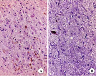
Figure 2. Locus niger. Case 2/III-20.
(A) Massive neuronal loss in
hematoxylin-eosin staining. (B) Dense
fibrillary gliosis. Holzer.
Similar findings were obtained with the various
anti-bodies used in the present study. Neurons, astrocytes, and
oligodendrocytes were stained equally with
anti-phospho-tauSer396 antibodies and with anti-phospho-tauSer422,
tauThr181,
tauSer199,
tauSer202,
tauSer231,
and
tauSer214. However, inclusions were not stained with
anti-phospho-tauSer262.
Most tau-positive inclusions in astrocytes and
oligoden-drocytes were decorated with anti-ubiquitin antibodies.
Some tau deposits in neurons were ubiquitinated, but
pre-tangle neurons and tau-immunoreactive inclusions in
neu-rons of the dentate gyrus were not stained with ubiquitin
antibodies. No ballooned neurons were observed in the
ce-rebral cortex, although
␣
B-crystallin antibodies stained
the cytoplasm of astrocytes in the cerebral cortex and
oli-godendrocytes in the white matter. No
␣
-synuclein
inclu-sions and no

A4-amyloid deposits were found. Pick’s
bodies were not observed.
Biochemical studies.
Biochemical studies of total
ho-mogenates and sarkosyl-insoluble fractions from the brain
of Case 1/III-21 disclosed a pattern of two bands of
phospho-tau of 68 kDa and 64 kDa. Interestingly, the same
results were obtained with the different antibodies used in
the present study, including anti-phospho-tau Ser262.
Fro-zen samples for dephosphorylation studies were not
available.
satellite markers in this region showed that a common
ancestral haplotype could be inferred for both pedigrees
(see table E-1).
Discussion.
We present the clinical, pathologic,
biochemical, and genetic data of two pedigrees, with
an autosomal dominant tauopathy due to a novel
missense mutation (K317M) in the
MAPT
gene, the
third known in exon 11.
15,16There are striking
differ-ences in many clinical, pathologic, and biochemical
features among the effects of the three mutations in
exon 11. From the clinical point of view, the
present-ing symptoms in the L315R mutation are anomia,
memory loss, personality changes, and behavioral
disorders, and in the S320F mutation the first
symp-tom is a memory decline. In both mutations the full
clinical picture is dominated by a global or frontal
type dementia. In contrast, in the K317M mutation,
dysarthria and tremor are the more frequent
pre-senting symptoms, and motor features such as
par-kinsonism, pyramidalism, and amyotrophy are in the
forefront of the developed clinical picture. The
be-havioral abnormalities observed in some of our
pa-tients (disinhibition, apathy, irritability, anxiety,
emotional lability) were mild or moderate and never
disturbed the patient’s familial or social
relation-ships. Severe disorders such as aggressiveness,
socio-pathic
behavior,
paranoia,
sexual,
or
feeding
misconduct were never observed.
From the pathologic side, Pick’s bodies were found
in both the L315R and S320F mutations,
15,16while in
the K317M mutation pre-tangles and tangles were
the characteristic intraneuronal phospho-tau
inclu-sions and Pick’s bodies were never seen. In the
S320F mutation
15only rare coiled bodies were found
in oligodendroglial cells, whereas astrocytic tau
in-clusions were present in the L315R mutations.
16In
contrast, all types of oligodendroglial and astrocytic
inclusions were particularly abundant in the K317M
mutation. Finally, the tau biochemical profile of both
the L315R and S320F mutations was a double band
of 60 and 64 kDa, while a 64 and 68 kDa banding
was found in the K317M mutation.
A wide range of interindividual variability in the
clinical phenotype was observed in the course of the
evolution in our patients. In some of them, a
levodopa-resistant parkinsonism associated with
su-pranuclear gaze palsy, brisk reflexes, and
pseudobul-bar
palsy
mimicked
the
clinical
picture
of
progressive supranuclear palsy (PSP) as it has been
observed in other tau mutations (R5l, N279K,
delN296, S305S).
22-25However, Cases 1/III-21 and
2/V-7 exhibited prominent signs of parietal
involve-ment and their clinical picture was for some time
reminiscent of corticobasal ganglionic degeneration
(CBD) like in other
MAPT
mutations (N296N,
P301S).
26,27CBD and PSP share many clinical,
pathologic, biochemical, and genetic traits. At the
pathologic level it has been postulated that
cytic plaques are typical of CBD while tufted
astro-cytes are a histologic hallmark of PSP. Some
authors
28stated that astrocytic plaques and tufted
astrocytes never coexist in the same brains.
How-ever, other researchers have found atypical
tauopa-thies with a mixed glial pathology as in our cases.
29,30While a CBD or a PSP phenotype are common
presentations of the tau mutations, overt motor
neu-ron features resembling a classic ALS are unusual
findings.
1,31,32However, pyramidalism was present in
all of our patients and amyotrophy was observed in
seven of them. Indeed, a typical ALS neuropathologic
pattern was observed in six of seven spinal cords
examined. Motor neuron loss in the spinal cord has
been reported only in some cases bearing a
MAPT
mutation, and corticospinal tract degeneration is
rarely mentioned.
33Few spinal cord studies have
been reported in the
MAPT
mutations. In the
origi-nal family with the
⫹
14 intronic mutation associated
with
the
disinhibition-dementia-parkinsonism-amyotrophy complex,
23 of 13 patients had a
moder-ate or mild pyramidal syndrome and only one had
fasciculations, amyotrophy, and denervation in the
EMG study, fulfilling the clinical criteria of ALS.
Two spinal cords were examined and motor neuron
loss in anterior horns with reactive gliosis was found
in an irregular distribution, but no mention is made
of a corticospinal tract degeneration. In one case
with the
⫹
3 intronic mutation, some neurofibrillary
tangles in the anterior horn in the upper cervical
spinal cord were found but no pyramidalism or
amy-otrophy were observed.
34Pyramidal signs are often
described in patients with
MAPT
mutations but
without neuropathologic correlation.
35,36Only in
sin-gle cases with the N296N
27,37and the N279K
muta-tions
38has a degeneration of the corticospinal tract
been neuropathologically confirmed. In the latter
mutation an involvement of the anterior horn
motor-neurons has been found in one family.
39However,
amyotrophy is not described in a comparative study
of the clinical features between the large original
family with the pallido-ponto-nigral degeneration
linked to the N279K mutation and a French family
with the same mutation.
40,41Thus, the K317M
muta-tion is the first familial tauopathy in which motor
neuron disease is a consistent component of the
clin-icopathologic picture.
tau
phosphorylation
at
Ser262
impairs
tau-microtubule interactions and aggregation of
abnor-mal tau-containing filaments.
43The biochemical study in our Case 1/III-21 has
shown two bands of phospho-tau of 68 kDa and 64
kDa in sarkosyl-insoluble fractions. This pattern is
most commonly encountered in PSP, CBD,
argyro-philic grain disease,
12,21and in missense and deletion
mutations of the exon 10 in the tau gene,
29yet the
mutation in the present case occurs in exon 11. It is
interesting to note that the biochemical study in the
other two known mutations in exon 11 is
character-ized by two bands of 64 kDa and 60 kDa of
phospho-tau reminiscent of those found in Pick’s disease.
15,16Therefore, the neuropathology, covering distribution
of cell vulnerability, neuronal inclusions, astroglial
tau deposits, as well as the clinical profiles, differs
between the S320F/L315R mutations and K317M
mutation, although all of them affect exon 11. To
date, most of the mutations found within exon 10 or
its surrounding regions have been shown to increase
the proportion of exon 10-containing isoforms
whereas those mutations affecting other exons seem
to act through a different mechanism.
43To our
knowledge, the mutation that we present here is the
first one in an exon other than exon 10 that seems to
increase the proportion of exon-10 containing tau in
the intracellular deposits observed in the brain. This
suggests that this particular mutation, or mutations
affecting at least this part of the gene, can also
pro-duce an increase in exon 10 containing PHF either
by interfering with the processing of the mRNA or by
inducing specifically these isoforms to form tangles.
Acknowledgment
The authors thank Professor José M
aRivera-Pomar and the
De-partment of Pathology in the Hospital de Cruces for help with the
pathologic studies.
References
1. Morris HR, Khan MN, Janssen JC, et al. The genetic and pathological classification of familial frontotemporal dementia. Arch Neurol 2001;58: 1813–1816.
2. Lynch T, Sano M, Marder KS, et al. Clinical characteristics of a family with chromosome 17-linked disinhibition-dementia-parkinsonism-amyotrophy complex. Neurology 1994;44:1878 –1884.
3. Wszolek ZK, Pfeiffer RF, Bhatt MH, et al. Rapidly progressive autoso-mal dominant parkinsonism and dementia with pallido-ponto-nigral degeneration. Ann Neurol 1992;32:312–320.
4. Heutink P, Stevens M, Rizzu P, et al. Hereditary frontotemporal de-mentia is linked to chromosome 17q21-q22: a genetic and clinicopatho-logical study of three Dutch families. Ann Neurol 1997;41:150 –159. 5. Spillantini MG, Goedert M, Crowther RA, Murrell JR, Farlow MR,
Ghetti B. Familial multiple system tauopathy with presenile dementia: a disease with abundant neuronal and glial tau filaments. Proc Natl Acad Sci USA 1997;94:4113– 4118.
6. Wijker M, Wszolek ZK, Wolters EC, et al. Localization of the gene for rapidly progressive autosomal dominant parkinsonism and dementia with pallido-ponto-nigral degeneration to chromosome 17q21. Hum Mol Genet 1996;5:151–154.
7. Wilhelmsen KC, Lynch T, Pavlou E, Higgins M, Nygaard TG. Localiza-tion of disinhibiLocaliza-tion-dementia-parkinsonism-amyotrophy complex to 17q21–22. Am J Hum Genet 1994;55:1159 –1165.
8. Foster NL, Wilhelmsen K, Sima AA, Jones MZ, D’Amato CJ, Gilman S. Frontotemporal dementia and parkinsonism linked to chromosome 17: a consensus conference. Conference Participants. Ann Neurol 1997;41: 706 –715.
9. Poorkaj P, Bird TD, Wijsman E, et al. Tau is a candidate gene for chromosome 17 frontotemporal dementia. Ann Neurol 1998;43:815– 825.
10. Goedert M, Crowther RA, Spillantini MG. Tau mutations cause fronto-temporal dementias. Neuron 1998;21:955–958.
11. Spillantini MG, Van Swieten JC, Goedert M. Tau gene mutations in frontotemporal dementia and parkinsonism linked to chromosome 17 (FTDP-17). Neurogenetics 2000;2:193–205.
12. Lee VM, Goedert M, Trojanowski JQ. Neurodegenerative tauopathies. Annu Rev Neurosci 2001;24:1121–1159.
13. Kobayashi T, Ota S, Tanaka K, et al. A novel L266V mutation of the tau gene causes frontotemporal dementia with a unique tau pathology. Ann Neurol 2003;53:133–137.
14. Stanford PM, Shepherd CE, Halliday GM, et al. Mutations in the tau gene that cause an increase in three repeat tau and frontotemporal dementia. Brain 2003;126:814 – 826.
15. Rosso SM, van Herpen E, Deelen W, et al. A novel tau mutation, S320F, causes a tauopathy with inclusions similar to those in Pick’s disease. Ann Neurol 2002;51:373–376.
16. van Herpen E, Rosso SM, Serverijnen LA, et al. Variable phenotypic expression and extensive tau pathology in two families with the novel tau mutation L315R. Ann Neurol 2003;54:573–581.
17. van Swieten JC, Stevens M, Rosso SM, et al. Phenotypic variation in hereditary frontotemporal dementia with tau mutations. Ann Neurol 1999;46:617– 626.
18. Rizzu P, Van Swieten JC, Joosse M, et al. High prevalence of mutations in the microtubule-associated protein tau in a population study of fron-totemporal dementia in the Netherlands. Am J Hum Genet 1999;64: 414 – 421.
19. Houlden H, Baker M, Adamson J, et al. Frequency of tau mutations in three series of non-Alzheimer’s degenerative dementia. Ann Neurol 1999;46:243–248.
20. Poorkaj P, Grossman M, Steinbart E, et al. Frequency of tau gene mutations in familial and sporadic cases of non-Alzheimer dementia. Arch Neurol 2001;58:383–387.
21. Ferrer I, Barrachina M, Tolnay M, et al. Phosphorylated protein ki-nases associated with neuronal and glial tau deposits in argyrophilic grain disease. Brain Pathol 2003;13:62–78.
22. Delisle MB, Murrell JR, Richardson R, et al. A mutation at codon 279 (N279K) in exon 10 of the tau gene causes a tauopathy with dementia and supranuclear palsy. Acta Neuropathol (Berl) 1999;98:62–77. 23. Stanford PM, Halliday GM, Brooks WS, et al. Progressive supranuclear
palsy pathology caused by a novel silent mutation in exon 10 of the tau gene: expansion of the disease phenotype caused by tau gene muta-tions. Brain 2000;123:880 – 893.
24. Pastor P, Pastor E, Carnero C, et al. Familial atypical progressive supranuclear palsy associated with homozigosity for the delN296 muta-tion in the tau gene. Ann Neurol 2001;49:263–267.
25. Poorkaj P, Muma NA, Zhukareva V, et al. An R5L tau mutation in a subject with a progressive supranuclear palsy phenotype. Ann Neurol 2002;52:511–516.
26. Bugiani O, Murrell JR, Giaccone G, et al. Frontotemporal dementia and corticobasal degeneration in a family with a P301S mutation in tau. J Neuropathol Exp Neurol 1999;58:667– 677.
27. Spillantini MG, Yoshida H, Rizzini C, et al. A novel tau mutation (N296N) in familial dementia with swollen achromatic neurons and corticobasal inclusion bodies. Ann Neurol 2000;48:939 –943.
28. Komori T, Arai N, Oda M, et al. Astrocytic plaques and tufts of abnor-mal fibers do not coexist in corticobasal degeneration and progressive supranuclear palsy. Acta Neuropathol (Berl) 1998;96:401– 408. 29. Ferrer I, Hernandez I, Boada M, et al. Primary progressive aphasia as
the initial manifestation of corticobasal degeneration and unusual tauopathies. Acta Neuropathol (Berl) 2003;106:419 – 435.
30. Katsuse O, Iseki E, Arai T, et al. 4-repeat tauopathy sharing patholog-ical and biochempatholog-ical features of corticobasal degeneration and progres-sive supranuclear palsy. Acta Neuropathol (Berl) 2003;106:251–260. 31. Heutink P. Untangling tau-related dementia. Hum Mol Genet 2000;9:
979 –986.
32. Goedert M. Relevance of mutations in tau for understanding the tauopathies. Curr Med Chem-Immun Endoc & Metab Agents 2003;3: 341–348.
33. Spillantini MG, Bird TD, Ghetti B. Frontotemporal dementia and Par-kinsonism linked to chromosome 17: a new group of tauopathies. Brain Pathol 1998;8:387– 402.
34. Tolnay M, Spillantini MG, Rizzini C, Eccles D, Lowe J, Ellison D. A new case of frontotemporal dementia and parkinsonism resulting from an intron 10 ⫹3-splice site mutation in the tau gene: clinical and pathological features. Neuropathol Appl Neurobiol 2000;26:368 –378. 35. Yasuda M, Takamatsu J, D’Souza I, et al. A novel mutation at position
⫹12 in the intron following exon 10 of the tau gene in familial fronto-temporal dementia (FTD-Kumamoto). Ann Neurol 2000;47:422– 429. 36. Lossos A, Reches A, Gal A, et al. Frontotemporal dementia and
parkin-sonism with the P301S tau gene mutation in a Jewish family. J Neurol 2003;250:733–740.
38. Arima K, Kowalska A, Hasegawa M, et al. Two brothers with fronto-temporal dementia and parkinsonism with an N279K mutation of the tau gene. Neurology 2000;54:1787–1795.
39. Yasuda M, Kawamata T, Komure O, et al. A mutation in the microtubule-associated protein tau in pallido-nigro-luysian degenera-tion. Neurology 1999;53:864 – 868.
40. Tsuboi Y, Baker M, Hutton ML, et al. Clinical and genetic studies of families with the tau N279K mutation (FTDP-17). Neurology 2002;59: 1791–1793.
41. Tsuboi Y, Uitti RJ, Delisle MB, et al. Clinical features and disease
haplotypes of individuals with the N279K tau gene mutation: a compar-ison of the pallidopontonigral degeneration kindred and a French fam-ily. Arch Neurol 2002;59:943–950.
42. Schneider A, Biernat J, von Bergen M, Mandelkow E, Mandelkow EM. Phosphorylation that detaches tau protein from microtubules (Ser262, Ser214) also protects it against aggregation into Alzheimer paired heli-cal filaments. Biochemistry 1999;38:3549 –3558.
43. Hutton M. Missense and splice site mutations in tau associated with FTDP-17: multiple pathogenic mechanisms. Neurology 2001;56(11 suppl 4):S21–25.
Neuro
Images
Multiple cervical artery dissections
M.S. Hussain, MD; T. Muaygil, MD; and
M. Saqqur, MD, Alberta, Canada
A 49-year-old man with a history of bilateral vertebral artery
dissections (figure, A) presented with a left internal capsular
in-farct. A new left internal carotid artery dissection was found on
angiography (figure, B, C). There were no findings of connective
tissue disease on examination. A skin biopsy with elastin staining
(figure, D) showed marked fragmentation. Patients with cervical
artery dissections (CAD) have a risk of recurrent dissection of 1%
per year.
1Skin biopsies in patients with spontaneous CAD may
show ultrastructural connective tissue abnormalities, even with
no clinical disease,
2but our patient demonstrated abnormalities
on light microscopy.
Copyright © 2005 by AAN Enterprises, Inc.
1. Schievink WI, Mokri B, O’Fallon WM. Spontaneous recurrent cervical-artery dissection. N Engl J Med 1994;330:393–397.
2. Brandt T, Orberk E, Weber R, et al. Pathogenesis of cervical artery dissections: association with other connective tissue abnormalities. Neu-rology 2001;57:24 –30.
Address correspondence and reprint requests to Dr. M. Saqqur, 9335 Aber-hart 1, University of Alberta, Edmonton, Alberta, Canada T6G 2J3; e-mail: [email protected]