1 Title: Decoding Drosophila circadian pacemaker circuit
Anatoly Kozlov1 and Emi Nagoshi1*
1Department of Genetics and Evolution, University of Geneva, Sciences III, 30 Quai
Ernest-Ansermet, CH-1211, Geneva-4, Switzerland
*Corresponding author (e-mail: [email protected])
Abstract
Drosophila circadian circuit is one of the best described neural circuits but is complex enough to obscure our understanding of how it actually works. Animals’ rhythmic behavior, the
seemingly simple outcome of their internal clocks, relies on the interaction of heterogeneous
clock neurons that are spread across the brain. Direct observations of their coordinated
network interactions can bring us forward in understanding the circuit. The current challenge
is to observe activity of each of these neurons over a long span of time –hours to days– in live
animals. Here we review the progress in circadian circuit interrogation powered by in vivo
calcium imaging.
Circadian pacemaker circuit
Circadian rhythms drive organism’s intrinsic capacity to anticipate daily and seasonal changes
of the environment and to accordingly adapt their behavior and physiology. Circadian clocks,
present in most species from protozoa to mammals, are built using a conserved principle of
transcriptional-translational feedback loops (TTFL) [1, 2]. Molecular studies of circadian
clocks were pioneered using Drosophila melanogaster [3–5] and continue to bring new
discoveries to this day. Major players of Drosophila TTFL are transcriptional activators CLOCK
2 (CLK) and CYCLE (CYC) and two genes encoding repressors, period (per) and timeless (tim).
CLK/CYC heterodimer activates the expression of per and tim. Following transcription, they
undergo a series of post-transcriptional regulations and phosphorylation, which allow PER
and TIM to accumulate in the cytoplasm with a delay of approximately 6-8 h after the
transcription, then dimerize and enter the nucleus and suppress the transcriptional activity of
CLK/CYC. CLK and CYC also activate transcription of genes encoding the basic-zipper
regulators PAR DOMAIN PROTEIN 1 ℰ (PDP-1ℰ) and VRILLE (VRI), which activates and
inhibits Clk gene expression, respectively. These interlocked positive and negative feedback
loops stabilize the main loop and ensure the generation of 24-h rhythms [2]. The molecular
clock rhythmically controls the expression of a large number of genes and ultimately produces
rhythms in diverse cellular functions, including neuronal activity [6].
Drosophila also offers a proven, powerful tool to study neuronal mechanism of
circadian behavior. Drosophila melanogaster belongs to the class of crepuscular animals, which
are physically active at dawn and dusk. In the laboratory conditions, this feature is observed
using automatic recording of infrared beam crossing of single flies. In 12h:12h light-dark
cycles (LD), flies show a stereotypic pattern of activity, in which the peaks of activity are
aligned with the moment of light/dark transitions in the morning and the evening. Gradual
increments of activities precede the light transition, which display the endogenous origin of
their behavior controlled by their internal clocks. When flies are released into constant
darkness (DD), they free-run with similar bimodal pattern of activity that repeats roughly
every 24 hr, showing another evidence of endogenous clocks [7]. Approximately 75 pairs of
neurons in the brain containing a full set of clock genes are the pacemaker neurons that
coordinate this circadian behavior. Circadian pacemaker neurons are classified into several
groups according to their location in the brain: the large- ventral lateral neurons (l-LNvs, four
per hemisphere), small-ventral lateral neurons (four s-LNvs that express neuropeptide
3 (LNds, 6 per hemisphere) and three groups of dorsal neurons (DN1s (anterior and posterior
subgroups, 16 in total), DN2s (two) and DN3 (circa 30)) [8, 9].
Back in the 70s it was proposed that morning and evening activity peaks of crepuscular
animals are produced by two coupled but independent morning (M) and evening (E) circadian
oscillators, which track dawn and dusk, respectively, throughout the season[10]. It took about
30 years until the physical existence of such oscillators was first demonstrated in flies. The
pioneering studies by the groups of M. Rosbash [11] and F. Rouyer [12] independently showed
that s-LNvs represent the M-oscillator that drives the morning anticipation and rhythmic
locomotor activity in DD. They also identified E-oscillator that drives the evening anticipation
activity, which consists of the pacemaker neurons expressing circadian photoreceptor
Cryptochrome (CRY) but not PDF. Subsequent studies by many groups have verified the basic
principle of this dual-oscillator model and later defined LNds and 5th s-LNv as the main
constituents of the E-oscillator [13, 14].
Decoding the pacemaker circuit
Whereas the essence of the dual-oscillator model stands the test of time, comprehension of
the circuit operation requires identifying individual components, drawing wiring diagram,
describing neurochemistry and the nature of communication between the components.
Pursuing these questions, a number of studies have added evidence to the complexity of the
pacemaker circuit organization. Several lines of evidence indicate that DN1ps, the posterior
subgroup of DN1, contribute to both morning and evening activity peaks [15–18]. DN1ps
control morning activity peak downstream of the s-LNvs [15, 18], whereas their role in
controlling evening peak runs parallel to the E-oscillator and is regulated by light, which
inhibits DN1p output [16]. DN1s make physical contact with the neurosecretory cells in the
pars intercerebralis (PI). Signaling from the s-LNvs via DN1s to the PI appears to be one of the
4 Neurochemistry of the pacemaker circuit adds another dimension in the circuit
organizational logic. At least several neuropeptides, including PDF, neuropeptide F (NPF),
short neuropeptide F (sNPF) and, ion transport peptide (ITP), and several neurotransmitters,
such as glutamate, acetylcholine, glycine [21] and GABA, are expressed within or act on
pacemaker neurons (reviewed in [22, 23]). Findings from several studies highlight the
importance of neuropeptidergic signaling in the fly circadian pacemaker circuit and its
evolutionary significance, as evidenced by the parallel discoveries on the critical roles of
neuropeptides in mammalian pacemaker circuit [24–26]. Works with flies in this domain have
consistently indicated that all its main functions of the M-oscillator, s-LNvs, are mediated by
PDF, as any manipulation altering its production or expression of its cognate receptor leads to
the same set of phenotypes: the advance of the evening peak in LD, absence of the morning
anticipation and very low rhythmicity in DD [27–32]. Manipulation of the s-LNvs neuronal
excitability, which presumably controls the rate of PDF secretion, leads to the phenotypes
similar to those induced by up or down regulation of PDF signaling [33–37]. sNPF is another
neuropeptide expressed in the s-LNvs, as well as in the LNds and l-LNvs [38, 39]. LNds
additionally produce NPF and ITP [9, 35]. The expanding panel of neuropeptides found within
the pacemaker circuit and the diversity of neuropeptide function in general pose further
challenges.
A breakthrough came in 2016 when the group of P. Taghert recorded for the first time
the Ca2+ dynamics of individual groups of pacemaker neurons over 24 h in head-fixed live flies
[40, 41]. Intracellular Ca2+ levels, a convenient surrogate of neural activity, provides the critical
information to decipher the functional interaction among pacemaker neuron subgroups. This
was made possible by the use of genetically encoded Ca2+ sensor GCaMP and the
Objective-Coupled Planar Illumination microscopy (OCPI), a type of light sheet microscopy, which
overcomes the limitations of the standard confocal or two-photon microscopes in temporal
5 How does the information flow through the pacemaker circuit, whose intrinsic
rhythmicity is genetically underpinned? How do the environmental (light) stimuli transform
the information to shape the behavioral output? The group addressed these questions in three
consecutive studies [20, 40, 41]. The first, perhaps unexpected, observation was that each
pacemaker subgroup produces circadian Ca2+ wave that peaks at a distinct temporal window.
This is strikingly different from the ticking of their molecular clocks, which are roughly
in-phase in normal conditions [31, 44, 45]. The timing of the Ca2+ peakof the s-and l-LNvs was in
a rough accordance with electrophysiological measurements shown in prior works [46–48],
validating their experimental setup. Ca2+ waves of thes-LNvs (M-oscillator) and LNds
(E-oscillator) keep track of the morning and evening light transitions whether it is in the
standard 12h: 12h LD cycles or in the photoperiod mimicking the short-day, winter condition
(8h: 16h LD) or the long-day, summer condition (16h: 8h LD). This property of Ca2+ activity
patterns of the M- and E- oscillators is concordant with the morning and evening behavioral
peaks. Ca2+ rhythms are abolished in the “clockless” per0 mutants and phase-advanced in the
short-period persmutants (Fig. 1). Therefore, Ca2+ rhythms are generated by the molecular
clocks and connect the clocks of the M- and E-oscillators to the behavioral output.
How do the molecular clocks ticking in phase produce Ca2+ rhythms that are up to 12 h
out of phase? The answer turned out to be the action of two neuropeptides, PDF and sNPF. In
the null mutant for Pdf (Pdf01) and severe hypomorphic mutant of PdfR (han5304), Ca+2 peaks of
the M- and E- oscillators and DN3s coincided around dawn. Bath application of synthetic PDF
to Pdf01 mutants restored the sequential order of the Ca+2 rhythms in the 24-h recordings. The
immediate response of PDF bath application at ZT7 (zeitgeber time 7, 7h after lights-on) was a
decrease of Ca2 levels at least in the s-LNVs and LNds. Therefore, PDF/PDFR signaling plays a
major role in delaying the Ca2+ peaks in the LNds and DN3s by suppressing their Ca2+ levels.
Strikingly, neuropeptide signaling not only adjusts the phase but can
6 oscillations in a rough accordance with their membrane excitability [49]. However, their Ca2+
rhythms are abolished when sNPF secretion from the s-LNvs and LNds is blocked. As shown in
[41], sNPF bath application decreases Ca2+ levels in the DN1s, consistent with the notion that
sNPF is generally inhibitory [50]. Therefore, analogous to the role of PDF in coordinating Ca2+
waves by inhibiting the onset of Ca2+ rise, sNPF imposes Ca2+ rhythms in the DN1s via an
inhibitory mechanism (Fig. 2).
The phase of free-running locomotor activity can be delayed or advanced by a light
pulse delivered at early or late night, respectively. In vivo Ca2+ imaging also helped advance the
study of neuronal mechanisms underlying this well-established but poorly understood
phenomenon. In the head-fixed flies, phase-delaying light pulses given at ZT17 delayed Ca2+
rhythms of all pacemaker neurons within the first day, although it took two more days to
re-establish the normal Ca2+ phase pattern of the pacemaker network. In contrast, ZT21
phase-advancing light pulses initially phase-advanced only the s-LNvs, and the rest of the pacemaker
neurons shifted their Ca2+ phase on the second day. The gradual phase advance of non-s-LNv
pacemakers may be mediated by PDF released from the s-LNvs, as all pacemaker neurons
shifted their Ca2+ phases at once in response to phase-advancing pulses in Pdf01 flies [41].
These results suggest an intrinsic capacity of all pacemaker neurons to rapidly respond to
light stimuli, which is modulated by PDF signaling.
Activities of pacemaker neurons should be relayed to the area controlling locomotion.
Extending in vivo Ca2+ imaging beyond circadian pacemakers, Taghert’s group recently
identified an output circuit of the M- and E-oscillators [20]. They found that Ring Neurons of
the Ellipsoid Body (RNs) and the PPM3-EB subclass of DA neurons presynaptic to the
EB-RNs exhibit Ca2 rhythms with two peaks, coinciding with those of the M - and E- oscillators.
The experimental set up in this study included one interesting addition, an infrared detector
that monitors leg movements during in vivo Ca2+imaging. Leg movements of the head-fixed
7 flies and matched the Ca2+ activitiesof theidentified output circuit. Together, these results
demonstrate that activities of the M- and E-oscillators are relayed by dopamine signaling to
the EB-RNs, a known pre-motor center, thereby generating morning and evening peaks of
locomotor behavior.
The series of discoveries from in vivo Ca2+ imaging brought us forward in mechanistic
understanding of pacemaker circuit, but not without conundrums. Whereas different
experiments in Taghert group’s papers have consistently shown that PDF/PDFR signaling
reduces Ca2 levels in pacemaker neurons, several previous studies demonstrated the elevation
in Ca2 and/or cAMP levels upon PDF signaling activation [32, 50–54]. PdfR is a G-protein
coupled receptor, which generally increases cAMP levels upon activation, leading to
depolarization, increased firing rates and elevated Ca2 [53]. Nonetheless, two opposing
findings are not necessarily a contradiction, as the timescale of these phenomena are quite
different, from seconds (in acute pharmacological experiments) to hours (in 24-h recordings).
It should be noted that Ca2+ levels were recorded from the cell bodies of pacemaker neurons.
Ca2+ concentration in the cell bodies represents more than just the neuronal activity; it results
from the combination of the processes involving membrane calcium channels and
intracellular calcium sources. Another point that should not be ignored is the heterogeneity
within the neuronal classes. Because of the technical limitation, in vivo Ca2+ imaging lacked
cellular resolution. The DN1s, LNds and DN3s contain subclasses of neurons with different
receptivity towards neuronal signals and environment (light and temperature) and produce
different pools of neuropeptides/transmitters [9, 31, 55]. This means that some important
features of Ca2+ activity may not have been detected. Therefore, studies using complementary
approaches, such as short-term in vivo Ca2+ imaging [56], recording of bioluminescence Ca2+
-reporter in freely moving flies [57], live-imaging of molecular clockwork in cultured brains
using bioluminescent [45] or fluorescent clock gene reporters [58–60], are warranted to fill
8 PDF/PDFR signaling triggers multiple downstream processes, which are
cell-type-dependent, temporally gated and occur at different time scales. It affects membrane electrical
status, calcium levels and molecular clocks. One pathway is through the increase in cAMP
levels and protein kinase A (PKA) activity, leading to the increase in the stability of PER and
TIM [53, 54, 58]. It was also shown that PDF signaling upregulates per transcription in ex vivo
brain culture [58]. The mechanism of the third downstream effect, inhibition of Ca2+ levels, is
currently unknown and should entail more complex processes than hyperpolarization.
Together with the new finding that sNPF can impose Ca2+ rhythms that are independent of the
molecular clocks, a large body of work centered on the role of PDF casts further challenges in
decoding the pacemaker circuit. Network communication and the molecular clockwork
intertwine at different time scales and modulate membrane electrical status and cellular
processes; only when we understand each of these components and mechanisms, can we get
how flies display seemingly simple two peaks of activity.
Acknowledgements
We thank the members of our laboratory for suggestions on the manuscript. This work was
supported by the research grant from the Swiss National Science Foundation
(31003A_169548).
References
[1] J. M. Hurley, J. J. Loros, and J. C. Dunlap, “Circadian Oscillators: Around the
Transcription–Translation Feedback Loop and on to Output,” Trends in Biochemical Sciences, vol. 41, no. 10, pp. 834–846, Oct. 2016.
[2] P. E. Hardin, “Molecular Genetic Analysis of Circadian Timekeeping in Drosophila,” in Advances in Genetics, vol. 74, Elsevier, 2011, pp. 141–173.
**[3] R. J. Konopka and S. Benzer, “Clock mutants of Drosophila melanogaster,” Proc. Natl. Acad. Sci. U.S.A., vol. 68, no. 9, pp. 2112–2116, Sep. 1971.
9 1984.
**[5] T. A. Bargiello, F. R. Jackson, and M. W. Young, “Restoration of circadian behavioural rhythms by gene transfer in Drosophila,” Nature, vol. 312, no. 5996, pp. 752–754, Jan. 1984. [6] K. C. Abruzzi et al., “Drosophila CLOCK target gene characterization: implications for circadian tissue-specific gene expression,” Genes Dev., vol. 25, no. 22, pp. 2374–2386, Nov. 2011. [7] A. Klarsfeld, J. C. Leloup, and F. Rouyer, “Circadian rhythms of locomotor activity in Drosophila,” Behav. Processes, vol. 64, no. 2, pp. 161–175, Sep. 2003.
[8] C. Helfrich-Förster, “Development of pigment-dispersing hormone-immunoreactive neurons in the nervous system of Drosophila melanogaster,” J. Comp. Neurol., vol. 380, no. 3, pp. 335–354, Apr. 2007.
[9] F. K. Schubert, N. Hagedorn, T. Yoshii, C. Helfrich-Förster, and D. Rieger,
“Neuroanatomical details of the lateral neurons of Drosophila melanogaster support their functional role in the circadian system,” J. Comp. Neurol., vol. 526, no. 7, pp. 1209–1231, 01 2018.
**[10] C. S. Pittendrigh and S. Daan, “A functional analysis of circadian pacemakers in nocturnal rodents,” J. Comp. Physiol., vol. 106, no. 3, pp. 333–355, Oct. 1976.
**[11] D. Stoleru, Y. Peng, J. Agosto, and M. Rosbash, “Coupled oscillators control morning and evening locomotor behaviour of Drosophila,” Nature, vol. 431, no. 7010, pp. 862–868, Oct. 2004. **[12] B. Grima, E. Chélot, R. Xia, and F. Rouyer, “Morning and evening peaks of activity rely on different clock neurons of the Drosophila brain,” Nature, vol. 431, no. 7010, pp. 869–873, Oct. 2004.
**[13] M. Picot, P. Cusumano, A. Klarsfeld, R. Ueda, and F. Rouyer, “Light activates output from evening neurons and inhibits output from morning neurons in the Drosophila circadian clock,” PLoS Biol., vol. 5, no. 11, p. e315, Nov. 2007.
**[14] P. Cusumano, A. Klarsfeld, E. Chélot, M. Picot, B. Richier, and F. Rouyer, “PDF-modulated visual inputs and cryptochrome define diurnal behavior in Drosophila,” Nat. Neurosci., vol. 12, no. 11, pp. 1431–1437, Nov. 2009.
*[15] L. Zhang et al., “DN1(p) circadian neurons coordinate acute light and PDF inputs to produce robust daily behavior in Drosophila,” Curr. Biol., vol. 20, no. 7, pp. 591–599, Apr. 2010. *[16] Y. Zhang, Y. Liu, D. Bilodeau-Wentworth, P. E. Hardin, and P. Emery, “Light and
temperature control the contribution of specific DN1 neurons to Drosophila circadian behavior,” Curr. Biol., vol. 20, no. 7, pp. 600–605, Apr. 2010.
[17] D. J. Cavanaugh et al., “Identification of a Circadian Output Circuit for Rest:Activity Rhythms in Drosophila,” Cell, vol. 157, no. 3, pp. 689–701, Apr. 2014.
*[18] A. Chatterjee et al., “Reconfiguration of a Multi-oscillator Network by Light in the Drosophila Circadian Clock,” Curr. Biol., vol. 28, no. 13, pp. 2007-2017.e4, Jul. 2018. [19] M. Cavey, B. Collins, C. Bertet, and J. Blau, “Circadian rhythms in neuronal activity propagate through output circuits,” Nature Neuroscience, vol. 19, no. 4, pp. 587–595, Apr. 2016. **[20] X. Liang et al., “Morning and Evening Circadian Pacemakers Independently Drive Premotor Centers via a Specific Dopamine Relay,” Neuron, Apr. 2019.
[21] L. Frenkel et al., “Organization of Circadian Behavior Relies on Glycinergic Transmission,” Cell Rep, vol. 19, no. 1, pp. 72–85, 04 2017.
[22] L. Frenkel and M. F. Ceriani, “Circadian plasticity: from structure to behavior,” Int. Rev. Neurobiol., vol. 99, pp. 107–138, 2011.
[23] N. I. Muraro, N. Pírez, and M. F. Ceriani, “The circadian system: plasticity at many levels,” Neuroscience, vol. 247, pp. 280–293, Sep. 2013.
**[24] S. J. Aton, C. S. Colwell, A. J. Harmar, J. Waschek, and E. D. Herzog, “Vasoactive intestinal polypeptide mediates circadian rhythmicity and synchrony in mammalian clock neurons,” Nat. Neurosci., vol. 8, no. 4, pp. 476–483, Apr. 2005.
[25] C. Beaulé, J. W. Mitchell, P. T. Lindberg, R. Damadzic, L. E. Eiden, and M. U. Gillette, “Temporally Restricted Role of Retinal PACAP: Integration of the Phase-Advancing Light Signal to the SCN,” J Biol Rhythms, vol. 24, no. 2, pp. 126–134, Apr. 2009.
10 rhythms in the SCN is governed by the interplay of two coupling factors,” PLoS Comput. Biol., vol. 14, no. 12, p. e1006607, 2018.
[27] S. C. P. Renn, J. H. Park, M. Rosbash, J. C. Hall, and P. H. Taghert, “A pdf Neuropeptide Gene Mutation and Ablation of PDF Neurons Each Cause Severe Abnormalities of Behavioral Circadian Rhythms in Drosophila,” Cell, vol. 99, no. 7, pp. 791–802, Dec. 1999.
[28] S. Hyun et al., “Drosophila GPCR Han is a receptor for the circadian clock neuropeptide PDF,” Neuron, vol. 48, no. 2, pp. 267–278, Oct. 2005.
[29] B. C. Lear, C. E. Merrill, J.-M. Lin, A. Schroeder, L. Zhang, and R. Allada, “A G protein-coupled receptor, groom-of-PDF, is required for PDF neuron action in circadian behavior,” Neuron, vol. 48, no. 2, pp. 221–227, Oct. 2005.
[30] O. T. Shafer and P. H. Taghert, “RNA-Interference Knockdown of Drosophila Pigment Dispersing Factor in Neuronal Subsets: The Anatomical Basis of a Neuropeptide’s Circadian Functions,” PLoS ONE, vol. 4, no. 12, p. e8298, Dec. 2009.
*[31] T. Yoshii et al., “The neuropeptide pigment-dispersing factor adjusts period and phase of Drosophila’s clock,” J. Neurosci., vol. 29, no. 8, pp. 2597–2610, Feb. 2009.
*[32] C. Choi et al., “Autoreceptor Control of Peptide/Neurotransmitter Corelease from PDF Neurons Determines Allocation of Circadian Activity in Drosophila,” Cell Reports, vol. 2, no. 2, pp. 332–344, Aug. 2012.
[33] M. N. Nitabach, J. Blau, and T. C. Holmes, “Electrical silencing of Drosophila pacemaker neurons stops the free-running circadian clock,” Cell, vol. 109, no. 4, pp. 485–495, May 2002. [34] M. N. Nitabach, “Electrical Hyperexcitation of Lateral Ventral Pacemaker Neurons Desynchronizes Downstream Circadian Oscillators in the Fly Circadian Circuit and Induces Multiple Behavioral Periods,” Journal of Neuroscience, vol. 26, no. 2, pp. 479–489, Jan. 2006. [35] Y. Wu, G. Cao, and M. N. Nitabach, “Electrical silencing of PDF neurons advances the phase of non-PDF clock neurons in Drosophila,” J. Biol. Rhythms, vol. 23, no. 2, pp. 117–128, Apr. 2008.
[36] Y. Wu, G. Cao, B. Pavlicek, X. Luo, and M. N. Nitabach, “Phase coupling of a circadian neuropeptide with rest/activity rhythms detected using a membrane-tethered spider toxin,” PLoS Biol., vol. 6, no. 11, p. e273, Nov. 2008.
[37] A. Depetris-Chauvin, J. Berni, E. J. Aranovich, N. I. Muraro, E. J. Beckwith, and M. F. Ceriani, “Adult-specific electrical silencing of pacemaker neurons uncouples molecular clock from circadian outputs,” Curr. Biol., vol. 21, no. 21, pp. 1783–1793, Nov. 2011.
[38] H. A. D. Johard et al., “Peptidergic clock neurons in Drosophila: ion transport peptide and short neuropeptide F in subsets of dorsal and ventral lateral neurons,” J. Comp. Neurol., vol. 516, no. 1, pp. 59–73, Sep. 2009.
[39] Y. Shang, N. C. Donelson, C. G. Vecsey, F. Guo, M. Rosbash, and L. C. Griffith, “Short neuropeptide F is a sleep-promoting inhibitory modulator,” Neuron, vol. 80, no. 1, pp. 171–183, Oct. 2013.
**[40] X. Liang, T. E. Holy, and P. H. Taghert, “Synchronous Drosophila circadian pacemakers display nonsynchronous Ca2+ rhythms in vivo,” Science, vol. 351, no. 6276, pp. 976–981, Feb. 2016.
**[41] X. Liang, T. E. Holy, and P. H. Taghert, “A Series of Suppressive Signals within the Drosophila Circadian Neural Circuit Generates Sequential Daily Outputs,” Neuron, vol. 94, no. 6, pp. 1173-1189.e4, Jun. 2017.
[42] T. F. Holekamp, D. Turaga, and T. E. Holy, “Fast Three-Dimensional Fluorescence Imaging of Activity in Neural Populations by Objective-Coupled Planar Illumination Microscopy,” Neuron, vol. 57, no. 5, pp. 661–672, Mar. 2008.
11 [45] L. Roberts et al., “Light evokes rapid circadian network oscillator desynchrony followed by gradual phase retuning of synchrony,” Curr. Biol., vol. 25, no. 7, pp. 858–867, Mar. 2015.
**[46] G. Cao and M. N. Nitabach, “Circadian control of membrane excitability in Drosophila melanogaster lateral ventral clock neurons,” J. Neurosci., vol. 28, no. 25, pp. 6493–6501, Jun. 2008. **[47] V. Sheeba, H. Gu, V. K. Sharma, D. K. O’Dowd, and T. C. Holmes, “Circadian- and Light-Dependent Regulation of Resting Membrane Potential and Spontaneous Action Potential Firing of Drosophila Circadian Pacemaker Neurons,” J Neurophysiol, vol. 99, no. 2, pp. 976–988, Feb. 2008. **[48] G. Cao, J. Platisa, V. A. Pieribone, D. Raccuglia, M. Kunst, and M. N. Nitabach,
“Genetically targeted optical electrophysiology in intact neural circuits,” Cell, vol. 154, no. 4, pp. 904–913, Aug. 2013.
*[49] M. Flourakis et al., “A Conserved Bicycle Model for Circadian Clock Control of Membrane Excitability,” Cell, vol. 162, no. 4, pp. 836–848, Aug. 2015.
*[50] C. G. Vecsey, N. Pírez, and L. C. Griffith, “The Drosophila neuropeptides PDF and sNPF have opposing electrophysiological and molecular effects on central neurons,” J. Neurophysiol., vol. 111, no. 5, pp. 1033–1045, Mar. 2014.
[51] I. Mertens et al., “PDF Receptor Signaling in Drosophila Contributes to Both Circadian and Geotactic Behaviors,” Neuron, vol. 48, no. 2, pp. 213–219, Oct. 2005.
[52] O. T. Shafer, D. J. Kim, R. Dunbar-Yaffe, V. O. Nikolaev, M. J. Lohse, and P. H. Taghert, “Widespread receptivity to neuropeptide PDF throughout the neuronal circadian clock network of Drosophila revealed by real-time cyclic AMP imaging,” Neuron, vol. 58, no. 2, pp. 223–237, Apr. 2008.
*[53] A. Seluzicki, M. Flourakis, E. Kula-Eversole, L. Zhang, V. Kilman, and R. Allada, “Dual PDF signaling pathways reset clocks via TIMELESS and acutely excite target neurons to control circadian behavior,” PLoS Biol., vol. 12, no. 3, p. e1001810, Mar. 2014.
[54] Y. Li, F. Guo, J. Shen, and M. Rosbash, “PDF and cAMP enhance PER stability in Drosophila clock neurons,” Proc. Natl. Acad. Sci. U.S.A., vol. 111, no. 13, pp. E1284-1290, Apr. 2014.
[55] Z. Yao and O. T. Shafer, “The Drosophila circadian clock is a variably coupled network of multiple peptidergic units,” Science, vol. 343, no. 6178, pp. 1516–1520, Mar. 2014.
[56] S. Yadlapalli, C. Jiang, A. Bahle, P. Reddy, E. Meyhofer, and O. T. Shafer, “Circadian clock neurons constantly monitor environmental temperature to set sleep timing,” Nature, vol. 555, no. 7694, pp. 98–102, Mar. 2018.
[57] F. Guo, X. Chen, and M. Rosbash, “Temporal calcium profiling of specific circadian neurons in freely moving flies,” Proc. Natl. Acad. Sci. U.S.A., vol. 114, no. 41, pp. E8780–E8787, 10 2017.
[58] V. Sabado, L. Vienne, J. M. Nunes, M. Rosbash, and E. Nagoshi, “Fluorescence circadian imaging reveals a PDF-dependent transcriptional regulation of the Drosophila molecular clock,” Sci Rep, vol. 7, p. 41560, 30 2017.
[59] V. Sabado, L. Vienne, and E. Nagoshi, “Evaluating the Autonomy of the Drosophila Circadian Clock in Dissociated Neuronal Culture,” Front Cell Neurosci, vol. 11, p. 317, 2017. [60] S. Mezan, J. D. Feuz, B. Deplancke, and S. Kadener, “PDF Signaling Is an Integral Part of the Drosophila Circadian Molecular Oscillator,” Cell Reports, vol. 17, no. 3, pp. 708–719, Oct. 2016.
Figure legends
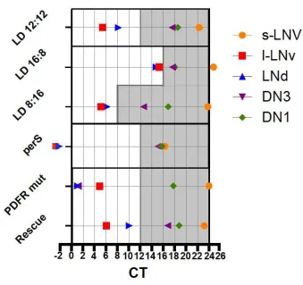
12 Peak times of pacemaker neuron subgroups in different experimental conditions, summarized
from the results in [40]. LD12:12, LD16:8 and LD8:16 are the profiles of wild-type flies in
different photoperiods. PerS, PDFR mut and Rescue indicate the Ca2+ peaks in perS, PdfR
mutants (han5304) and PdfR mutants with genetic rescue in 12h:12h LD photoperiod,
respectively. Each neuronal group has its distinct peak phase. Ca2+ peaks of the M-oscillator
(s-LNvs) and E-oscillator (LNds) track morning and evening light-dark transitions, respectively.
Long-and short-day photoperiod experiments suggest that circadian pacemaker circuit indeed
can work as a seasonal adaptation system. Ca2+ rhythms in perS are phase-advanced and have a
shorter period. In pdfR mutants, LNd and DN3 Ca2+ peaks crowd around dawn, in phase with
the s-LNvs. Restoration of PDFR re-establishes the proper order of the peaks.
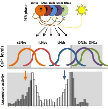
Figure 2. Synchronous circadian pacemakers produce asynchronous Ca2+ waves.
Molecular clocks in all subgroups tick roughly in sync (arrows in the colored circles indicate
PER phase). The s-LNvs enforce delays in Ca2+ waves of the LNds and DN3s through
PDF-mediated inhibition (orange lines). Similar mechanism shortens the time of their own active
phase. The s-LNvs and LNds sequentially secrete sNPF and induce circadian variation of the
DN1 Ca2+ levels, which are otherwise constantly upregulated. In coordination with PDF, light
delays Ca2+ rhythms in the LNds (yellow dotted line). Blue dotted line indicates sNPF released
from the LNds, orange dotted line indicates sNPF from the LNvs. As a consequence, the