der Fakultät für Chemie und Pharmazie
der Ludwig-Maximilians-Universität München
Surface modification
of metal-organic framework nanoparticles
for biomedical applications
Andreas Zimpel
aus
Rosenheim, Deutschland
Erklärung
Diese Dissertation wurde im Sinne von § 7 der Promotionsordnung vom 28. November 2011 von Herrn Prof. Dr. Thomas Bein betreut.
Eidesstattliche Versicherung
Diese Dissertation wurde eigenständig und ohne unerlaubte Hilfe bearbeitet.
München, den 03. August 2018
____________________________ Andreas Zimpel
Dissertation eingereicht am 28.06.2018
1. Gutachter: Prof. Dr. Thomas Bein
2. Gutachter: Dr. Stefan Wuttke
Danksagung
Zu Beginn meiner Dissertation möchte ich einigen Leuten danken, ohne die das Erlangen meines Doktorgrades niemals zu Stande gekommen wäre.
Als erstes möchte ich mich bei meinem Chef, Mentor und Freund Dr. Stefan Wuttke bedanken, ohne den ich ziemlich sicher nie zu diesem, sehr interessanten Forschungsgebiet der „Metall-organischen Gerüstverbindungen“ gekommen wäre. Danke, dass du mich auf
meinem wissenschaftlichen Weg, von unserer ersten Begegnung und Zusammenarbeit als F-Praktikant von Christian Dietl in der organischen Chemie, über das PC-F-Praktikum und die Masterarbeit bis hin zu meiner Doktorarbeit, immer angetrieben und unterstützt hast.
Besonders herzlicher Dank gilt meinem Doktorvater, Prof. Dr. Thomas Bein für die Möglichkeit meine Dissertation in seiner Arbeitsgruppe anfertigen zu dürfen, für all die finanzielle und wissenschaftliche Unterstützung über die Jahre meiner Promotion, sowie bereits während meiner Masterarbeit.
Bei beiden möchte ich mich auch speziell für die Chancen bedanken, meine Forschungsarbeiten auf diversen nationalen und internationalen Konferenzen präsentieren zu dürfen. Diese haben mich nicht nur durch interessante Diskussionen mit anderen Doktoranden wissenschaftlich neu inspiriert, sondern auch menschlich gefestigt, indem ich gezwungen war dadurch meine Kontaktscheu Fremden gegenüber abzulegen.
Vielen Dank auch an meine diversen Kooperationspartner, besonders an Ulrich Lächelt, Hanna Engelke und Michael Peller. Ohne eurer Engagement und wissenschaftlichen Input zu den gemeinsamen Projekten wäre diese Arbeit in ihrer jetzigen Form nie zustande gekommen. Dafür möchte ich mich auch speziell bei meinen Kollegen in den verschiedenen Forschungslaboren bedanken, die zusammen mit mir die Vielzahl an Experimenten durchgeführt haben (u.a. Tobias Preiß, Ruth Röder, Konstantin Böll, Miriam Höhn, Nader Danaf, Simone Braig, Michael Ingrisch, Waldemar Schrimpf,...). Danke auch an die Gruppenleiter und Professoren Ernst Wagner, Joachim Rädler, Angelika Vollmar, Don Lamb und Silke Meiners für ihre Ideen, Kommentare und Korrekturen an den gemeinsamen Publikationen.
All meinen Praktikanten (namentlich Linda, Aylin, Nadine, Luca, Kate und Marina) möchte ich an dieser Stelle für die gute und erfolgreiche Zusammenarbeit bedanken. Durch eure Betreuung habe ich viel für eine spätere Position mit Führungsverantwortung gelernt.
Danke an all meine Bürokollegen (Alesja, Andi, Andre, Noggi, Patrick, Sabrina, Stefan und Tina) für die angenehme Büroatmosphäre. Ganz besonderer Dank gilt dabei Noggi, Stefan und Sabrina, welche im Laufe meiner Promotion zu sehr guten Freunden geworden sind. Außerdem danke ich Hans, Mona und Fabi für ihr „Mentoring“ auf den unterschiedlichsten
Gebieten während meiner Promotion.
Für alles Organisatorische möchte ich mich ganz herzlich bei Tina, Regina und Corinna bedanken.
Mein besonderer Dank gilt natürlich auch allen anderen Personen im Arbeitskreis Bein, welche hier nicht namentlich erwähnt worden sind. Ein großes Dankeschön an alle jetzigen und ehemaligen Kollegen und Kolleginnen mit denen ich diese interessanten und spaßigen Jahre verbringen konnte.
Meiner Ehefrau Marion danke ich ganz herzlich für all die bisherigen gemeinsamen Jahre, für die aufbauenden Worte und die volle Unterstützung während meiner Promotion. Danke für dein Verständnis für meine schlechte Laune wenn es mal nicht so gut lief (eher selten) und für späteres Heimkommen wenn es mal wieder was zu feiern gab (häufiger). Ich liebe dich!
In diesem Zuge möchte ich mich auch bei meiner Schwägerin Andrea bedanken, die durch das mehrmalige Bereitstellen eines Schlafplatzes in ihrer Wohnung in Großhadern auch sehr viel zum Erfolg von Einstands-, Doktor-, Weihnachts- und sonstigen Feiern beigetragen hat.
Weiterhin möchte ich mich noch bei meinen Schwiegereltern Renate und Reiner für die nette Aufnahme in die Familie bedanken. Danke vor allem für die mentale Unterstützung in Prüfungszeiten und an „Kapuzentagen“.
Meinen beiden Brüdern Fredi und Stefan danke ich für den tollen geschwisterlichen Zusammenhalt, der sehr viel dazu beiträgt, dass ich mich zu Hause sehr wohl fühle und eine sehr große Heimatverbundenheit spüre. In diesem Zusammenhang auch danke an all meine Freunde in Rott am Inn!
Abstract
Nowadays, cancer is one of the most challenging diseases on earth. Since the 1970s, the number of patients almost doubled, due to the demographic development of mankind. Cancer researcher Robert A. Weinberg put it in a nutshell by claiming, "If we lived long enough, sooner or later we all would get cancer." Therefore, effective and targeted treatment of affected tissue is of immense interest as common chemotherapy suffers from severe side effects. One way towards selective cancer treatment is the implementation of porous nanocarrier systems for the targeted delivery of chemotherapeutics into tumor tissue to minimize side effects. To fulfil all of its ambitious tasks, the nanocarrier has to provide several different properties such as long circulation lifetimes in the bloodstream, a stimuli-responsive capping system which allows drug release at the desired location or targeting ligands on their external surface to enhance preferential uptake in cancer cells. All these properties can be addressed by the functionalization of the external surface of the designated nanocarrier system. In recent years, metal-organic frameworks (MOFs) have attracted great interest in the field of drug delivery. The ability to adjust their pore sizes and to implement functionalities within the pores as well as on their external surface makes this material class a promising candidate.
This thesis focuses on the surface modification of MOF nanoparticles (NPs) with regard to prospective biomedical applications. In this context, the uptake potential of porous MOF NPs for guest molecules and the in vitro toxicity of the MOF NPs used in this study are investigated in detail. Further, the possibility for external surface functionalization using different approaches is an important focus of this work. The resulting MOF NPs were fully characterized by various methods to ensure their expected morphology, composition and structure. The final achievement of the work is to evaluate the MOF NPs in the biological context. The work aims at determining how the MOF NP structure and their responsiveness to the surrounding biological environment are related to each other and how this behaviour can be correlated to their toxicity.
the chemical and colloidal stability and to provide fluorescence properties by using dye-labeled polymers. The functionalization of the MOF NPs with fluorescent-dye-labeled polymers enables the investigation by fluorescence-based techniques, as demonstrated by fluorescence correlation spectroscopy (FCS) and confocal fluorescence microscopy. Furthermore, the influence of the polymer shell on the intrinsic magnetic resonance imaging (MRI) activity of MIL-100(Fe) is investigated in detail.
As already demonstrated in the third chapter, the effective bio-application of MOF NPs is still hampered by limited control of their surface chemistry and insuffucient understanding of their interactions at the biointerface. Using a self-assembly approach, the fourth chapter of the thesis shows that coating of MOF NPs (Zr-fum) with polymers, frequently used for biomedical applications, is a convenient way for peripheral surface functionalization. Detailed investigation of the binding reveals the mechanism to be a self-assembly modulator replacement by the coordinating group-containing polymers. This strong coordinative binding is further used to attach the shielding polymer polysarcosine onto the MOF surface, which results in an exceptionally high colloidal stability of the NPs. The effect of the polymer coatings on the biointerface is investigated with regard to cell interactions and protein binding.
An important feature of MOF NPs for their use as nanocarriers is the high loading capacity for cargo molecules in their porous scaffold. Therefore, the molecular transport of the model-cargo fluorescein into two MOF NPs, MIL-100(Fe) and MIL-101(Cr) is studied in detail in the second main part (Chapter 5). The equilibrium dissociation constants and maximum number of adsorbed molecules per NP are determined via fluorescence spectroscopy. The resulting maximum payload capacity of 65 wt% MIL-100(Fe) and 41 wt% MIL-101(Cr) is shown to be in agreement with the internal area estimated from nitrogen sorption measurements. Kinetic studies show that release and loading rates are pH dependent. Theoretical modeling of diffusion to target, slowed internal diffusion and equilibrium binding reproduce the observed loading and release times. This study helps to optimize payload and release rates of MOF NPs under varying pH conditions as for example encountered in medical drug delivery applications.
NPs (MIL-100(Fe) and MIL-101(Cr)). It is shown that the created MOF@Lipid system can effectively store dye molecules inside the porous scaffold of the MOF while the lipid bilayer prevents the premature release. Efficient uptake of the MOF@Lipid NPs by cancer cells makes these nanocarriers promising for drug delivery and diagnostic purposes.
The study presented in chapter seven comprehensively analyzes the nanosafety of different MOF NPs used so far in this thesis, namely bare Zr-fum NPs, MIL-100(Fe) NPs and MIL-101(Cr) NPs as well as their MOF@DOPC NP analogs (see chapter six) with regard to diverse medical applications such as drug delivery via blood or lung to multifunctional surface coatings of medical implants. For that purpose, biocompatibility of the MOF NPs on different effector cells (e.g., primary human gingiva fibroblasts) which are defined as those cells that directly interact with NPs when these are introduced into the biological system are tested. Nanosafety of tested MOF NPs strongly varies with the effector cell types revealing their differential suitability as nanomedical agents for drug delivery and implant coatings. These results thus demonstrate the requirement for thorough testing of nanomaterials for their nanosafety with respect to their particular medical application and their interacting primary cell type, respectively.
Finally, chapter eight deals with a modified lipid-coating procedure for MIL-100(Fe) NPs using the same lipid (DOPC). It shows the applicability of such Lip-MOF NPs as effective anti-cancer agents, without loading of any toxic chemotherapeutics into the framework. The toxicity of the particles is thereby triggered by a slightly acidic pH of the extracellular medium (pH = 7.2). These results are promising for a selective treatment of tumor tissue, which provides lower extracellular pH due to an increased lactic acid fermentation of cancer cells (Warburg effect).
Table of contents
1. Introduction 13
1.1. Metal-organic frameworks (MOFs) 13
1.2. Nanoparticles 13
1.3. Metal-organic framework nanoparticles (MOF NPs) 18
1.4. Synthesis of MOF NPs 19
1.5. Engineering MOF NPs 24
1.6. Biomedical application of MOF NPs 29
1.7. Toxicology of MOF NPs 34
1.8. References 37
2. Characterization techniques 45
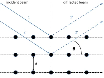
2.1. X-ray diffraction (XRD) 45
2.2. Dynamic light scattering (DLS) 47
2.3. Zeta potential measurement 48
2.4. Sorption measurement 50
2.5. Thermogravimetric analysis (TGA) 53
2.6. Infrared spectroscopy (IR) 53
2.7. Raman spectroscopy 54
2.8. Ultraviolet-Visible spectroscopy (UV-Vis) 55
2.9. Fluorescence spectroscopy (FS) 56
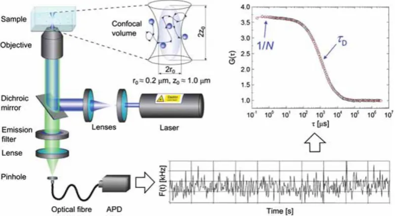
2.10. Fluorescence correlation spectroscopy (FCS) 57
2.11. Fluorescence microscopy 58
2.12. Electron microscopy 60
2.13. Nuclear molecular resonance (NMR)
Magnetic resonance imaging (MRI) 62
3. Imparting functionality to MOF nanoparticles by external surface selective
covalent attachment of polymers 65
3.1. Introduction 65
3.2. Results and Discussion 70
3.3. Conclusion 80
3.4. Materials and Methods 81
3.5. References 87
3.6. Appendix 91
4. Self-assembly of different polymers on MOF nanoparticles for better control of interactions at the biointerface 107
4.1. Introduction 107
4.2 Results and Discussion 111
4.3. Conclusion 121
4.4. Materials and Methods 122
4.5. References 127
4.6. Appendix 132
5. Kinetic analysis of the uptake and release of fluorescein by
metal-organic framework nanoparticles 145
5.1. Introduction 145
5.2. Results and Discussion 148
5.3. Conclusion 157
5.4. Materials and Methods 158
5.5. References 161
5.6. Appendix 164
6. MOF nanoparticles coated by lipid bilayers and their uptake by
cancer cells 173
6.1. Introduction 173
6.2. Results and Discussion 175
6.3. Conclusion 180
6.4. Materials and Methods 181
6.5. References 186
7. Validating metal-organic framework nanoparticles for their nanosafety in diverse biomedical applications 197
7.1. Introduction 197
7.2. Results and Discussion 201
7.3. Conclusion 214
7.4. Materials and Methods 215
7.5. References 221
7.6. Appendix 225
8. pH-selective toxicity of lipid-coated MOF nanoparticles for
use as chemotherapeutics 235
8.1. Introduction 235
8.2. Results and Discussion 237
8.3. Conclusion 245
8.4. Materials and Methods 246
8.5. References 252
8.6. Appendix 254
9. Conclusion and Outlook 261
10. Curriculum vitae 265
1. Introduction
This chapter is based on the following book chapter:
Beetz, M.; Zimpel, A.; Wuttke, S. (August 2016); Nanoparticles. In: Kaskel, S. (Ed.); The Chemistry of Metal-Organic Frameworks: Synthesis, Characterization, and Applications.
Weinheim: Wiley-VCH
1.1. Metal-organic frameworks (MOFs)
Metal-organic frameworks are a class of materials, which came up by the end of the 20th century. They consist of organic linker molecules and inorganic metal clusters acting as nodes in between. The two different building blocks are connected via coordinative bonds forming a rigid porous scaffold which is accessible e.g. for small molecules. Both, the choice of organic linker and metal strongly determine the properties of the resulting framework (structure, pore size, pore environment…). Due to the variety of possible combinations of metal and linker, the material class of MOFs offers a nearly endless number of different compounds.
1.2. Nanoparticles
Introduction into the nanoworld
The term “nano” (Greek for dwarf) became an important notion in science and technology in the last two decades. The prefix “nano“ stands for the order of magnitude of 10-9
. On a scale of length one nanometer correspond to 0.000,000,001 m = 1·10-9 m. In a vivid size comparison, a bacterium, one of the smallest forms of life on earth, is thousand times larger than a nanometer. A human hair in average has a diameter of around 50,000 nm.
fires.3 Furthermore, virus particles are naturally nanostructured particles, consisting of nucleic acids and a protein shell.4
There are different ways in which nanomaterials can be classified. The degree of structural order in nanoparticles can be either crystalline or amorphous. The crystalline nanoparticles are referred to as nanocrystals. They are mostly single-crystalline and hence, have different optical and electrical properties compared to them polycrystalline or bulk form.5 They can be composed of either one material or distinctly different components, the latter are denoted as nanocomposites. Another class of nanoparticles is found in so-called nanostructured materials. The main focus of such materials lies on the shape, surface structure or the superstructure, which give them characteristic abilities with respect to nano-properties. The structure of these particles causes them to have different properties compared to the bulk material due to nano-relevant effects. It is important to say that strong agglomeration or aggregation of this kind of particles mostly leads to a loss of their specific nano-properties and they act like the macroscopic bulk material.5 Nevertheless, sometimes agglomeration is intentionally used to adjust particles sizes and surface-structures. Colloidal nanoclusters and nanoparticle aerogels, for example, have interesting optical and magnetic properties, and are used as catalysts.
Size depending forces between particles
Nanoparticles are so tiny that some effects and forces vanish while others strongly increase. This leads to a shift in the balance of forces influencing the nanoparticle itself. The impact of gravitational force on nanoparticles is strongly reduced due to their lower weight per particle, leading to a more flexible particles’ behavior: they act like molecules. With smaller particle
sizes, the influence of the force of surface charges increases, resulting in strong interactions (Coulomb-attraction/repulsion) between the particles themselves or towards counter ions. Another dominant effect is the strong increase of the surface energy. Reducing the particle size, the surface area to volume ratio increases drastically, leading to a high surface energy. Therefore, nanoparticles have a strong tendency to agglomerate between each other, as it is energetically favored.
Figure 1-1. Different size-depending properties of silicon dioxide.
As a rock, silicon dioxide is hard and brittle. The smaller the particles of silicon dioxide are, for example like in sand, the softer and more flexible the material gets, and it acts rather like a fluid than a rock. Very small SiO2 particles like fumed silica have a very fluffy appearance.
They are soft and can be fluidized due to the increasing impact of electrostatic repulsion and the very low effect that the gravitational force has on the particles. This trend continues towards silicon dioxide nanoparticles, where the influence of electrostatic and gravitational forces further increases.
Nanoscale-effects
The importance of size-relevant effects increases with decreasing particle sizes. Especially when particles are observed in the dimensions of about 1–100 nm (nanometer-scale) the properties of the material change significantly. In this scale, properties like melting point,6 color,7 electrical conductivity,8 magnetic permeability,9 catalytic activity10 and chemical reactivity11 are a function of both particle size and shape.
Effects that play a role in nanoparticles or nanostructures are mostly surface-,12 optical- 13 or quantum-effects14. One well-known example for a surface effect is the Lotus-effect.15 Materials using the Lotus-effect are commonly used as self-cleaning surfaces due to their highly hydrophobic properties. On normal surfaces, adhesive effects ensure water droplets to cover the whole surface-area. Superhydrophobic-Lotus-like materials have a nanostructured surface that minimizes the contact area of the droplets. This causes the droplet to form its most stable form, the sphere. Due to the strong surface curvature of a sphere, there is almost no contact area and hence very little adhesive force of the material. This causes the water droplet to roll down along this surface until it falls off.16
composition, size and shape, specific resonant wavelengths resulting in a color or other optical properties differ from those of the bulk-material.17 A commercial example for such materials are the so-called quantum dots. These are semiconductor-nanoparticles, whose properties can be adjusted to a high degree by composition, doping, size distribution or interactions with each other.14
Another interesting example, where optical properties can be adjusted by nanoscaled structures, is the so-called Vantablack-material (Vanta stands for Vertically Aligned Nano Tube Arrays). It is absorbing 99.965% of the radiation in the visible spectrum and appears therefore as the blackest substance known at the moment. This material has vertically aligned nano-tubes on its surface which scatter the light very effectively between one another and the energy of light is finally converted to heat.18
Synthesis
For synthesis, processing and analytics, it is important to obtain and manipulate nanostructures at the nanometer-scale. In general, the used methods can be classified in the so-called bottom-up and top-down approaches. Examples for bottom-up methods are the liquid phase synthesis (hydrothermal/solvothermal synthesis, the sol-gel processing, etc.) or gas phase methods (chemical vapor deposition, laser ablation deposition, sputtering techniques, etc.). Most top-down methods are based on milling or grinding processes.
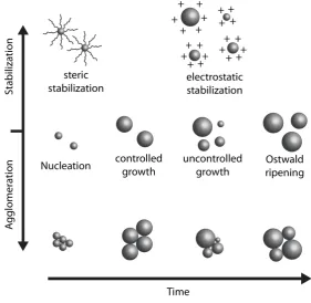
Figure 1-2. Time-dependence of nanoparticle growth and agglomeration/Ostwald-ripening.
To form nanoparticles with a narrow size distribution, the nucleation and growth should be controlled precisely and adjusted as accurately as possible. However, some MOFs preferentially form nanoparticles in the regular synthesis. Another important aspect is the suppression of agglomeration processes. Nanomaterials lose or change their very specific properties when they agglomerate. Additionally, most agglomerated particle-clusters cannot be separated into single particles. The agglomeration behavior in liquid-phase synthesis can be controlled by functionalizing the surface of the nanoparticles immediately after nucleation. Typically, steric demanding organic molecules like long-chain alkyl compounds, surfactants or polymers are used. These can influence the growth direction of the particle and can prevent particles from interacting with each other. Another way of stabilizing single particles is the electrostatic stabilization, where polar molecules on the particle surface prevent agglomeration through electrostatic repulsion. Both strategies prevent also the Ostwald ripening – the process where small particles are merged into larger ones.
1.3. Metal-organic framework nanoparticles (MOF NPs)
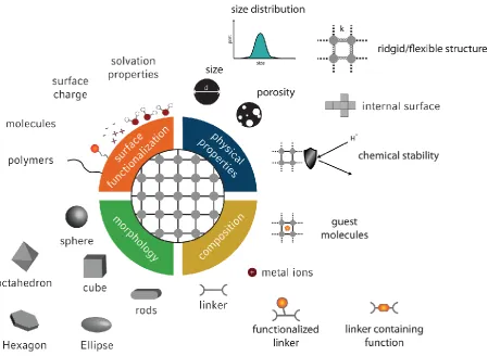
Metal-organic framework materials stand for crystalline materials, a huge number of inorganic building blocks combined with almost endless organic linkers, a tunable pore structure, ultrahigh porosity and different functionalization concepts. The combination of these properties with the nano-world offers manifold perspectives for the synthesis of well-defined multifunctional nanoparticles with novel properties. Operating at a length scale of one-billionth of a meter, the properties of MOF nanoparticles differ significantly from their bulk substances due to the high surface-to-volume ratio and quantum size effects. By combining the inorganic and organic chemistry worlds, MOF nanoparticles will possibly display novel and enhanced properties compared to the already established nanomaterials such as gold nanoparticles, iron oxide nanoparticles, quantum dots, polymers, carbon nanotubes, liposomes, mesoporous silica etc. Further, they could be integrated in well-established systems for enhancing diffusion paths in catalysis19 or as MOF membrane20. Establishing synthesis protocols for precisely tuning the composition, morphology and the physical properties of MOF nanoparticles (Fig. 1-3) is a huge task but at the same time a chance for synthesis chemists to develop new creative ideas.
In addition, for different applications the internal and external surface can be functionalized with MOF specific functionalization concepts. However, the appropriate characterization and evaluation of the MOF nanoparticle properties are a tremendous challenge as it requires different and very expensive analytic instruments. Here, the MOF community is confronted with the challenge to prove the crystallinity of a MOF nanoparticle and with this the underlying MOF structure. Due to peak broadening in powder X-ray diffraction analysis and MOFs being beam sensitive for transmission electron microscopy (TEM) this challenge can hopefully be solved with the new versions of TEM instruments operating at low voltage. Once all the issues mentioned above have being met, well-defined and precisely functionalized MOF nanoparticles can possibly bring new fundamental understanding for the nanoscience area.
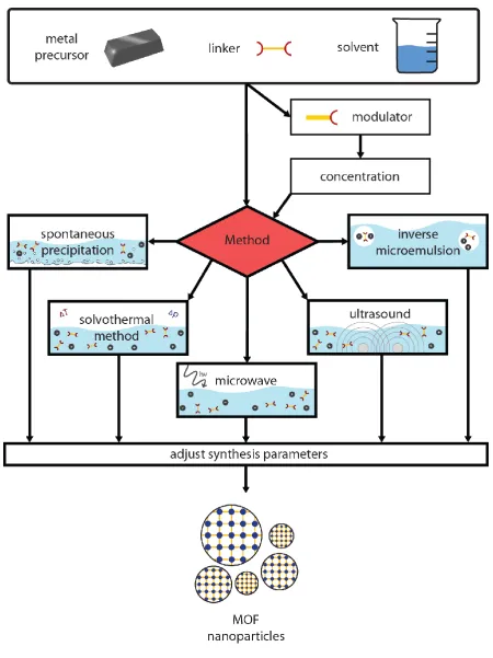
1.4. Synthesis of MOF NPs
Nanoparticles made of metal-organic frameworks (NMOFs) can have versatile applications. In general, these applications require narrow particle size distributions and uniformly shaped crystallites. Therefore, the controlled synthesis of well-defined MOF nanoparticles is of huge interest. Several techniques have been developed in recent years - this chapter deals with procedures which are most commonly applied in chemistry laboratories.
Spontaneous precipitation method
The simplest way to synthesize MOF NPs is their precipitation after mixing metal and linker in a stirring solution. The spontaneous assembly of linker and metal clusters can lead, if controlled by time, temperature and concentration of the precursors, to the formation of NMOFs. Two different techniques have been applied for amorphous coordination polymers first and then have found their way into the synthesis of crystalline MOF NPs. The first one was published by Sun et al. in 2005, where they showed a successful preparation of amorphous spherical colloids, consisting of coordinatively bounded p-phenylenediamine (PPD) and platinum ions, by combining H2PtCl6 and PPD in aqueous solution.23 The second
technique was introduced by the group of Chad A. Mirkin also for amorphous particles. A solution of M(OAc)2 (M = Cu, Zn, Ni) and carboxylate-functionalized binaphthyl
bis-metallotridentate Schiff base (BMSB) in pyridine was diluted with diethylether to induce the precipitation of nanospheres. They also showed the reversibility of this system by dissolving the particles in an excess of pyridine.24 Due to the versatility of these methods, the techniques have been extended to a wide range of coordination particles as well as NMOFs. HKUST-1 nanoparticles, for instance, have been synthesized by pouring an aqueous solution of copper nitrate into a preliminary prepared aqueous solution of trimesic acid.25-26 Further, Pan et al. showed a precipitation of a zeolitic imidazolate framework (ZIF-8) out of an aqueous solution of Zn(NO3)2 ∙ 6 H2O and 2-methylimidazole at ambient temperature.27
Solvo-thermal method
Microwave synthesis
Using microwave irradiation for the production of nano-scaled MOFs turned out to be a useful type of solvo-thermal synthesis. Compared to the classical route, microwave synthesis provides the advantages of fast heating within the reaction mixture. Local superheating provides a huge amount of hot spots which can serve as nucleation seeds for crystal growth.31 This allows short reaction times towards other techniques and narrow particle size distributions. Various MOF NPs have been synthesized by microwave synthesis.32 Jhung et al., for instance, were able to produce very homogeneous MIL-101(Cr) nanoparticles in high yield and uniform shape.33
Preparation by ultrasonic sound
This approach is based on the interaction of high-energetic ultrasonic sound with the reaction solvent, which is followed by cyclic alternating areas of compression and rarefaction. In rarefaction areas, occurring pressures below the vapor pressure of the solvent lead to cavitation. After reaching a maximum size, the cavitation collapses under rapid release of energy, leading to so-called hot spots were MOF formation can take place.34
The ultrasonic method was established for MOF nanoparticle synthesis in 2008, when Qui et al. investigated the formation of Zn3(BTC)2 ∙ 12 H2O by combining zinc acetate dihydrate
and trimesic acid in an ethanol/water mixture. Spontaneous precipitation did not occur by just mixing the precursors at room temperature, but sonochemical synthesis resulted in MOF nanoparticles with approx. 100 nm in diameter in a high yield.35 A few month later, Son et al. successfully prepared MOF-5 particles at least at the micrometer scale by applying ultrasonic sound. They were able to significantly reduce the reaction time compared to conventional solvothermal synthesis from 24 hours to about 30 minutes.36
Reverse microemulsion method
by Mann for the synthesis of Prussian Blue nanoparticles37 or an isooctane/1-hexanol/water mixture, which was applied by the group of Lin for the formation of [Gd(1,2,4-BTC)(H2O)3] ∙
H2O nanocrystals.38
Morphology modulation using additives
Furthermore, addition of modulators to the reaction medium can lead to an improved crystal shape as well as to a narrow size distribution. The idea behind this approach is to limit the particle growth by adding capping molecules to the reaction media. Different types of additives have been approved in literature and showed a confinement effect leading to homogeneously shaped MOF nanocrystals.
One possibility is the usage of small molecules with the same chemical functionality as the linker molecule. In contrast to the linker, these molecules possess only one coordinating functional group and which allow for the coordination to metal centers of the framework, but do not provide a chemical functionality for a further crystal growth. The group of Kitagawa originally introduced the modulation approach, using acetic acid as a monovalent modulator molecule. By taking advantage of the competitive interaction between modulator and linker molecules, they were able to obtain small and homogeneous [{Cu2(ndc)2-(dabco)}n]
nanoparticles 39 as well as bigger, heterogeneous [Cu3(btc)2] nanoparticles40. Further
investigations on this synthesis approach have been performed by Behrens and co-workers, focusing on benzoic acid, acetic acid or formic acid as modulator for the synthesis of Zr-based metal-organic frameworks. They provided control over the nucleation rate of the nanocrystals by changing the concentration of the modulator molecule.41-42
Kitagawa’s group also used the confinement effect of polyvinyl- pyrrolidine (PVP) for the
synthesis of MOF nanoparticles for the first time.43 Prussian Blue nanoparticles were synthesized in an aqueous solution of FeCl3 and K3Fe(CN)6 in the presence of PVP. Without
PVP, large particles (>300 nm) with a broad size distribution were formed. They attributed the latter to a steric stabilization effect of the PVP due to a weak coordination of its amide moiety to the Fe ions during the nucleation and growth process of the particles. Using the same technique, Kerbellec et al. stabilized ultra-small luminescent Tb2(bdc)3(H2O)4
nanoparticles with sizes below 10 nm.44
1-hexanol/n-heptane/water microemulsion.45 They showed that microemulsion synthesis without addition of surfactant resulted in an amorphous material.
Top down processing and combination of different techniques
The synthesis of high-quality nanoparticles, namely those with a defined diameter, almost monodispersed size distribution, and small degree of agglomeration, is often realized by starting from a homogeneous liquid phase solution. Especially in MOF chemistry downsizing by milling is detrimental causing often surface area loss and amorphization. However, some elaborations have recently focused on the formation of superstructures made of MOF nanoparticles 46 (see chapter “Engineering MOF NPss”). Disassembly of these hierarchical structured architectures can lead to nanoparticles. Maspoch and co-workers, for instance, developed a spray-drying strategy to build up MOF hollow spheres. After sonication in methanol, they obtained a colloidal dispersion of homogeneous HKUST-1 nanoparticles.47 Due to the diversity of different synthetic methods towards MOF nanoparticles, researchers started to combine those methods for a further improved control over size and shape of the particles. As an example, Tanaka et al. used reversed microemulsion technique in combination with ultrasonic sound for the preparation of {[Zn(ip)(bpy)]}n (ip = isophthalate,
bpy = 4,4′-bipyridyl; CID-1) nanoparticles.48
1.5. Engineering MOF NPs
External surface functionalization of MOF NPs
The functionalization of MOF NPs on their external surface (Fig. 1-1) is of immense interest, especially with regard to their possible application as drug delivery vehicle (see chapter “Applications of MOF NPs”). Towards an explicit attachment of molecules on the particle
shell, depending on the surface appearance, functionalization can be done effectively in different ways.
fluorescence microscopy.50 This method might as well be a promising approach for the functionalization of MOF NPs. Furthermore, the linking groups of MOFs can also be used as anchoring point for modifications on the outer surface of nanoparticles. In fact, typical organic linkers of the MOFs contain carboxylate groups, for instance. Park and co-workers assumed that a certain amount of carboxylate groups is exposed on the surface and they confirmed this assumption by covalent attachment of enhanced green fluorescent protein (EGFP). They demonstrated their results on a one- dimensional indium-based coordination polymer, the two dimensional [Zn(bpydc)(H2O)]∙(H2O)n, and IRMOF-3 as a model system of
a three dimensional MOF. Activation of the carboxylates was achieved with 1-ethyl-3-(3-dimethylaminopropyl) carbodiimide (EDC) or dicyclohexyl carbodiimide (DCC), respectively.51
Structuring MOF NPs at the macroscopic scale
In order to enrich the overall performance of MOF NPs, researchers started to focus on the construction of hierarchical MOF superstructures. The specific arrangement can have versatile advantages, considering the desired application. In general, there are four different possibilities for the structuring at the macroscopic scale (Fig. 1-5).
Figure 1-5. Illustration of the different types of MOF superstructures made of nanoparticles.
applications,55 as membrane for gas separation 56 or proton and electronic conduction.57 The advantage of a MOF nanoparticle arrangement in three dimensional (3D) frameworks is mainly its contribution to an increased diffusion rate of the guest molecules into the nanoparticle pores, compared to a packed bed system.58 Fast diffusion rates can be crucial in applications such as gas sorption, gas separation or chemical sensing.
The assembly of these superstructures is based on two general methods, “top-down” and “bottom-up” approaches. “Top-down” means the pre-synthesis of MOF NPs and their further
structuring by coating, etching or aligning techniques. As an example, the rearrangement of a dispersion of ZIF-8 crystals into a one-dimensional superstructure by applying an electric field was investigated.59-60 Ostermann et al. obtained MOF nanofibers with an adjustable diameter between 150 nm and 300 nm, adding PVP to a dispersion of ZIF-8 in methanol and injecting the mixture into a reaction chamber, where a voltage of 5 kV was applied.59, 61 “Bottom-up” means the direct synthesis of MOF NPs in an oriented superstructure. Therefore,
different strategies have been developed in recent years. Adding a pre-shaped macrostructural (hard) template or a molecular (soft) template to the reaction, can lead to controlled particle crystallization on the template surface. Subsequent removal of the structure directing material provides hierarchical architectures of the metal organic framework. Common hard templates which have already found their way into the synthesis of MOF superstructures, are e.g. carbons 62, silica 63 or organic polymers 64-65.
Cao et al. showed the use of block-copolymers as soft template for the formation of three-dimensional superstructures of ZIF-8 and HKUST-1 nanoparticles.66
Interfacial reactions provide another possibility for the formation of hierarchically structured metal-organic frameworks. An important feature of this technique is the confinement of metal and precursor at different compartments of the reaction mixture. Thus, MOF formation can only occur at the border between those compartments.
hierarchical structure of MOF NPs with same architecture, reacting the alumina in an aqueous solution of 1,4-naphtalenedicarboxylic acid.
A liquid-liquid interfacial reaction for the generation of hollow spheres, consisting of [Cu3(BTC)2] nanocrystals was published by Ameloot et al. in 2011.69 Injecting droplets of an
aqueous cupper acetate solution in a flow of trimesic acid in 1-octanol yielded homogenous MOF capsules in a range of 300–400 µm in size.
Reaction confinement by evaporation has already been explored in recent years for the structuring of MOF NPs. Thereby, metal source and organic linker have been well stabilized in the reaction medium, so that reaction cannot take place spontaneously. The formation of the MOF is induced by the evaporation of the solvent. This strategy opens the possibility to “print” MOF particles on desired surfaces, which was published by de Vos and co-workers.70
A stable solution of copper nitrate trihydrate and trimesic acid in DMSO was patterned by a stamp onto a glass substrate. After solvent evaporation, they obtained micro-sized MOF crystals in an ordered manner. A route towards zero-dimensional MOF hollow spheres by a reaction confinement approach was shown by Carné-Sánchez et al. in 2013.47 Spray-drying of the precursor solution of different MOFs resulted in spherical capsules, consisting of highly crystalline and homogenous nanoparticles.
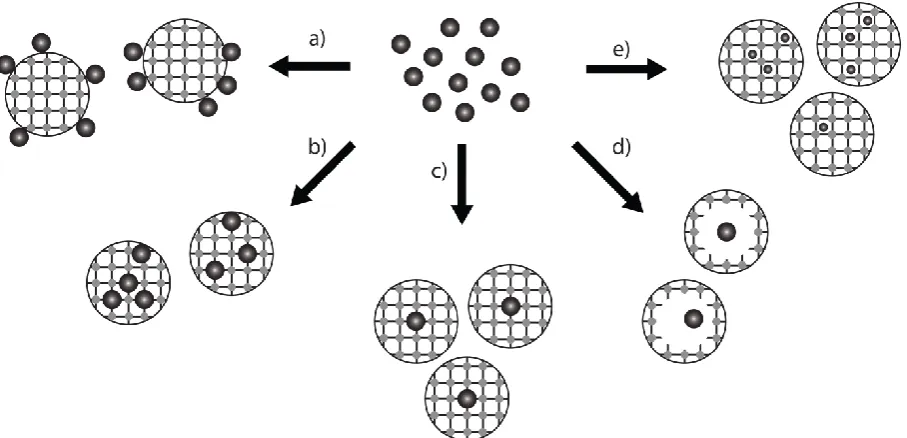
Core-shell MOF NPs
The fascination of polyfunctional nanoparticles and their use in different fields of application leads to the synthesis of core-shell particles, which exhibit new or enhanced properties in comparison to individual units. However, rising the degree of functionality goes along with an increased level of synthesis complexity. Controlling the size of the core and shell, the composition, the dispersed nature, the colloidal stability, the spatial distribution as well as confinement of the core-shell nanoparticles is a huge and exciting challenge.71
Figure 1-6. Synthesis of different core-shell MOF NPs: a) metal nanoparticles attached on the external surface of a MOF nanoparticle; b) multiple metal core materials covered by a MOF shell; c) one core metal material covered by a MOF shell; d) metal nanoparticle yolk and MOF shell nanoparticle; e) small metal core nanoparticles synthesized inside the pores of the MOF.
This synthesis concept is known as “ship-in-bottle” approach.72
The MOF scaffold is loaded with a metal precursor solution in a first step, followed by a reduction step. In this way, different metals, e.g. Pd, Au, Pt, could be synthesized inside the MOF pores.73-79 Another synthesis approach was developed by Fischer and co-workers based on metal organic chemical vapor deposition (MOCVD).80-85 The main challenge of these core-shell synthesis approaches is the formation step in the pores of MOF NPs, which tend to agglomerate during this step.72
controlling the spatial distribution of the core nanoparticles Hupp, Huo and co-workers reported an interesting work.99 They achieved the spatial distribution of polyvinylpyrrolidone (PVP) modified metal nanoparticles within the MOF matrix by simply controlling the moment of addition. Tsung and co-workers could even demonstrate the synthesis of yolk-shell nanoparticles (Fig. 1-6).100 Last but not least, MOFs and metal nanoparticles can be synthesized both separately and the metal nanoparticles can be attached to the MOF nanoparticle surface (Fig. 1-6).72
1.6. Biomedical application of MOF NPs
MOFs NPs as drug carrierThe delivery of drugs is an area of immense importance for human health. Main challenges in drug delivery include: low drug solubility, drug stability and toxicity, rapid metabolism and clearance, and most importantly a lack of selectivity. Nanocarriers hold the key to addressing these challenges. Incorporating drugs into nanoparticles offers exciting opportunities to redefine the pharmacokinetic properties, improving therapeutic efficiency and reducing side effects.101-103 However, the key challenge to realize this potential is to advance the methodologies for the enhanced design of nanoparticles with the following prerequisites:104
Biocombatibility
High loading and protection of the drug molecules
Zero premature release before reaching the target
Efficient cellular uptake
Efficient endosomal escape
Controllable rate of release to achieve an effective local concentration
Cell targeting
In the past decades, several strategies have been developed to design drug delivery materials to accomplish the above mentioned goals. Several drug delivery nanocarriers based on organic platforms such as liposomes, polymers, and dendrimers have been used as “smart”
synthesis of functional nanocarriers, the goals for the targeted release of drugs in the context of treating severe diseases with nanomaterials are still far from being met.
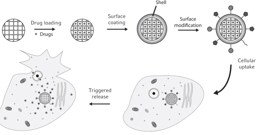
In this respect, MOFs are a unique class of porous hybrid solids with a wide range of compositions, structures, tunable pore sizes, and pore volumes. Their ability to combine both organic and inorganic design principles is one key advantage. Therefore, they could bridge the gap of purely inorganic and organic nanocarriers. The challenging task is the design of site-specific, stimuli-responsive controlled MOF drug delivery systems that - in addition - are biocompatible. Their successful application for medical purposes requires the development of MOF NPs with inner pore functionalization for controlled interaction with biologically active molecules as well as outer functionality for targeted cell uptake, triggered drug release, and with surface shielding against unwanted interactions inside the physiological environment (Fig. 1-7).
Figure 1-7. Synthesis path of a multifunctional MOF nanocarrier as well as the cell uptake of the nanocarrier and triggered drug release inside the cell.
drugs do not require additional external stimuli escape from the nanocarrier, but are rather released from the nanoparticle based on a triggered opening mechanism.
This approach has been extended in the following years by different researchers. Horcajada and co-workers tested different carboxylate-linked MOF NPs for their loading capacities of drugs as well as the release kinetics.109 They could demonstrate that especially iron carboxylate MOF NPs are suitable for encapsulation and controlled delivery of a large number of drugs, such as busulfan, cidofovir, doxorubicin or azidothymidine triphosphate, but being at the same time biocompatible.28, 110-111 Due to the high surface area of MOF NPs, new records of loading capacity for certain drugs in comparison to other nanosystems could be found.
Lin and co-workers reported an approach based on MOF decomposition behavior in physiological medium.112-113
Silica has been used to cover and to control the MOF NP degradation and the core-shell nanoparticle could further be functionalized by postsynthetic covalent attachment of targeting ligands.113 The same group recently published their results of using UiO MOF NPs for the co-delivery of cisplatin and pooled siRNAs.114 The interior of the particles was loaded with cisplatin and afterwards the external surface with siRNA. The efficiency of this multi-functionalized system compared to the individual compartments could be demonstrated. Another functional system was reported by Zhang and co-workers using magnetic porous MOF for drug delivery.115 A core-shell system was synthesized with a Fe3O4 nanorod core
and HKUST-1. The shell material could be loaded with the anticancer drug Nimesulide. One challenge by encapsulating sophisticated drugs into MOF NP structures is the small window size of the pores. Normally, MOF pores are like a space with a small entrance (pore window diameter) in comparison to the void itself (pore diameter). Weerapana, Tsung and co-workers proposed a ship-in-the-bottle strategy to address this challenge by simply forming the MOF structure around the drug.116 This trapping strategy was demonstrated for ZIF-8 nanoparticles by encapsulating fluorescein or the anticancer drug campthothecin. The drawback of the ship-in-the-bottle strategy is that the stability of many drugs does not match with the condition of the MOF synthesis.
suitable for the MOF synthesis and, at the same time, does not change its bioactivity. Since the contergan-scandal we know that even two enantiomers can have totally different biological effects and we should keep this in mind when changing the structure of bioactive molecules.
Recently, Lin’s group could report the synthesis of Hf-porphyrin MOF NPs (named as
DBP-UiO) being able to generate cytotoxic reactive oxygen species.119 Instead of encapsulating a drug, a photosensitizer was used as linker molecule for the MOF NPs, which can be applied for photodynamic therapy. The high in vivo efficacy could be demonstrated by 50-fold tumor volume reduction in half of the mice and complete tumor eradication in the other half of the mice that were treated with the Hf-porphyrin MOF NPs.
The same group lately reported the in vivo performance of so-called nanoscale coordination polymers (NCPs).120 They are built up from metal ions and organic bridging ligands – same design principle like MOFs - but in contrast to MOFs they are not crystalline. However, using cisplatin and oxaliplatin as linkers for NCPs and applying them against different cancer tumors, reveal, that NCPs could be a new promising nanocarrier class and should therefore be mentioned here.
Last but not least, MOF NPs can be used to transport gasotransmitters such as nitric oxide (NO) or hydrogen sulfide (H2S). Morris’ group of St. Andrews is the leading group for storing
and release gasotransmitter molecules inside MOFs.121, 109 The occurrence of coordinatively unsaturated metal sites (CUSs) and the high surface area of MOFs ensure a high uptake of the various gases. First results show promising bioactivity of such systems, which will be further improved and biologically tested in the future 109. In this respect, Furukawa and co-workers recently reported a strategy for controllable NO release based on photoactive MOFs.122
MOF NPs for Diagnostics
Figure 1-8. Schematic illustration of the theranostic idea (MRI = magnetic resonance imaging, PET = positron emission tomography, SPECT = single-photon emission computed tomography, NIR = near-infrared).
Magnetic resonance imaging (MRI) is a non-invasive method of mapping the internal structure and certain aspects of function within the body. It is based on the detection of nuclear spin reorientations in a magnetic field. To improve the visibility and with this the clinical diagnostics MRI contrast agents are used. They can be classified into two groups: (i) MRI contrast agents with positive signal enhancement by shortening the value of the T1 relaxation time or (ii) MRI contrast agents with negative signal enhancement by reducing the T2 signals.
The most commonly used compounds for contrast enhancement are Gadolinium-based contrast agents for positive signal enhancement. Lin and co-workers reported first the effectiveness of Gd3+-based MOF NPs as T1-weighted contrast agents.38 Other MOFs based on Gd3+ or even on Mn2+ have been published afterwards.45, 49, 123-124 The main problem of these materials for their practical use is the poor chemical stability resulting in toxicity. Another approach was proposed by Horcajada and co-workers by using iron-based MOF NPs as a negative signal enhancement contrast agent.28 The key advantage of this strategy is, that the combination of the MRI diagnostic capability with drug delivery properties is straightforward.28 The disadvantage of the lower visibility of these MRIs in comparison with gadolinium-based contrast agent images should be improved in the future.
incorporation into the MOF.125-127 Such an example was reported by Lin and co-workers who functionalized MIL-101(Fe)-NH2 with a BODIPY dye.128
The dye was not fluorescent due to quenching but exhibited strong turn-on fluorescence based on the decomposition of the MOF NPs in the cells, which induces the release of the BODIPY dyes. If this degradation of the MOF is specific to one analyte, the development of a sensitive optical MOF sensor is possible.125 Lin and co-workers reported the design of a real-time pH MOF sensor for cells by covalent attachment of fluorescein isothiocyanate (FITC) to a UiO NMOF.114 4 wt% of FITC loading was chosen for the calibration curve due to the absence of FITC self-quenching at such amount. Incubation of fixed cells with these functionalized NPs revealed a pH change over time from 6.4 to 5.6, showing the intracellular endocytosis of the nanoparticles.
1.7. Toxicology of MOF NPs
Nanotoxicology refers to the toxicity of nanomaterials and is a very important but at the same time a complex research field. Health effects of nanoparticles have attracted considerable and increasing concern of the public society. With nanomaterials offering completely different properties than the bulk-material, the toxicity of these materials cannot be broken down to the chemical composition. They can be easily incorporated into organisms due to their small diameter and taken up by our smallest building block unit of life – the cell.129 But not only direct incorporation is a problem since plants can take up nanoparticles from the soil and translocate them to organisms by the food chain.130
Nanotoxicology is a relative new field of interest. There are no results from long-term studies about the toxicology of materials in the nanometer scale. Long-term studies are needed especially for cancerogenious and statistically relevant investigations. There are short-term investigations which show that even inert materials like gold can have toxic effects on cells in the nanometer scale.130 This compels us to handle each material in different nanometer-size and shape as a new material with unknown effects on living tissue. Furthermore, it has to be distinguished between acute and chronic exposure of the nanoparticles.
The toxicity of nanoparticles is depending on their size 131, shape 132, surface-area 133, material
134
proteins to work unspecifically or inhibit their function.137 This can cause irreversible damages to cells or tissues. Another important factor is the size depending toxicity of the particles, which determines the distribution of them in the human body. It could be shown that ultra-fine titanium dioxide particles were taken up significantly faster and had a faster clearance rate than fine titanium dioxide particles in the lung. Moreover, the shape of these materials can have an important influence in their toxicological effect.
Shape depending toxicology is already known from asbestos which then was a commonly used thermal insulator and robust material. Its dangers were not taken seriously first, due to its long-term cancer hazard. The long and very thin fibers of which asbestos is built up, can find their way into the alveoli of the lung and cause a chronic inflammatory. The lung’s alveolar macrophages (dust cells) have the task to wrap around external particles like dust or soot and remove them from the very sensitive alveoli. In the case of asbestos fibers, it is not possible for these macrophages to remove the fibers if they have a width/length ratio larger than 3:1. This leads to the chronic inflammatory of the lung’s tissue which can result in lung cancer for
the long term. Similar size and shape depending effects can be expected for a variety of nanomaterials. Further, with nanomaterials having very active structured surfaces their influence on special tissues is widely unknown.
1.8. References
[1] J. F. Gomes, R. M. Miranda, T. J. Santos, P. A. Carvalho, Journal of toxicology and environmental health. Part A 2014, 77, 924-930.
[2] B. M. Simonet, M. Valcárcel, Anal Bioanal Chem 2009, 393, 17-21.
[3] N. Tepe, M. Bau, The Science of the total environment 2014, 488-489, 243-251. [4] J. E. Johnson, W. Chiu, Current Opinion in Structural Biology 2000, 10, 229-235. [5] G. Nichols, S. Byard, M. J. Bloxham, J. Botterill, N. J. Dawson, A. Dennis, V. Diart,
N. C. North, J. D. Sherwood, Journal of Pharmaceutical Sciences 2002, 91, 2103-2109.
[6] K. Dick, T. Dhanasekaran, Z. Zhang, D. Meisel, Journal of the American Chemical Society 2002, 124, 2312-2317.
[7] W. L. Barnes, A. Dereux, T. W. Ebbesen, Nature 2003, 424, 824-830.
[8] S. Makhlouf, M. Kassem, M. A. Abdel-Rahim, J Mater Sci 2009, 44, 3438-3444. [9] H. Yun, X. Liu, T. Paik, D. Palanisamy, J. Kim, W. D. Vogel, A. J. Viescas, J. Chen,
G. C. Papaefthymiou, J. M. Kikkawa, M. G. Allen, C. B. Murray, ACS nano 2014, 8, 12323-12337.
[10] X. Zhou, W. Xu, G. Liu, D. Panda, P. Chen, Journal of the American Chemical Society 2010, 132, 138-146.
[11] C. Carlson, S. M. Hussain, A. M. Schrand, L. K. Braydich-Stolle, K. L. Hess, R. L. Jones, J. J. Schlager, The Journal of Physical Chemistry B 2008, 112, 13608-13619. [12] A. Albanese, P. S. Tang, W. C. Chan, Annual Review of Biomedical Engineering
2012, 14, 1-16.
[13] C. C. D. Wang, W. C. H. Choy, C. Duan, D. D. S. Fung, W. E. I. Sha, F.-X. Xie, F. Huang, Y. Cao, Journal of Materials Chemistry 2012, 22, 1206-1211.
[14] A. P. Alivisatos, Science 1996, 271, 933-937.
[15] H. J. Ensikat, P. Ditsche-Kuru, C. Neinhuis, W. Barthlott, Beilstein Journal of Nanotechnology 2011, 2, 152-161.
[16] A. Marmur, Langmuir 2004, 20, 3517-3519.
[17] S. Link, M. A. El-Sayed, The Journal of Physical Chemistry B 1999, 103, 8410-8426. [18] E. Theocharous, R. Deshpande, A. C. Dillon, J. Lehman, Appl. Opt. 2006, 45,
1093-1097.
[20] Y. Liu, N. Wang, J. H. Pan, F. Steinbach, J. Caro, Journal of the American Chemical Society 2014, 136, 14353-14356.
[21] W. Lin, W. J. Rieter, K. M. L. Taylor, Angewandte Chemie (International ed. in English) 2009, 48, 650-658.
[22] T. Chalati, P. Horcajada, R. Gref, P. Couvreur, C. Serre, Journal of Materials Chemistry 2011, 21, 2220-2227.
[23] X. Sun, S. Dong, E. Wang, Journal of the American Chemical Society 2005, 127, 13102-13103.
[24] M. Oh, C. A. Mirkin, Nature 2005, 438, 651-654.
[25] J. Huo, M. Brightwell, S. El Hankari, A. Garai, D. Bradshaw, Journal of Materials Chemistry A 2013, 1, 15220-15223.
[26] L. H. Wee, M. R. Lohe, N. Janssens, S. Kaskel, J. A. Martens, Journal of Materials Chemistry 2012, 22, 13742-13746.
[27] Y. Pan, Y. Liu, G. Zeng, L. Zhao, Z. Lai, Chemical Communications 2011, 47, 2071-2073.
[28] P. Horcajada, T. Chalati, C. Serre, B. Gillet, C. Sebrie, T. Baati, J. F. Eubank, D. Heurtaux, P. Clayette, C. Kreuz, J.-S. Chang, Y. K. Hwang, V. Marsaud, P.-N. Bories, L. Cynober, S. Gil, G. Ferey, P. Couvreur, R. Gref, Nat Mater 2010, 9, 172-178. [29] A. Sonnauer, F. Hoffmann, M. Fröba, L. Kienle, V. Duppel, M. Thommes, C. Serre,
G. Férey, N. Stock, Angewandte Chemie International Edition 2009, 48, 3791-3794. [30] P. Horcajada, H. Chevreau, D. Heurtaux, F. Benyettou, F. Salles, T. Devic, A.
Garcia-Marquez, C. Yu, H. Lavrard, C. L. Dutson, E. Magnier, G. Maurin, E. Elkaim, C. Serre, Chemical Communications 2014, 50, 6872-6874.
[31] Z. Ni, R. I. Masel, Journal of the American Chemical Society 2006, 128, 12394-12395.
[32] A. García Márquez, A. Demessence, A. E. Platero-Prats, D. Heurtaux, P. Horcajada, C. Serre, J.-S. Chang, G. Férey, V. A. de la Peña-O'Shea, C. Boissière, D. Grosso, C. Sanchez, European Journal of Inorganic Chemistry 2012, 2012, 5165-5174.
[33] S. H. Jhung, J. H. Lee, J. W. Yoon, C. Serre, G. Férey, J. S. Chang, Advanced Materials 2007, 19, 121-124.
[34] N. Stock, S. Biswas, Chemical Reviews 2011, 112, 933-969.
[35] L.-G. Qiu, Z.-Q. Li, Y. Wu, W. Wang, T. Xu, X. Jiang, Chemical Communications 2008, 3642-3644.
[37] S. Vaucher, M. Li, S. Mann, Angewandte Chemie International Edition 2000, 39, 1793-1796.
[38] W. J. Rieter, K. M. L. Taylor, H. An, W. Lin, W. Lin, Journal of the American Chemical Society 2006, 128, 9024-9025.
[39] T. Tsuruoka, S. Furukawa, Y. Takashima, K. Yoshida, S. Isoda, S. Kitagawa, Angewandte Chemie International Edition 2009, 48, 4739-4743.
[40] S. Diring, S. Furukawa, Y. Takashima, T. Tsuruoka, S. Kitagawa, Chemistry of Materials 2010, 22, 4531-4538.
[41] A. Schaate, P. Roy, A. Godt, J. Lippke, F. Waltz, M. Wiebcke, P. Behrens, Chemistry – A European Journal 2011, 17, 6643-6651.
[42] G. Wißmann, A. Schaate, S. Lilienthal, I. Bremer, A. M. Schneider, P. Behrens, Microporous and Mesoporous Materials 2012, 152, 64-70.
[43] T. Uemura, S. Kitagawa, Journal of the American Chemical Society 2003, 125, 7814-7815.
[44] N. Kerbellec, L. Catala, C. Daiguebonne, A. Gloter, O. Stephan, J.-C. Bunzli, O. Guillou, T. Mallah, New Journal of Chemistry 2008, 32, 584-587.
[45] K. M. L. Taylor, A. Jin, W. Lin, Angewandte Chemie International Edition 2008, 47, 7722-7725.
[46] S. Furukawa, J. Reboul, S. Diring, K. Sumida, S. Kitagawa, Chemical Society Reviews 2014.
[47] A. Carné-Sánchez, I. Imaz, M. Cano-Sarabia, D. Maspoch, Nat Chem 2013, 5, 203-211.
[48] D. Tanaka, A. Henke, K. Albrecht, M. Moeller, K. Nakagawa, S. Kitagawa, J. Groll, Nat Chem 2010, 2, 410-416.
[49] M. D. Rowe, D. H. Thamm, S. L. Kraft, S. G. Boyes, Biomacromolecules 2009, 10, 983-993.
[50] B. Liu, M. Ma, D. Zacher, A. Bétard, K. Yusenko, N. Metzler-Nolte, C. Wöll, R. A. Fischer, Journal of the American Chemical Society 2011, 133, 1734-1737.
[51] S. Jung, Y. Kim, S.-J. Kim, T.-H. Kwon, S. Huh, S. Park, Chemical Communications 2011, 47, 2904-2906.
[52] X. W. Lou, Y. Wang, C. Yuan, J. Y. Lee, L. A. Archer, Advanced Materials 2006, 18, 2325-2329.
[54] S. E. Skrabalak, J. Chen, Y. Sun, X. Lu, L. Au, L. M. Cobley, Y. Xia, Accounts of chemical research 2008, 41, 1587-1595.
[55] F. M. Hinterholzinger, A. Ranft, J. M. Feckl, B. Ruhle, T. Bein, B. V. Lotsch, Journal of Materials Chemistry 2012, 22, 10356-10362.
[56] A. Huang, N. Wang, C. Kong, J. Caro, Angewandte Chemie International Edition 2012, 51, 10551-10555.
[57] G. Xu, K. Otsubo, T. Yamada, S. Sakaida, H. Kitagawa, Journal of the American Chemical Society 2013, 135, 7438-7441.
[58] R. Krishna, J. R. Long, The Journal of Physical Chemistry C 2011, 115, 12941-12950. [59] R. Ostermann, J. Cravillon, C. Weidmann, M. Wiebcke, B. M. Smarsly, Chemical
Communications 2011, 47, 442-444.
[60] N. Yanai, M. Sindoro, J. Yan, S. Granick, Journal of the American Chemical Society 2012, 135, 34-37.
[61] M. Rose, B. Böhringer, M. Jolly, R. Fischer, S. Kaskel, Advanced Engineering Materials 2011, 13, 356-360.
[62] P. Pachfule, B. K. Balan, S. Kurungot, R. Banerjee, Chemical Communications 2012, 48, 2009-2011.
[63] N. Liu, Y. Yao, J. Cha, M. McDowell, Y. Han, Y. Cui, Nano Res. 2012, 5, 109-116. [64] H. J. Lee, W. Cho, M. Oh, Chemical Communications 2012, 48, 221-223.
[65] T. Ben, C. Lu, C. Pei, S. Xu, S. Qiu, Chemistry – A European Journal 2012, 18, 10250-10253.
[66] S. Cao, G. Gody, W. Zhao, S. Perrier, X. Peng, C. Ducati, D. Zhao, A. K. Cheetham, Chemical Science 2013, 4, 3573-3577.
[67] W.-w. Zhan, Q. Kuang, J.-z. Zhou, X.-j. Kong, Z.-x. Xie, L.-s. Zheng, Journal of the American Chemical Society 2013, 135, 1926-1933.
[68] J. Reboul, S. Furukawa, N. Horike, M. Tsotsalas, K. Hirai, H. Uehara, M. Kondo, N. Louvain, O. Sakata, S. Kitagawa, Nat Mater 2012, 11, 717-723.
[69] R. Ameloot, F. Vermoortele, W. Vanhove, M. B. J. Roeffaers, B. F. Sels, D. E. De Vos, Nat Chem 2011, 3, 382-387.
[70] R. Ameloot, E. Gobechiya, H. Uji-i, J. A. Martens, J. Hofkens, L. Alaerts, B. F. Sels, D. E. De Vos, Advanced Materials 2010, 22, 2685-2688.
[73] Y. K. Hwang, D.-Y. Hong, J.-S. Chang, S. H. Jhung, Y.-K. Seo, J. Kim, A. Vimont, M. Daturi, C. Serre, G. Férey, Angewandte Chemie International Edition 2008, 47, 4144-4148.
[74] D. Esken, X. Zhang, O. I. Lebedev, F. Schroder, R. A. Fischer, Journal of Materials Chemistry 2009, 19, 1314-1319.
[75] A. Aijaz, T. Akita, N. Tsumori, Q. Xu, Journal of the American Chemical Society 2013, 135, 16356-16359.
[76] A. Aijaz, A. Karkamkar, Y. J. Choi, N. Tsumori, E. Rönnebro, T. Autrey, H. Shioyama, Q. Xu, Journal of the American Chemical Society 2012, 134, 13926-13929. [77] Q.-L. Zhu, J. Li, Q. Xu, Journal of the American Chemical Society 2013, 135,
10210-10213.
[78] M. Sabo, A. Henschel, H. Frode, E. Klemm, S. Kaskel, Journal of Materials Chemistry 2007, 17, 3827-3832.
[79] S. Opelt, S. Türk, E. Dietzsch, A. Henschel, S. Kaskel, E. Klemm, Catalysis Communications 2008, 9, 1286-1290.
[80] S. Hermes, M.-K. Schröter, R. Schmid, L. Khodeir, M. Muhler, A. Tissler, R. W. Fischer, R. A. Fischer, Angewandte Chemie International Edition 2005, 44, 6237-6241.
[81] M. Muller, O. I. Lebedev, R. A. Fischer, Journal of Materials Chemistry 2008, 18, 5274-5281.
[82] F. Schröder, D. Esken, M. Cokoja, M. W. E. van den Berg, O. I. Lebedev, G. Van Tendeloo, B. Walaszek, G. Buntkowsky, H.-H. Limbach, B. Chaudret, R. A. Fischer, Journal of the American Chemical Society 2008, 130, 6119-6130.
[83] F. Schröder, S. Henke, X. Zhang, R. A. Fischer, European Journal of Inorganic Chemistry 2009, 2009, 3131-3140.
[84] M. Meilikhov, K. Yusenko, A. Torrisi, B. Jee, C. Mellot-Draznieks, A. Pöppl, R. A. Fischer, Angewandte Chemie International Edition 2010, 49, 6212-6215.
[85] D. Esken, S. Turner, O. I. Lebedev, G. Van Tendeloo, R. A. Fischer, Chemistry of Materials 2010, 22, 6393-6401.
[86] J. H. Cavka, S. Jakobsen, U. Olsbye, N. Guillou, C. Lamberti, S. Bordiga, K. P. Lillerud, Journal of the American Chemical Society 2008, 130, 13850-13851.
[87] H. Furukawa, F. Gándara, Y.-B. Zhang, J. Jiang, W. L. Queen, M. R. Hudson, O. M. Yaghi, Journal of the American Chemical Society 2014, 136, 4369-4381.
[89] P. Hu, J. Zhuang, L.-Y. Chou, H. K. Lee, X. Y. Ling, Y.-C. Chuang, C.-K. Tsung, Journal of the American Chemical Society 2014, 136, 10561-10564.
[90] R. Pérez, B. Albero, J. Tadeo, E. Molero, C. Sánchez-Brunete, Anal Bioanal Chem 2015, 1-12.
[91] K. Khaletskaya, J. Reboul, M. Meilikhov, M. Nakahama, S. Diring, M. Tsujimoto, S. Isoda, F. Kim, K.-i. Kamei, R. A. Fischer, S. Kitagawa, S. Furukawa, Journal of the American Chemical Society 2013, 135, 10998-11005.
[92] O. Shekhah, H. Wang, S. Kowarik, F. Schreiber, M. Paulus, M. Tolan, C. Sternemann, F. Evers, D. Zacher, R. A. Fischer, C. Wöll, Journal of the American Chemical Society 2007, 129, 15118-15119.
[93] O. Shekhah, H. Wang, D. Zacher, R. A. Fischer, C. Wöll, Angewandte Chemie International Edition 2009, 48, 5038-5041.
[94] O. Shekhah, H. Wang, M. Paradinas, C. Ocal, B. Schupbach, A. Terfort, D. Zacher, R. A. Fischer, C. Woll, Nat Mater 2009, 8, 481-484.
[95] F. Ke, L.-G. Qiu, Y.-P. Yuan, X. Jiang, J.-F. Zhu, Journal of Materials Chemistry 2012, 22, 9497-9500.
[96] F. Ke, J. Zhu, L.-G. Qiu, X. Jiang, Chemical Communications 2013, 49, 1267-1269. [97] T.-H. Park, K. J. Lee, S. Hwang, J. Yoon, C. Woell, J. Lahann, Advanced Materials
2014, 26, 2883-2888.
[98] M. E. Silvestre, M. Franzreb, P. G. Weidler, O. Shekhah, C. Wöll, Advanced Functional Materials 2013, 23, 1210-1213.
[99] G. Lu, S. Li, Z. Guo, O. K. Farha, B. G. Hauser, X. Qi, Y. Wang, X. Wang, S. Han, X. Liu, J. S. DuChene, H. Zhang, Q. Zhang, X. Chen, J. Ma, S. C. J. Loo, W. D. Wei, Y. Yang, J. T. Hupp, F. Huo, Nat Chem 2012, 4, 310-316.
[100] C.-H. Kuo, Y. Tang, L.-Y. Chou, B. T. Sneed, C. N. Brodsky, Z. Zhao, C.-K. Tsung, Journal of the American Chemical Society 2012, 134, 14345-14348.
[101] M. Ferrari, Nat Rev Cancer 2005, 5, 161-171.
[102] D. Peer, J. M. Karp, S. Hong, O. C. Farokhzad, R. Margalit, R. Langer, Nat Nano 2007, 2, 751-760.
[103] M. E. Davis, Z. Chen, D. M. Shin, Nat Rev Drug Discov 2008, 7, 771-782.
[104] J. L. Vivero-Escoto, I. I. Slowing, B. G. Trewyn, V. S. Y. Lin, Small 2010, 6, 1952-1967.