Development of ABF and Fuzzy Based
Techniques for detection of heart blocks in
CT-Scan image
M.Ramu
1, B.Sridhar
2, K.Mounika
3, M.Sreelekha
4,K.Aravind babu
51,2,3,4 Department of ECE, Lendi Institute of Engineering &Technology, Vizianagaram
Andrapradesh, India.
Abstract—CT scan imagings is one of the fastest and accurate methods for examining heart and plays a vital role in detection of size of the blocks and identify its precise region in heart. Physicians often prefer the CT scan examination to quickly identify the blocks in heart and assess the results for monitor response. The proposed method mainly consists of three blocks, they are pre-processing, segmentation of blocks in CT scan image of heart, evaluation. Pre-processing has done by using adaptive bilateral filter that which removes a high robust noise on the image and highlights the boundaries of the regions in the image. In the process of segmentation an image subtraction is applied on the CT scan image to extract the high intensity areas followed a fuzzy logic based detection process to highlight the blocks in heart and rest of the regions are being suppressed. The proposed method is to be simulated on MATLAB and to be tested with patient CT scan data sets. The results of the proposed method are compared with expert radiologists detection, which shows proposed method is efficient and gives a high performance results.
Keywords —1. CT scan, 2.Fuzzy technique, 3.Image segmentation, 4.Bi lateral filtering.
I. INTRODUCTION
Heart is a muscular organ in humans that pumps blood through the blood vessels to circulatory system. Fig. 1 shows Blood provides the body with oxygen and nutrients, and assists in the removal of metabolic wastes. The heart is located in the middle component of the mediastinum in the chest.
Fig. 1: Heart muscular system
1. TECHNIQUES USED TO DETECT HEART PROBLEMS
Heart diseases can be diagnosed using different techniques, some of the recommended techniques are used to detect the heart problems.
ECG (Electrocardiogram)
An ECG is a simple, painless test for detect and records the heart electrical activity. The test shows the fastness of the heart beat and the rhythm. An ECG also records strength and timing of electrical signals that which pass through the heart. Echocardiography
Electrocardiography uses sound waves to create a moving picture of the heart. The test provides information of the size and shape of the heart and its working condition of the chambers. Chest X Ray
A chest x-ray creates pictures of the organs and structures inside the chest such as heart, lungs and blood vessels. It can reveal signs of heart failure.
2. CT SCAN
A CT scanner uses digital geometry processing to generate a 3-dimensional (3D) image of the inside of an object. The 3D image is made after many 2-dimensional (2D) X-ray images are taken around a single axis of rotation. In other words, many pictures of the same area are taken from many angles and then placed together to produce a 3D image.
Fig.2: CT scan device
Fig.3: CT scan image of heart Related work “Four-chamber heart modeling and automatic segmentation for 3D cardiac CT volumes using marginal space learning and steerable features” by Yefeng Zheng, Adrian Barbu, Bogdan Georgescu, Michael scheuering and Dorin comanicie [1] employees a method for learning in detail about the heart and its four-chambers to analyze the dimensions of the heart during heart beat based on its CT volumes the images are obtained by highlighting their boundaries based on segmentation method. As a consequence, new techniques must be explored to identify the blocks in the CT scan images so to understand this we have learned the concept of CT scan technique by considering “Automatic Heart Isolation in 3D CT Images” by Hua Zhong, Yefeng Zheng, Gareth Funka-Lea, and Fernando Vega-Higuera [7] and Calcification Detection in Coronary Arteries Using Image Processing [3] from international journals.
“Enhancement of Image Sharpness with Bilateral and Adaptive Filter” [11] In this paper, we present the adaptive bilateral filter (ABF) for sharpness enhancement and noise removal. The ABF sharpens an image by increasing the slope of the edges without producing overshoot or undershoot. It is an approach to sharpness enhancement that is fundamentally different from the un sharp mask. This new approach to slope restoration also differs previous slope restoration algorithms. In the ABF, the edge slope is enhanced by transforming the histogram via a range filter with adaptive offset and width. The ABF is able to smooth the noise, while enhancing edges and textures in the image. The parameters of the ABF are optimized with a training procedure.
“A Novel Approach of Fuzzy Logic Based Edge Detection for Coronary Angiogram Using Virtual Instrumentation” by Dr.A.Umarani, N.Vishnudevi, R. Naveeth Kumar, 2015 [8] as this can result in a myocardial infarction (heart attack) when a blood clot develops at the site of the plaque and suddenly cuts off most or all of the blood supply causing permanent damage to the heart muscle. It is necessary to find the border of coronary artery to determine the stenosis and we go for edge detection algorithm using fuzzy logic.
“Fuzzy Techniques for Image Segmentation” by Laszlo, G.Nyul, Department of Image Processing and Computer Graphics, University of Szeged, 2008 [10]. In this thesis the algorithm is proposed using fuzzy membership function. This algorithm enhances image contrast very effectively. If the observed data are distributed by random noise then the intensifier operator should convert the probabilistic data into fuzzy data. Some images are not available to good quality, so proposed fuzzy algorithm can be used for image enhancement to improve the quality of images. All the implementation work has been done in MATLAB image processing tool box. An experimental result shows that the quality of image is improved.
3. FUZZY SET
Fuzzy systems and models are capable of representing diverse, inexact, and inaccurate information. Fuzzy logic provides a method to formalize reasoning when dealing with vague terms. Not every decision is either true or false. Fuzzy logic allows for membership functions, or degrees of truthfulness and falsehoods.
Let X be the universal set. For (sub) set A of X
µA(x) = (1 if x ∈ A 0 if x 6∈ A)
For crisp sets µA is called the characteristic function of A.
A fuzzy subset A of X is A = {(x, µA(x))|x ∈ X}
Where µA is the membership function of A in X
µA: X → [0, 1]
FUZZY MEMBERSHIP FUNCTIONS:
membership in those categories.
Fig.4 Fuzzy membership function
FUZZY IMAGE PROCESSING
Fig.9 is a Collection of all approaches that understand, represent and process the images, their segments and features as fuzzy sets. The representation and processing depend on the selected fuzzy technique and on the problem to be solved.
Fig.5: Fuzzy image processing
APPLICATION AREA FOR FUZZY SYSTEMS • Quality control
• Error diagnostics • Control theory • Pattern recognition
4. IMAGE SEGMENTATION
The division of an image into meaningful structures of image segmentation, is often an essential step in image analysis, object
representation, visualization, and many other image processing tasks. Here we focussed to analyse and represent an object, but we assumed the group of pixels that identified the object was known beforehand. In this chapter, we will focus on methods that find the particular pixels that make up an object.
Threshold based segmentation
Histogram thresholding and slicing
techniques are used to segment the image. They may be applied directly to an image, but can also be combined with pre- and post-processing techniques. Edge based segmentation
With this technique, detected edges in an image are assumed to represent object boundaries, and used to identify these objects.
Region based segmentation
Where an edge based technique may attempt to find the object boundaries and then locate the object itself by filling them in, a region based technique takes the opposite approach, by (e.g.) starting in the middle of an object and then “growing” outward until it meets the object boundaries.
5. MATLAB SOFTWARE
MATLAB is a high-level technical computing language and interactive environment for algorithm development, data visualization, data analysis, and numeric computation. Using the MATLAB product, you can solve technical computing problems faster than with traditional programming languages, such as C, C++.
1.
High-level language for technical computing2.
Development environment for managing code,files, and data
3.
Interactive tools for iterative exploration, design, and problem solving4.
Mathematical functions for linear algebra, statistics, Fourier analysis, filtering, optimization, and numerical integration5.
2-D and 3-D graphics functions for visualizingdata
6.
Tools for building custom graphical user interfaces7.
Functions for integrating MATLAB based algorithms with external applications and languages, such as C, C++, Fortran, Java™.6. PROPOSED METHOD
The propsed method has shown in the fig:10 gives a FLOW GRAPH of the process
Young person
Cold beer
tri
a
ngl
e
trapezoi
d
ga
u
ssia
n
Fig.6: Flow graph
The above flow graph describes the concept of the proposed method in sequential step by step procedure of an algorithm. By following this algorithm we are going to implement the concept of proposed method based on segmentation technique and segregated into different block diagrams.
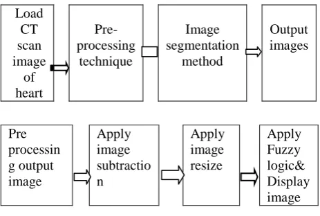
7. BLOCK DIAGRAM
This is the main block diagram of our project in which majorly we follow the four steps: Step 1: To load the CT scan image of heart into the MATLAB environment using code the image should be saved in .jpg format. We have designed our code in such a way that even we have an image which is not saved in the format of .jpg those kind of images can also be loaded into the MATLAB environment but after loading the image into the code and to perform the simulation operations simply specify (re-write) the format of the loaded image in the code. Step 2: After loading the image into the MATLAB environment initially the pre-processing technique is performed. In pre-processing technique we undergo some filtering methods and mathematical operations.
Step 3: In this step we follow the image
segmentation method, to perform the operations like image analysis, object representation, visualization and many other processing tasks.
Step 4: The final resultant output images are obtained in this step by highlighting the detected blocks in CT scan image of heart based on fuzzy technique. Pre-processing technique: Load heart image Introduce pseudo noise Blur the image(PSF) Apply histogram Set binary filter paramet er Apply adaptiv e bilatera l filter Display grey image filter output Display histogra m after filtering
Above block diagram describes pre-processing technique after loading the image into the proposed method the pseudo random noise is introduced to blur the loaded image (as in practical case naturally the noise interferes with the original image) and applies histogram to convert the original image into grey level image by following the binary filter parameters and adaptive bilateral filter processing. Adaptive bilateral filter is a filter that which can allow both low frequencies and high frequencies based on the requirement and the image of histogram after filtering is obtained to calculate the mean square error and PSNR ratio.
Image segmentation:
The right After pre-processing technique the obtained resulted image is fed to the image segmentation process here the image subtraction and resize is done to filter the final output image based on fuzzy logic.
8. RESULT AND ANALYSIS
We consider CT scan image of heart and performed pre-processing, segmentation and evaluation methods. Pre-processing has done by using adaptive bilateral filter which removes a high robust noise on the image and highlights the boundaries of the regions in the image. In the process of segmentation an image subtraction is applied on the CT scan image to extract the high intensity areas followed a fuzzy logic based detection process is applied to highlight the blocks in the hearts and rest of the regions are being suppressed. The proposed method is to be simulated on MATLAB and to be tested with patient CT scan data sets. The result of the proposed method is compared with expert radiologists detection, which shows proposed method is efficient and gives a high performance results.
The top left image in Fig. 9 is the intially loaded CT scan image of bloked heart and the top right image is the result of adaptive bilateral filtering.
Fig.12: Histogram of original image The Fig.12 is obtained by applying histogram to the image.
Fig.13: Log responce of gray image The Fig.13 is obtained by applying laplacian of Gaussian response of grey image.
Fig.14: Histogram of gray scale image The Fig.14 is obtained from histogram of grey image after filtering.
Fig.15: Output of image subtraction and resize
The Fig.15 is obtained after applying image subtraction and resize .
Fig.16: Fuzzy output
The Fig.16 is the final resultant image obtained after applying fuzzy logic and the detected blocks are highlited along with the bones.
9. CONCLUSION
The detection of blocks in CT scan image of heart has been done successfully by following Pre-processing, Image-segmentation and Evolution. The final output of the proposed method is obtained by calculating mean square error and PSNR ratio. We have also compared our project obtained results with radiologists plotted images and they are perfectly matched. Hence we say that the project is most efficient and accurate. In future this project develops the method in 3-dimensional way and also suppresses the bones in the CT scan images of heart.
10. REFERENCES
[1] Yefeng Zheng, Adrian Barbu, Bogdan Georgescu, Michael scheuering and Dorin comaniciu, “Four-chamber heart modelling and automatic segmentation for 3D cardiac CT volumes using marginal space learning and steerable features”, 2013, IEEE paper.
[2] Jorge Larrey-Ruiz, Juan Morales-Sánchez, María C Bastida-Jumilla, Rosa M Menchón-Lara,Rafael Verdú-Monedero and José L Sancho-Gómez, “Automatic image-based segmentation of the heart from CT scans”, Larrey-Ruiz et al. EURASIP Journal on Image and Video Processing 2014, Research paper.
[3] International Journal of Advanced Research in Computer Science and Software engineering, Research paper, “Calcification Detection in Coronary Arteries Using Image Processing”, Volume 3, Issue 8, August 2013.
[4] Gonzalez woods, Digital image processing, 3rd edition, India, pearson education, 2011.
[5] Dr. Mukesh K Goyal, Dr Rajesh Verma, Dr Shiv R Kochar, Dr Shrikant S Asawa, Correlation of CT Scan with Postmortem findings of Acute Head Trauma cases at SMS Hospital, Jaipur J Indian Acad Forensic Med, 32(3), 1990. [6] The use of Image processing techniques for the analysis of