I
IJJMMCCMM Original Article A
Auuttuummnn22001144,,VVooll33,,NNoo44
Adult Stem Cells Properties in Terms of Commitment, Aging and
Biological Safety of Grit-Blasted and Acid-Etched Ti Dental
Implants Surfaces
Chiara Gardin1, Letizia Ferroni1, Eriberto Bressan2, José L. Calvo - Guirado3, Marco Degidi4, Adriano Piattelli5, Barbara Zavan1∗
1. Department of Biomedical Sciences, University of Padua, Padua, Italy.
2. Department of Neurosciences, University of Padua, Padua, Italy.
3. Department of General Dentistry, Faculty of Medicine and Dentistry, University of Murcia, Murcia, Spain.
4. Private Practice, Bologna, Italy.
5. Department of Medical, Oral and Biotechnological Sciences, University of Chieti, Italy.
Titanium (Ti) is one of the most widely used biomaterials for manufacturing dental implants. The implant
surface properties strongly influence osseointegration. The aim of the present study was to in vitro investigate
the characteristics of Ti dental implants in terms of mutagenicity, hemocompatibility, biocompatibility,
osteoinductivity and biological safety. The Ames test was used to test the mutagenicity of the Ti dental implants,
and the hemolysis assay for evaluating their hemocompatibility. Human adipose - derived stem cells (ADSCs)
were then seeded onto these implants in order to evaluate their cytotoxicity. Gene expression analyzing with
real-time PCR was carried out to investigate the osteoinductivity of the biomaterials. Finally, the genetic stability
of the cells cultured onto dental implants was determined by karyotyping. Our results demonstrated that Ti dental
implants are not mutagenic, do not cause hemolysis, and are biocompatible. The MTT assay revealed that
ADSCs, seeded on Ti dental implants, proliferate up to 30 days in culture. Moreover, ADSCs loaded on Ti
dental implants show a substantial expression of some osteoblast specific markers, such as COL1A1, OPN,
ALPL, and RUNX2, as well as chromosomal stability after 30 days of culture in a medium without osteogenic
factors. In conclusion, the grit-blasted and acid-etched treatment seems to favor the adhesion and proliferation of
ADSCs and improve the osteoinductivity of Ti dental implant surfaces.
Key words: Titanium dental implants, surface properties, adipose- derived stem cells, biocompatibility, osteogenic differentiation
∗
Corresponding author: Department of Biomedical Sciences, University of Padua, Via Ugo Bassi 5, 35100Padua, Italy. Email: [email protected]
itanium (Ti) is one of the most widely used
biomaterials for dental implants (1, 2) because
of its excellent mechanical strength and chemical
stability (3). In addition, the low-toxicity and the
low rate of ion release from its surface make Ti a
highly biocompatible material (4, 5).
T
Submmited 5 September 2014; Accepted 18 October 2014; Published 2 November 2014
The clinical success of Ti dental implants is their
osseointegration, which is the formation of a strong
connection between the implant surface and the
surrounding host bone (6, 7). It is now well
documented that the surface properties of Ti
implants, such as wettability, charge, chemistry and
topography, are the most influencing factors in the
establishment of cell-biomaterial contacts and in the
improvement of osseointegration (8-11). In
particular, cell attachment, proliferation and
differentiation into an osteoblastic phenotype seem
to be strongly regulated by the surface roughness of
dental implants (12-14). Plasma- spray coatings,
grit- blasting, acid- etching, electrochemical
processes or a combination of them are the most
frequently used techniques to obtain Ti rough
surfaces (15, 16). Grit- blasting is usually achieved
by treating the implant surface with hard ceramic,
such as alumina, titanium oxide and calcium
phosphate particles (17-19). Various sizes of these
ceramic particles generate different roughness on Ti
implants surfaces. Another method for obtaining
rough surfaces consists in treating Ti dental
implants with strong acids, such as HCl, H2SO4,
HNO3 and HF (20). This chemical process, known
as acid- etching, improves the osteoconductive
properties of implants enhancing osteoblasts
adhesion, thus resulting in bone formation directly
on the surface of the implant (21). However, the
effects of acid- etching on the long- term stability
of the Ti dental implant are rather limited. Indeed,
the acid- etching technique causes hydrogen
embrittlement, which leads to microcracks on the
surface of the titanium dental implant. Such cracks
compromise the good mechanical properties,
especially fatigue resistance, of the Ti implant (22).
To avoid this drawback, acid- etching is used in
combination with grit- blasting: the result is an
implant surface both macrotopographically wavy
and rough at the microlevel (23). In vitro and in
vivo studies demonstrated that grit-blasted and acid-
etched surfaces show great biomechanical stability,
high mechanical resistance, low risk of clinical
failures, and high bond between implant and bone
(24, 25).
Although research is investing significantly on
developing new Ti modified surfaces, a detailed
understanding of the molecular and cellular
mechanisms of osseointegration is still lacking.
Traditionally, bone regeneration around Ti dental
implants is considered a process comparable to
healing after a fracture (26). The healing process
always occurs through a series of three overlapping
events: inflammation, proliferation, and remodeling
(27). In all these events, an important role is carried
out by mesenchymal stem cells (MSCs), which
have self- renewal capacity and multi-lineage
potential. For example, MSCs are able to
differentiate into osteoblasts, which are the cells
responsible of bone growth (28). In the presence of
an implant, it is crucial that these cells adhere to the
dental implant surface in order to develop a bone-
specific extracellular matrix (ECM), which later
mineralizes to form an integrated bone- implant
interface (23).
The aim of this study was to investigate the
influence of the grit- blasted and acid- etched Ti
implants surface on the biological response of
human MSCs derived from adipose tissue (ADSCs)
by means of in vitro tests. Initially, the
mutagenicity and the hemocompatibility of Ti
dental implants were investigated. Then, their
cytotoxicity towards human ADSCs, as well as the
chromosomal stability of the cells seeded onto these
surfaces, were evaluated.
Material and Methods
Biomaterials
In this study, Ti dental implants crew shaped
and with grit- blasted and acid- etched surfaces (3-
4 mm diameter and 11 mm length; XiVE® S plus Screw Implant, Friadent®, Dentsply, Mannheim, Germany) were used. All dental implants used were
sterilized by γ- rays.
Ames test
The mutagenic potential of Ti implants was
evaluated by the Ames test performed with the
Salmonella mutagenicity complete test kit (Moltox,
Molecular toxicology Inc., Boone, NC, USA).
Nutrient Broth (blank) was used as the extraction
vehicle; aluminium oxide ceramic rod (VITA In-
Ceram Alumina CA-12, CE 0124, lot 15320) was
used as negative control; ICR 191 acridine (Moltox,
60- 101) and sodium azide (Moltox, 60- 103) were
used as positive controls. Extraction conditions
were (24± 2 h at 37± 1°C). Three replicates were
performed for each sample. The bacteria plates
were incubated with the different extracts for 48 h
at 37°C, then the number of revertant colonies per
plate was counted. Interpretation of results was as
follows: negative (not mutagenic) if the number of
reverted colonies was equivalent to those observed
with blank and negative controls; positive
(mutagenic) if the number of reverted colonies was
equivalent to those observed with positive controls.
Hemolysis assay
The blood compatibility of Ti implants was
evaluated by the hemolysis assay performed
following standard practices set forth in ASTM
F756. Blood was obtained from three healthy New
Zealand rabbits, pooled, then diluted in PBS to a
total hemoglobin concentration of 10± 1 mg/ ml.
One ml of diluted rabbit blood was added to 7 ml of
the following PBS extracts. For the extraction of
the test material, triplicate 2 gr portions of Ti
implants were covered with 10 ml PBS. For the
negative control, triplicate 30 cm2 portions of high density polyethylene (HDPE) were covered with 10
ml of PBS. For the positive control, triplicate 10 ml
portions of sterile water for injection (SWFI) were
used. Extraction conditions were 50 °C for 72 h for
all samples. Each tube was incubated for 3 h at 37
°C with periodic inversions. Following incubation,
the tubes were centrifuged for 15 min at 800 g. A 1
ml aliquot of the resulting supernatant from test
materials, negative and positive controls was added
to 1 ml of Drabkin’s reagent (Sigma- Aldrich) and
incubated at room temperature for 15 min. The
reaction product between hemoglobin and
Drabkin’s reagent is a cyanoderivative that was
quantified by measuring absorbance at 540 nm with
a multilabel plate reader (Victor 3 Perkin Elmer,
Milano, Italy). The hemolysis index (HI) was then
calculated using the mean absorbance value (OD)
for each group as follows:
HI (%) = OD (test material)- OD (negative
control) / OD (positive control)- OD (negative
control)× 100.
The implant was considered as non- hemolytic
if the HI was 2% or less.
Human stem cells isolation
Human adipose- derived stem cells (ADSCs)
were isolated from the adipose tissue of healthy
patients (age: 21-36 years; BMI: 30-38) undergoing
cosmetic surgery procedures according to the
guidelines of the plastic surgery clinic at the
University of Padova. Written informed consent
was obtained from all patients, in accordance with
the Helsinki Declaration, before their inclusion in
this study. The Ethical Committee of Padua
Hospital approved the research protocol.
The adipose tissues were digested and the
cells isolated, expanded and seeded as previously
described (29). Briefly, the adipose tissue was
washed with phosphate buffered saline (PBS,
EuroClone, Milan, Italy) and digested using a
solution of 0.075% collagenase from Clostridium
histolyticum type II (Sigma- Aldrich, St. Louis,
MO, USA) in Hank's balanced salt solution (HBSS,
Lonza S.r.l., Milano, Italy), for 3 h at room
temperature and under slow agitation. At the end of
the digestion, the collagenase activity was blocked
with an equal volume of cDMEM which consisted
of Dulbecco’s modified Eagle’s medium (DMEM,
Lonza S.r.l.) supplemented with 10% fetal bovine
serum (FBS, Bidachem S.p.A., Milano, Italy) and
1% Penicillin/ Streptomycin (P/S, EuroClone).
After centrifugation for 4 min at 1200 rpm, the
pellet was washed in PBS and filtered with a 70 µM
cell strainer (BD Biosciences, Mississauga, Ontario,
Canada). The cell suspension was resuspended in
cDMEM, transferred to a 25 cm2 tissue culture flask, then incubated at 37 °C and 5% CO2. After 3
days, floating cells were discarded and fresh
medium was added on the adherent cells. At
confluence, ADSCs were harvested by trypsin
treatment, then cultivated up to passage 3 (p3). At
this point, flow cytometry analyzes were performed
for evaluating the stemness of these cells: ADSCs
resulted positive for CD 73, CD 90 and CD 105
antibodies; negative for CD 34 antibody (data not
shown).
Cells seeding onto Ti implants
ADSCs at p4 were seeded onto the Ti implants
at a density of 2x 10⁶ cells/ implant in a 12- well plate. The cells were cultured in cDMEM without
any osteogenic differentiation factor at 37 °C with
5% CO2 up to 30 days, and the medium was
changed twice a week.
At the same time, 1x 10⁴ cells were seeded on a polystyrene 24- well plate in the presence of
cDMEM or osteogenic differentiation medium
(EuroClone) and cultured for 15 days. These cells
were used as control for normalization of gene
expression data.
MTT assay
To determine the proliferation rate of cells
grown on Ti implants, the MTT- based (methyl
thiazolyl- tetrazolium) cytotoxicity assay was
performed according to the method of Denizot and
Lang with minor modifications (30). The test is
based on mitochondria viability, i.e., only
functional mitochondria can oxidize an MTT
solution, giving a typical blue- violet end product.
After harvesting the culture medium, the cells were
incubated for 3 h at 37 °C in 1 mL of 0.5 mg/ mL
MTT solution prepared in PBS solution. After
removal of the MTT solution by pipette, 0.5 mL of
10% dimethyl sulfoxide in isopropanol (iDMSO)
was added for 30 min at 37 °C. For each sample,
absorbance values at 570 nm were recorded in
duplicate on 200 µL aliquots deposited in 96- well
plates using a multilabel plate reader (Victor 3
Perkin Elmer). All samples were examined after 15
and 30 days of culture.
RNA extraction and first strand cDNA synthesis Total RNA was extracted with RNeasy Mini
Kit (Qiagen GmbH, Hilden, Germany), including
DNase digestion with the RNase- free DNase set
(Qiagen), from ADSCs seeded onto Ti implants for
15 and 30 days. The RNA quality and concentration
of the samples were measured using the
NanoDropTM ND-1000 (Thermo Scientific).
For the first strand cDNA synthesis, 200 ng of
total RNA of each sample was reverse transcribed
with M-MLV Reverse Transcriptase (Invitrogen,
Carlsbad, CA, USA), following the manufacturer’s
protocol.
Real- time PCR
Human primers were selected for each target
gene with Primer 3 software (Table 1). Real-time
PCRs were carried out using the designed primers
at a concentration of 300 nM and FastStart SYBR
Green Master (Roche Diagnostics, Mannheim,
Germany) on a Rotor- Gene 3000 (Corbett
Research, Sydney, Australia). Thermal cycling
conditions were as follows: 15 min denaturation at
95 °C; followed by 40 cycles of denaturation for 15
sec at 95 °C; annealing for 30 sec at 60 °C; and
elongation for 20 sec at 72 °C. Differences in gene
expression were evaluated by the 2∆∆Ct method
(31) using ADSCs cultured in cDMEM onto tissue
culture polystyrene as control. The expression level
of the selected genes were also evaluated for
ADSCs seeded onto tissue culture polystyrene in
the presence of osteogenic differentiation medium
(EuroClone). Values were normalized to the
expression of the glyceraldehyde- 3-phosphate
dehydrogenase (GAPDH) internal reference, whose
abundance did not change under our experimental
conditions.
Karyotype analysis
Table 1. Human primers sequences.
gene
symbol forward primer (5’→ 3’) reverse primer (5’→ 3’)
product length (bp)
ALPL GGCTTCTTCTTGCTGGTGGA CAAATGTGAAGACGTGGGAATGG 181
COL1A1 TGAGCCAGCAGATCGAGA ACCAGTCTCCATGTTGCAGA 178
GAPDH TCAACAGCGACACCCAC GGGTCTCTCTCTTCCTCTTGTG 203
OCN GCAGCGAGGTAGTGAAGAGAC AGCAGAGCGACACCCTA 193
ON TGCATGTGTCTTAGTCTTAGTCACC GCTAACTTAGTGCTTACAGGAACCA 183
OPN TGGAAAGCGAGGAGTTGAATGG GCTCATTGCTCTCATCATTGGC 192
PPARG CAGGAGATCACAGAGTATGCCAA TCCCTTGTCATGAAGCCTTGG 173
RUNX2 AGCCTTACCAAACAACACAACAG CCATATGTCCTCTCAGCTCAGC 175
ALPL, alkaline phosphatase, liver/bone/kidney,COL1A1, collagen, type I, alpha 1,GAPDH, glyceraldehyde-3-phosphate dehydrogenase, OCN, osteocalcin,ON, osteonectin, OPN, osteopontin ,PPARG, peroxisome proliferator-activated receptor gamma, RUNX2, runt- related transcription factor 2
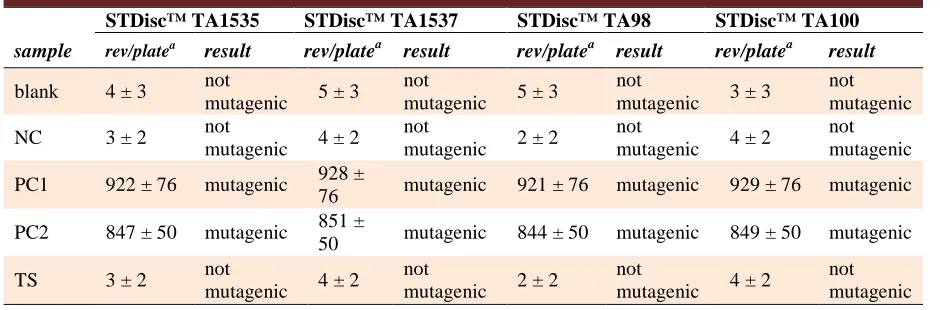
Table 2. Mutagenicity evaluation by the Ames test.
STDisc™ TA1535 STDisc™ TA1537 STDisc™ TA98 STDisc™ TA100
sample rev/platea result rev/platea result rev/platea result rev/platea result
blank 4 ± 3 not
mutagenic 5 ± 3
not
mutagenic 5 ± 3
not
mutagenic 3 ± 3
not mutagenic
NC 3 ± 2 not
mutagenic 4 ± 2
not
mutagenic 2 ± 2
not
mutagenic 4 ± 2
not mutagenic
PC1 922 ± 76 mutagenic 928 ±
76 mutagenic 921 ± 76 mutagenic 929 ± 76 mutagenic
PC2 847 ± 50 mutagenic 851 ±
50 mutagenic 844 ± 50 mutagenic 849 ± 50 mutagenic
TS 3 ± 2 not
mutagenic 4 ± 2
not
mutagenic 2 ± 2
not
mutagenic 4 ± 2
not mutagenic
aNumber of revertants/plate: mean of three independent experiments ± SD, NC, negative control: aluminium oxide ceramic rod, PC1,
positive control 1: ICR 191 Acridine, PC2, positive control 2: Sodium Azide, TS, tested sample: Ti implant
After 30 days of culture on Ti implants, cells
were exposed to colchicine (Sigma-Aldrich, St.
Louis, MO, USA) for 6 h, washed in PBS,
dissociated with trypsin (Lonza S.r.l), and
centrifuged at 300 g for 5 min. The pellet was
carefully resuspended and incubated in 1% sodium
citrate for 15 min at 37 °C, then fixed and spread
onto -20 °C cold glass slides. Metaphases of cells
were Q-banded and karyotyped in accordance with
the international system for human cytogenetic
nomenclature recommendations. Twenty five meta-
phases were analyzed for three expansions.
Statistical analyzes
One- way analysis of variance (ANOVA)
was used to analyze the data. Repeated measures
ANOVA with a post- hoc analysis using
Bonferroni’s correction for multiple comparisons
was performed, and t-tests were used to determine
signifycant differences P<0.05). Repeatability was
calculated as the stan-dard deviation of the
difference between measurements. All testings were
performed using SPSS 16.0 software (SPSS Inc,
Chicago, Illinois, USA) (licensed by the university
of Padova).
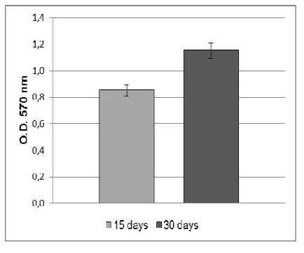
Fig. 1. MTT assay of ADSCs cultured on the Ti dental implants. ADSCs proliferation rate increase during the culturing time, reaching the maximum value at 30 days.
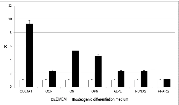
Fig. 2. Osteoblast markers expression in ADSCs cultured in cDMEM on the Ti implants. The results are reported as ratios (R) with respect to the mRNA expression of ADSCs seeded in tissue culture on polystyrene for 15 days in cDMEM.
Results
Evaluation of the mutagenicity of Ti dental implants
The Ames test was performed in order to
assess the mutagenic potential of Ti implants. Four
different histidine dependent mutant strains
(TA1535, TA1537, TA98 and TA100) of
Salmonella typhimurium were used. As reported in
table 2, no mutagenic activity has been revealed.
Evaluation of the hemocompatibility of Ti discs The hemolysis assay was performed in order
to evaluate the blood compatibility of the Ti
implants, which are intended for blood contacting
applications. The HI was less than 2%, indicating
the absence of any hemolytic activity of the tested
material (Table 3).
Biocompatibility of Ti implants
In order to evaluate the biocompatibility of Ti
implants, ADSCs were seeded and cultivated onto
these surfaces up to 30 days. The results of MTT
assay show that the cells were able to adhere and
proliferate onto the Ti implants (Fig. 1).
Expression of osteoblast markers
The gene expression level of some osteoblast
markers were analyzed at day 15 and 30 by means
of real- time PCR in order to verify the
osteoinductive properties of the Ti implants used in
the present study. The expression of selected genes
(ALPL, COL1A1, OCN, ON, OPN, RUNX2, and
PPARG) were evaluated in relation to the
expression of a reference gene (GAPDH). Cells
seeded on tissue culture polystyrene in cDMEM for
15 days were used as control for data normalization.
As shown in figure 2, the expression of some
osteoblast markers in ADSCs seeded onto the Ti
dental implants is higher compared to the control
condition. In particular, high gene expression levels
were observed for COL1A1, OPN, ALPL, and
RUNX2.
Similar results were obtained when comparing
the expression level of the same markers in ADSCs
seeded on tissue culture plates in the presence of
Table 3. Blood compatibility evaluation by the hemolysis assay
sample ODa HIb result
PC1 0.8762 ± 0.012 100% hemolytic
NC 0.0143 ± 0.002 0% nonhemolytic
TS 0.0144 ± 0.002 0,046% nonhemolytic
OD, absorbance value at 540 nm: mean of three independent experiments ± SD, HI, hemolysis index, PC, positive control: Sterile Water for Injection (SWFI), NC, negative control: High Density PolyEthylene (HDPE), TS, tested sample: Ti implant
Fig. 3.Effect of osteogenic differentiation medium on osteoblast markers expression in 15 days cultured ADSCs. The results are reported as ratios (R) with respect to the mRNA expression of ADSCs seeded in tissue culture polystyrene for 15 days in the presence of cDMEM.
Fig. 4.Karyotype analysis of ADSCs seeded on the Ti implants for 30 days. No chromosomal alterations are present. osteogenic differentiation medium to the control
(Fig.3). Also in this case, the expression of
COL1A1, OPN, ALPL, and RUNX2 was higher in
cells cultivated with osteogenic factors as opposed
to the control condition (cDMEM). In addition, in
ADSCs treated with the osteogenic medium an
increase in OCN and ON mRNA expression was
also detected.
Cytogenetic analysis
The chromosomal stability of ADSCs seeded
on the Ti implants was analyzed by means of
karyotyping. As reported in figure 4, no
chromosomal alterations are present in ADSCs
seeded onto these surfaces for 30 days.
Discussion
Ti and its alloys are the most commonly used
biomaterials in dental implantology. Nevertheless, a
question that remains to be answered is how
molecular and cellular events are influenced by the
material surface properties. In this study, we have
analyzed the effects of Ti dental implants with grit-
blasted and acid- etched surfaces on the behavior of
MSCs isolated from human adipose tissue
(ADSCs). Preliminary analyses were performed to
test the mutagenicity and hemocompatibility of the
Ti dental implants. Subsequently, ADSCs were
seeded onto these surfaces to evaluate their
biocompatibility and osteoinductive properties.
Finally, the safety of the biomaterials was
investigated by means of karyotyping.
The first experiments were carried out to
assess whether the treatments of Ti implants have
mutagenic potential. There is considerable evidence
that gene mutations are involved in cancer
formation in humans. The mutagenic potential of Ti
implants was examined with the Ames test (32). In
the present study, four Salmonella typhimurium
strains were used: TA1535 and TA100, which
result from a base-pair substitution; TA1537 and
TA98, products of a frameshift mutation. In this
way, it was possible to identify mutagens acting
with different mechanisms. The four Salmonella
strains were incubated with extracts deriving from
Ti implants for 48 h. The mutagenicity of a
substance is proportional to the number of colonies
observed. The low number of histidine revertant
colonies indicates that Ti implants lack mutagenic
activity at the conditions tested.
At this point, we performed the hemolysis
assay which is considered to be a very simple and
reliable test for estimating blood compatibility of
materials. The test relies on the measurement of
free hemoglobin released into the plasma when
blood cells are damaged. Generally, the smaller the
HI, the better the blood compatibility of the
biomaterial. The material extract tested in this study
induced less than 2% of contacting erythrocytes to
hemolyze over 3 h of contact with blood. These
results indicate that Ti implants have no hemolytic
effects and meet the requirements for clinical
application.
In the process of bone healing and implant
osseointegration, MSCs are the key repair cells, and
their cellular response is important because
successful osseointegration of implants depends on
the adhesion of MSCs onto the implant surface
(33). In this study, human ADSCs were used to
evaluate the cytotoxicity of Ti implants. The results
of the MTT assay indicate that ADSCs are able to
attach and grow on Ti implants and that cell
proliferation rate increases during the culturing
time, reaching the maximum value after 30 days. It
seems that grit- blasted and acid- etched treatment
of Ti surfaces positively affects cell proliferation.
As explained before, the regeneration of bone
is regulated by a series of complex events that
involve the sequential cascade of ECM proteins
production and its subsequent controlled
calcification (34). These proteins include collagens
as well as non- collagenous proteins (35). In order
to evaluate the osteoinductivity of Ti implants on
osteoblast differentiation of ADSCs, the expression
of osteogenic specific markers were evaluated with
real- time PCR. Collagen type I (COL1A1)
represent 90% of the total bone protein content
(36). When ADSCs are cultured on the Ti implants,
the gene expression of COL1A1 is found to be
significantly up- regulated. Such a result is very
interesting since COL1A1 synthesis is known to be
a prerequisite for ECM formation and
minerali-zation in bone (37).
Osteocalcin (OCN), osteonectin (ON) and
osteopontin (OPN) are the non- collagenous
proteins of bone, which collectively contribute to
the bone mineralization. OCN, a specific osteoblast
protein, is the most abundant non- collagenous
protein found in bone ECM after collagens. It is
thought that OCN is implicated in bone
mineralization and calcium ion homeostasis (38).
ON is a glycoprotein that binds calcium (39). It is
secreted by osteoblasts during bone formation,
initiating mineralization and promoting mineral
crystal deposition. ON also shows affinity for
collagen in addition to bone mineral calcium. In this
study, the expression levels of both OCN and ON
are similar at 15 and 30 days of culture.
Although no significant changes are found in
the expression of OCN and ON, other markers
associated with the osteogenic differentiation are
up-regulated. For example, the gene expression of
OPN and alkaline phosphatase (ALPL) is strongly
increased in ADSCs cultured onto Ti implants both
at 15 and 30 days. OPN is an important factor in
bone remodeling (40), and different studies have
shown that it plays a role in anchoring osteoclasts
to the mineral matrix of bones (41). Alkaline
phosphatase (ALPL) is a membrane- bound protein
with the catalytic domain on the osteoblastic
plasmalemma. It is a marker of early osteogenic
development and has probably an initiator and
regulator role in calcification (42). The elevated
OPN and ALPL expression observed in this study
supports the success of the osteoblastic
differentiation of ADSCs and may be an indication
of the osteoinductive properties of the scaffolds
used.
The expression of transcription factor genes
are essential for cellular commitment to a specific
differentiation lineage (43-45). Many studies have
confirmed the existence of an inverse reciprocal
relationship between adipogenesis and osteogenesis
(46-49). Osteoblast differentiation requires
expression of the osteoblast- specific transcription
factor runt- related transcription factor 2 (RUNX2)
(50-52). Likewise, adipogenic differentiation is
regulated by peroxisome proliferator- activated
receptor gamma (PPARG), which also possesses
anti-osteoblastogenic effects (53, 54). In this study,
ADSCs seeded onto Ti implants showed high
expression level of RUNX2 both at 15 and 30 days.
On the contrary, PPARG expression did not change
over time. Such a result might indicate that Ti
dental implants are able to stimulate the
differentiation of ADSCs towards the osteogenic
phenotype while suppressing the adipogenic
commitment of these cells. This is in line with the
hypothesis that increased expression of one
transcription factor is typically associated with
down- regulation of the other (47-49).
At the same time, high mRNA expression of
osteogenic markers were obtained when ADSCs
were cultured on tissue culture plates in the
presence of a differentiation medium supplemented
with osteogenic factors. Indeed, the gene expression
level of COL1A1, OCN, ON, OPN, ALPL and
RUNX2 was significantly higher compared to the
control condition, that is ADSCs seeded in
monolayer with cDMEM. On the contrary, the
expression of PPARG did not change under these
culture conditions.
Taken together, our results demonstrate that
the osteogenic differentiation of ADSCs may be
dependent on the Ti implant surface characteristics,
which have effects similar to the addition of
osteogenic growth factors in monolayer ADSCs
cultures.
In order to evaluate the chromosomal stability
of ADSCs maintained in culture 30 days on the Ti
implants, we performed karyotyping. This method
consisted in the analysis of metaphases of cells for
testing the presence of chromosomes alterations
following their proliferation and differentiation onto
the Ti implants. No chromosomal alterations were
found in the karyotype of ADSCs seeded on Ti
implants for 30 days. This confirms that the cells
are able to maintain their chromosomal stability, an
extremely important fact when considering possible
clinical use (55).
In conclusion, our results indicate that Ti
implants are not mutagenic and do not cause
hemolysis. Moreover, their surfaces are found to be
biocompatible and not toxic when seeded with
human ADSCs. Rather, the grit- blasted and acid-
etched treatment seem to favor the adhesion and
proliferation of these cells. The osteoinductivity of
Ti implants has been determined by the osteogenic
commitment of ADSCs in absence of a
differen-tiation medium. Finally, the maintenance of
chro-mosomal stability by ADSCs seeded on the Ti
implants ensures the biological safety of these
materials.
Acknowledgment
This research was supported by funds from
University of Padua, Progetto di Ateneo awarded to
Barbara Zavan.
Conflict of interests
The author declared no conflict of interests.
References
1. Brunette DM, Tengvall P, Textor M, et al. Titanium in
medicine: material science, surface science, engineering,
biological response and medical applications. 1 ed. Berlin:
Springer Verlag; 2001.
2. Long M, Rack HJ. Titanium alloys in total joint replacement-a
materials science perspective. Biomaterials 1998;19:1621-39.
3. Kasemo B. Biocompatibility of titanium implants: surface
science aspects. J Prosthet Dent 1983;49:832-7.
4. Keller JC, Stanford CM, Wightman JP, et al.
Characterizations of titanium implant surfaces. III. J Biomed
Mater Res 1994;28:939-46.
5. Eliades T. Passive film growth on titanium alloys:
physicochemical and biologic considerations. Int J Oral
Maxillofac Implants 1997;12:621-7.
6. Albrektsson T, Branemark PI, Hansson HA, et al.
Osseointegrated titanium implants. Requirements for ensuring a
long-lasting, direct bone-to-implant anchorage in man. Acta
Orthop Scand 1981;52:155-70.
7. Boyan BD, Lohmann CH, Dean DD, et al. Mechanisms
involved in osteoblast response to implant surface morphology.
Annu Rev Mater Res 2001;31:357-71.
8. Boyan BD, Batzer R, Kieswetter K, et al. Titanium surface
roughness alters responsiveness of MG63 osteoblast-like cells to
1 alpha,25-(OH)2D3. J Biomed Mater Res 1998;39:77-85.
9. Schwartz Z, Martin JY, Dean DD, et al. Effect of titanium
surface roughness on chondrocyte proliferation, matrix
production, and differentiation depends on the state of cell
maturation. J Biomed Mater Res 1996;30:145-55.
10. Ponsonnet L, Reybiera K, Jaffrezica N, et al. Relationship
between surface properties (roughness, wettability) of titanium
and titanium alloys and cell behaviour. Mater Sci Eng C
2003;23:551-60.
11. Le Guehennec L, Soueidan A, Layrolle P, et al. Surface
treatments of titanium dental implants for rapid osseointegration.
Dent Mater 2007;23:844-54.
12. Cochran DL, Schenk RK, Lussi A, et al. Bone response to
unloaded and loaded titanium implants with a sandblasted and
acid-etched surface: a histometric study in the canine mandible. J
Biomed Mater Res 1998;40:1-11.
13. Wennerberg A, Hallgren C, Johansson C, et al. A
histomorphometric evaluation of screw-shaped implants each
prepared with two surface roughnesses. Clin Oral Implants Res
1998;9:11-9.
14. Anselme K, Bigerelle M, Noel B, et al. Qualitative and
quantitative study of human osteoblast adhesion on materials
with various surface roughnesses. J Biomed Mater Res
2000;49:155-66.
15. Eisenbarth E, Linez P, Biehl V, et al. Cell orientation and
cytoskeleton organisation on ground titanium surfaces. Biomol
Eng 2002;19:233-7.
16. Aparicio C, Gil FJ, Planell JA, et al. Human-osteoblast
proliferation and differentiation on grit-blasted and bioactive
titanium for dental applications. J Mater Sci Mater Med
2002;13:1105-11.
17. Aparicio C, Gil FJ, Fonseca C, et al. Corrosion behaviour of
commercially pure titanium shot blasted with different materials
and sizes of shot particles for dental implant applications.
Biomaterials 2003;24:263-73.
18. Ivanoff CJ, Hallgren C, Widmark G, et al. Histologic
evaluation of the bone integration of TiO(2) blasted and turned
titanium microimplants in humans. Clin Oral Implants Res
2001;12:128-34.
19. Piattelli M, Scarano A, Paolantonio M, et al. Bone response
to machined and resorbable blast material titanium implants: an
experimental study in rabbits. J Oral Implantol 2002;28:2-8.
20. Shi GS, Ren LF, Wang LZ, et al. H2O2/HCl and heat-treated
Ti-6Al-4V stimulates pre-osteoblast proliferation and
differentiation. Oral Surg Oral Med Oral Pathol Oral Radiol
Endod 2009;108:368-75.
21. Park JY, Davies JE. Red blood cell and platelet interactions
with titanium implant surfaces. Clin Oral Implants Res
2000;11:530-9.
22. Yokoyama K, Ichikawa T, Murakami H, et al. Fracture
mechanisms of retrieved titanium screw thread in dental implant.
Biomaterials 2002;23:2459-65.
23. Guo CY, Tang ATH, Matinlinna JP. Insights into surface
treatment methods of titanium dental implants. J Adhes Sci
Technol 2012;26:189-205.
24. Wennerberg A, Albrektsson T. Effects of titanium surface
topography on bone integration: a systematic review. Clin Oral
Implants Res 2009;20 Suppl 4:172-84.
25. Herrero-Climent M, Lazaro P, Vicente Rios J, et al.
Influence of acid-etching after grit-blasted on osseointegration of
titanium dental implants: in vitro and in vivo studies. J Mater Sci
Mater Med 2013;24:2047-55.
26. Palmquist A, Omar OM, Esposito M, et al. Titanium oral
implants: surface characteristics, interface biology and clinical
outcome. J R Soc Interface 2010;7 Suppl 5:S515-27.
27. Singer AJ, Clark RA. Cutaneous wound healing. N Engl J
Med 1999;341:738-46.
28. Caplan AI. Mesenchymal stem cells. J Orthop Res
1991;9:641-50.
29. Gardin C, Bressan E, Ferroni L, et al. In vitro concurrent
endothelial and osteogenic commitment of adipose-derived stem
cells and their genomical analyses through comparative genomic
hybridization array: novel strategies to increase the successful
engraftment of tissue-engineered bone grafts. Stem Cells Dev
2012;21:767-77.
30. Denizot F, Lang R. Rapid colorimetric assay for cell growth
and survival. Modifications to the tetrazolium dye procedure
giving improved sensitivity and reliability. J Immunol Methods
1986;89:271-7.
31. Pfaffl MW. A new mathematical model for relative
quantification in real-time RT-PCR. Nucleic Acids Res
2001;29:e45.
32. Mortelmans K, Zeiger E. The Ames Salmonella/microsome
mutagenicity assay. Mutat Res 2000;455:29-60.
33. Razzouk S, Schoor R. Mesenchymal stem cells and their
challenges for bone regeneration and osseointegration. J
Periodontol 2012;83:547-50.
34. Rosset P, Deschaseaux F, Layrolle P. Cell therapy for bone
repair. Orthop Traumatol Surg Res 2014;100:S107-12.
35. Fisher LW, Termine JD. Noncollagenous proteins
influencing the local mechanisms of calcification. Clin Orthop
Relat Res 1985:362-85.
36. Bilezikian JP, Raisz LG, Rodan GA. Principles of Bone
Biology. New York: Academic Press; 1996.
37. Franceschi RT, Iyer BS, Cui Y. Effects of ascorbic acid on
collagen matrix formation and osteoblast differentiation in
murine MC3T3-E1 cells. J Bone Miner Res 1994;9:843-54.
38. Lee NK, Sowa H, Hinoi E, et al. Endocrine regulation of
energy metabolism by the skeleton. Cell 2007;130:456-69.
39. Termine JD, Kleinman HK, Whitson SW, et al. Osteonectin,
a bone-specific protein linking mineral to collagen. Cell
1981;26:99-105.
40. Choi ST, Kim JH, Kang EJ, et al. Osteopontin might be
involved in bone remodelling rather than in inflammation in
ankylosing spondylitis. Rheumatology (Oxford) 2008;47:1775-9.
41. Reinholt FP, Hultenby K, Oldberg A, et al. Osteopontin--a
possible anchor of osteoclasts to bone. Proc Natl Acad Sci U S A
1990;87:4473-5.
42. Peng F, Yu X, Wei M. In vitro cell performance on
hydroxyapatite particles/poly(L-lactic acid) nanofibrous
scaffolds with an excellent particle along nanofiber orientation.
Acta Biomater 2011;7:2585-92.
43. Wagner EF, Karsenty G. Genetic control of skeletal
development. Curr Opin Genet Dev 2001;11:527-32.
44. Komori T. Regulation of osteoblast differentiation by Runx2.
Adv Exp Med Biol 2010;658:43-9.
45. Karsenty G. Minireview: transcriptional control of osteoblast
differentiation. Endocrinology 2001;142:2731-3.
46. Lin YF, Jing W, Wu L, et al. Identification of osteo-adipo
progenitor cells in fat tissue. Cell Prolif 2008;41:803-12.
47. Valenti MT, Garbin U, Pasini A, et al. Role of ox-PAPCs in
the differentiation of mesenchymal stem cells (MSCs) and
Runx2 and PPARgamma2 expression in MSCs-like of
osteoporotic patients. Plos One 2011;6:e20363.
48. Zhang L, Su P, Xu C, et al. Melatonin inhibits adipogenesis
and enhances osteogenesis of human mesenchymal stem cells by
suppressing PPARgamma expression and enhancing Runx2
expression. J Pineal Res 2010;49:364-72.
49. Zhang X, Yang M, Lin L, et al. Runx2 overexpression
enhances osteoblastic differentiation and mineralization in
adipose--derived stem cells in vitro and in vivo. Calcif Tissue Int
2006;79:169-78.
50. Ducy P, Zhang R, Geoffroy V, et al. Osf2/Cbfa1: a
transcriptional activator of osteoblast differentiation. Cell
1997;89:747-54.
51. Thirunavukkarasu K, Halladay DL, Miles RR, et al. The
osteoblast-specific transcription factor Cbfa1 contributes to the
expression of osteoprotegerin, a potent inhibitor of osteoclast
differentiation and function. J Biol Chem 2000;275:25163-72.
52. Nakashima K, Zhou X, Kunkel G, et al. The novel zinc
finger- containing transcription factor osterix is required for
osteoblast differentiation and bone formation. Cell 2002;
108:17-29.
53. Tzameli I, Fang H, Ollero M, et al. Regulated production of
a peroxisome proliferator-activated receptor-gamma ligand
during an early phase of adipocyte differentiation in 3T3-L1
adipocytes. J Biol Chem 2004;279:36093-102.
54. Schopfer FJ, Lin Y, Baker PR, et al. Nitrolinoleic acid: an
endogenous peroxisome proliferator-activated receptor gamma
ligand. Proc Natl Acad Sci U S A 2005;102:2340-5.
55. Angelo PC, Ferreira AC, Fonseca VD, et al.
Cryopreservation does not alter karyotype, multipotency, or
NANOG/SOX2 gene expression of amniotic fluid mesenchymal
stem cells. Genet Mol Res 2012;11:1002-12.