Daiva Gorczyca
1, A–F, Daria Augustyniak
2, A–D, F,
Barbara Basiewicz-Worsztynowicz
1, A, B, D, F, Wiesława Karnas-Kalemba
1, A–C, FSerum and Urinary MIP-1α and IP-10 Levels
in Children with Urinary Tract Infections*
1 3rd Department and Clinic of Pediatrics, Immunology and Rheumatology of Developmental Age,
Wroclaw Medical University, Poland
2 Department of Pathogen Biology and Immunology, Institute of Genetics and Microbiology,
Wroclaw Medical University, Poland
A – research concept and design; B – collection and/or assembly of data; C – data analysis and interpretation;
D – writing the article; E – critical revision of the article; F – final approval of article; G – other
Abstract
Objectives. Urinary tract infection (UTI) is a common bacterial disease in infants and children, with potentially serious complications, including kidney damage. The aim of this study was to test whether serum and urinary lev-els of interleukin-6 (IL-6), macrophage inflammatory protein-1α (MIP-1α) and interferon-γ-inducible protein-10
(IP-10) can be used as biomarkers in children with urinary tract infections.
Material and Methods. The study group consisted of 22 children with UTI and 20 controls. Blood and urine samples were collected in the acute phase and the convalescent phase, on the eighth day after the onset of antibiotic therapy. Serum and urine levels of MIP-1α, IP-10 and IL-6 were measured.
Results. In children with UTI in the acute phase, serum MIP-1α and IL-6 levels were significantly higher compared
to the controls (p < 0.05 and p < 0.005, respectively). A correlation between the serum levels of the chemokines MIP-1α and IP-10 in the acute phase was found.
Conclusions. The findings suggest that the chemokines MIP-1α or IP-10 respond to infection, but they cannot be used as biomarkers for UTI in childhood (Adv Clin Exp Med 2014, 23, 6, 933–938).
Key words: chemokines, children, urinary tract infection.
Adv Clin Exp Med 2014, 23, 6, 933–938 ISSN 1899–5276
ORIGINAL PAPERS
© Copyright by Wroclaw Medical University
Urinary tract infection (UTI) is a common bacterial disease in infants and children, with po-tentially serious complications, including kidney damage and chronic renal failure. UTI is diagnosed in approximately 5% of infants who are brought to hospital emergency departments because of a fever without any obvious cause [1].
Recent European standards regarding the inves-tigation and treatment of UTI are based on the Na-tional Institute for Health and Clinical Excellence (NICE) guidelines, which require a clinical exam-ination, urinalysis, urine culture and imaging tests to diagnose UTI [2]. Some authors have attempted to identify markers of inflammation in urine and to use them in the diagnosis of infection of the urinary tract and in monitoring response to therapy [3–6].
Escherichia coli is the most common cause of UTI, but Klebsiella, Proteus, Enterobacter, Staph-ylococcus saprophyticus and Enterococcus also grow in the urine culture [7]. Bacteria that enter the urinary tract cause an activation of the host inflammatory response. In the results of bacterial stimulation, renal epithelial cells have been shown to produce a number of cytokines and chemo-kines, among other substances [8]. Cytochemo-kines, es-pecially interleukin-6 (IL-6), participate in the lo-cal inflammatory response to infections [4, 9–11]. Chemokines (chemotactic cytokines) medi-ate this process locally and systemically; they are important mediators of leukocyte extravasa-tion and chemotaxis [12]. Chemokine concentra-tion results in an influx of leukocytes including
neutrophils, natural killer cells, macrophages and lymphocytes.
The importance of chemokines during kidney inflammation has been described in various stud-ies [5, 13–15], but little is known about the role of each chemokine in the kidney. Chemokine mac-rophage inflammatory proteins-1alpha (MIP-1α, CCL3), produced by macrophages, may be in-volved in the development of cellular crescents in the acute phase of kidney pathology in bacterial in-fections [8]. Interferon-gamma-induced protein 10 (IP-10, CXCL10) is produced by several cell types, such as fibroblasts, endothelium cells and mono-cytes, and its secretion is stimulated by tumor ne-crosis factor alpha (TNFα), interleukin-1 beta (IL-1β) and macrophage colony-stimulating fac-tor (M-CSF) [12]. Plasma IP-10 is considered to be a predictor of serious bacterial infection in in-fants [16] and has recently been associated with acute kidney injury [13]. Both chemokines have been detected in urine [17, 18]. Evaluating the re-sponse of chemokines MIP-1α and IP-10 to in-fection could help to explain whether they can be markers in a febrile urinary tract infection in children.
The aim of this pilot study was to investigate and compare serum and urinary levels of MIP-1α, IP-10 and IL-6 in children with urinary tract infec-tions on admission, in the acute phase, and after eight days of parenteral antibiotic therapy. This in-formation could be useful for diagnosing UTI.
Material and Methods
Patients with Urinary Tract
Infections
The study involved 22 children of both sexes (aged from 4 to 17 years; median age 10 years; 18 fe-male, 4 male) diagnosed with a UTI at the Depart-ment of Pediatric Immunology and Rheumatology, Wroclaw Medical University (Wrocław, Poland) be-tween 2006 and 2009. To qualify for the study a diag-nosis of UTI according to NICE clinical guideline [2] and significant bacteriuria (>105 colony-forming
units (CFU)/L of a single uropathogenic bacteria in a clean catch sample) were required. Children who have bacteriuria and a fever of 38°C or higher, and children presenting a fever lower than 38°C with loin pain/tenderness and bacteriuria were considered to have acute pyelonephritis/upper urinary tract in-fection [19]. All other children with bacteriuria but no systemic symptoms or signs (a peripheral white blood cell [WBC] count of > 15,000 cells/μL, or C-re-active protein [CRP] level of ≥ 6.0 g/dL) were consid-ered to have cystitis/lower urinary tract infection.
Healthy Control Group
The study also included 20 healthy controls (aged from 4 to 17 years; median age 11.5; 35 fe-male, 7 male) without any symptoms of UTI and with one negative urinary culture. These controls were selected from 2 distinct populations: outpa-tients seen at the hospital for routine medical fol-low-up, and children whose parents (or guardians) volunteered them as participants in the study.
Exclusion Criteria for the Group
with UTI and the Control Group
Patients with a history of previous UTIs, those suffering from vesicoureteral reflux or severe re-nal function impairment, those who had under-gone bladder catheterization within the previ-ous two weeks, those who had had sexual activity within the previous 2 weeks, those using immu-nosupression or being treated with nephrotoxic medications, and those who had signs of any oth-er infection requiring antibiotic treatment within 2 weeks prior to the screening were excluded from the study.Detection of Inflammatory
Mediators
In the group with UTI, blood and urine sam-ples were obtained twice:
– in the acute phase, while admitting the pa-tients to the hospital, while the symptoms were presenting; and
– in the convalescent phase, on the 8th day
af-ter the onset of antibiotic therapy.
In the control group, blood and urine samples were collected one time only.
All the blood samples were drawn between 8 and 9 am. The sera received were aliquoted and stored at a temperature of –70oC until the assay.
Urine samples obtained by clean catch sampling were centrifuged (7000 g, 15 min, 4oC), divided
in-to portions and immediately frozen at –70oC.
Serum levels of MIP-1α and IP-10 were
ana-lyzed with Duo-Set ELISA systems (R&D, Abing-don, UK). Briefly, flat-bottom 96-well MaxiSorp microtiter plates (Nunc) were coated overnight at room temperature with 100 µL per well of relevant capture antibodies diluted in PBS buffer (pH 7.4). The wells were blocked with 300 µL of 1% BSA in PBS for 1 h at 37oC. Dilutions of the serum
sam-ples in the 1:2–1:16 range (for MIP-1αand IP-10),
100 µL of the relevant biotinylated detection anti-bodies were added and the plate was incubated for 2 hrs at room temperature. After subsequent wash-ing with TPBS, the wells were filled with strepta-vidin-HRP (diluted ×200) and the plate was in-cubated for 20 min at room temperature and in darkness. After washing, 100 µL per well of TMB substrate (R&D Abingdon, UK) was added and the plate was incubated 20 min at room tempera-ture and in darkness. The enzymatic reaction was stopped with 50 µL 1M H2SO4. The optical
densi-ty was read at 450 nm with a Dynatech 5000 pho-tometric reader.
Serum IL-6 levels were measured using a com-mercial kit according to the manufacturer’s in-structions (R&D, Abingdon, UK) with a detection limit of 1 pg/mL.
Additionally, the following markers of inflam-mation were measured in the UTI group: serum CRP level, peripheral WBC, urine dipstick analysis and microscopic exam, using standardized meth-ods. Pyuria was defined as ≥ 5 WBC/high power field (hpf) or ≥ 10 WBC/mm3 on a
hemocytom-eter with an enhanced urinalysis, and bacteriuria was defined as the presence of any bacteria per hpf [19].
The study protocol was approved by the Re-search Ethics Committee of Wroclaw Medical University. The parents (or guardians) of the par-ticipants and parpar-ticipants older than 16 years signed written consent forms before participating in the study.
Statistical Analysis
The results are expressed as median values and interquartile ranges. The statistical analyses were performed using non-parametric tests for data without a normal distribution. The Mann- -Whitney U-test was used for independent vari-ables and the Wilcoxon paired rank test was used for dependent variables. Spearman’s rank cortion coefficient was used to investigate any rela-tionship between the parameters. The level of sta-tistical significance was assumed to be p < 0.05. The statistical analyses were carried out using Stat-Soft software (STATISTICA 9).
Results
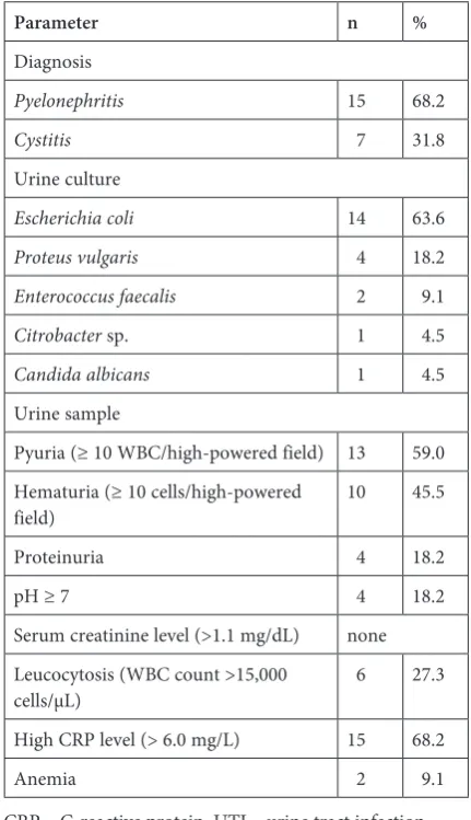
The group of patients with UTI and the control group were similar in terms of age and gender. The results of the urinalysis and of urine collections as well as selected results of the blood tests are pre-sented in Table 1. Most patients (n = 15, 68.2%) were suffering from pyelonephritis. The results
of the urine cultures revealed that E. coli was the cause of UTI in most cases (n = 14, 63.6%).
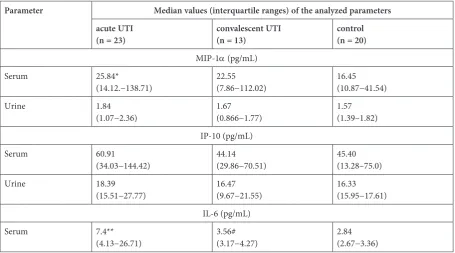
The serum and urine concentrations of the chemokines MIP-1α and IP-10 and the cytokine IL-6 in the acute and convalescent phases of UTI compared with the healthy controls are present-ed in Table 2. The mpresent-edian serum MIP-1α and IL-6 level was found to be significantly higher in the acute phase of UTI as compared to the control group (p < 0.05 and p < 0.005, respectively) but no such difference was observed in the convales-cent phase. Although a similar trend was observed for IP-10 levels, it was not statistically significant (p = 0.058). No statistically significant differenc-es in the urinary levels of MIP-1α and IP-10 were found in the patients in the acute or convalescent phases, or in the healthy controls.
A correlation was found between the serum level of MIP-1α and IP-10 in the acute phase (Sr = 0.522, p = 0.01, Fig. 1). No correlation
be-tween the urinary levels of MIP-1α and IP-10 was observed.
Table 1. Clinical data of the patients with urine tract infection, n = 22
Parameter n %
Diagnosis
Pyelonephritis 15 68.2
Cystitis 7 31.8
Urine culture
Escherichia coli 14 63.6
Proteus vulgaris 4 18.2
Enterococcus faecalis 2 9.1
Citrobacter sp. 1 4.5
Candida albicans 1 4.5
Urine sample
Pyuria (≥ 10 WBC/high-powered field) 13 59.0 Hematuria (≥ 10 cells/high-powered
field) 10 45.5
Proteinuria 4 18.2
pH ≥ 7 4 18.2
Serum creatinine level (>1.1 mg/dL) none Leucocytosis (WBC count >15,000
cells/μL) 6 27.3
High CRP level (> 6.0 mg/L) 15 68.2
Anemia 2 9.1
No correlation was found between the serum and urinary levels of the chemokines MIP-1α and IP-10 and leukocytosis, CRP level, serum IL-6 lev-el, a positive urine nitrite test or leucoctyuria (da-ta not present).
Discussion
Early diagnosis and treatment of acute UTI in children is of particular importance since the ma-jority of children examined by the authors (68%) had already developed pyelonephritis, which can
result in serious complications such as renal scar-ring, hypertension and renal failure. Diagnosis of urinary infection in patients with acute urinary tract symptoms is usually done on the basis of uri-nalysis, pyuria and urine collection [20]. Screening methods including commonly used inflammatory markers such as leukocyte count, procalcitonin and C-reactive protein, as well as serum IL-6 levels, are helpful. IL-6 promotes the growth and differentia-tion of T and B lymphocytes. Kassir et al. report-ed that elevatreport-ed IL-6 confirmreport-ed the inflammatory process, and that IL-6 levels rapidly decreased af-ter appropriate antibiotic treatment [6]. The find-ings in the current study are not consistent with this observation. In the current study, elevated se-rum levels of IL-6 in children were already present at the time of diagnosis; they decreased on the 8th
day of antibiotic treatment, but were still signifi-cantly higher than in the control group. These re-sults suggest that the inflammatory process does not cease as rapidly as described in the report by Kassir et al.
Recently, several immune mediators pres-ent in the peripheral circulation have been detect-ed in the urine of patients with kidney dysfunc-tions. Some studies have focused on a number of proteins and enzymes normally present in urine or released to urine as a direct response to infec-tion, which have been suggested as potential bio-markers [21, 22]. Efforts to identify novel, sensitive urinary biomarkers, however, have led to the con-clusion that a combination of mediators could be even more useful.
Table 2. The median values and interquartile ranges of the examined parameters in UTI patients and the control group
Parameter Median values (interquartile ranges) of the analyzed parameters acute UTI
(n = 23) convalescent UTI(n = 13) control(n = 20)
MIP-1α (pg/mL) Serum 25.84*
(14.12.−138.71) 22.55(7.86−112.02) 16.45(10.87−41.54)
Urine 1.84
(1.07−2.36) 1.67(0.866−1.77) 1.57(1.39–1.82) IP-10 (pg/mL)
Serum 60.91
(34.03−144.42) 44.14(29.86−70.51) 45.40(13.28–75.0)
Urine 18.39
(15.51−27.77) 16.47(9.67−21.55) 16.33(15.95−17.61) IL-6 (pg/mL)
Serum 7.4**
(4.13−26.71) 3.56#(3.17−4.27) 2.84(2.67−3.36) * p < 0.05 acute UTI vs. control; ** p < 0.0001 acute UTI vs. control; # p < 0.005 convalescent UTI vs. control.
Fig. 1. Correlation between the serum concentration of MIP-1α and IP-10 in children with urinary tract infec-tions during the acute phase of the disease (Sr = 0.522,
Febrile urinary tract infections are accompa-nied by a complex chemokine response [23]. Ac-cording to Godolay et al. E. coli strains isolated from patient with pyelonephritis caused an in-crease in vitro of epithelial secretion of the chemo-kines CXC (CXCL1, CXCL8, CXCL9, CXCL10) and CC (CCL2, CCI3, CCL5) [24, 25].
In this study the relationship between the che-mokine level in plasma and urine of children with UTI was investigated. The results revealed that se-rum MIP-1α levels are significantly higher in the acute phase of UTI than in the convalescent phase and in the control group. There was no significant difference between the median serum level of MIP-1α in the convalescent phase and the control group. Elevated MIP-1α in serum at the time of diagnosis suggests a chemokine release in response to UTI, and is followed by a decrease in serum MIP-1α af-ter treatment. The proinflammatory activities of
MIP-1α induced chemotaxis of CD8+ T
lympho-cytes [26], while CD4+ T cells migrated in response to IP-10 [27]. In the field of respiratory infection, the evidence suggests that the response to microor-ganism invasion is regulated by a distinct chemo-kine expression profile involving MIP-1α [28].
Unfortunately, in the current study no such relationship was found in the urine in the pa-tients with UTI. The results of this study indicate
that the chemokine MIP-1α is not a good
mark-er in urine samples. Othmark-er authors have report-ed that chemokines – but not MIP-1α − were el-evated in infected uro-epithelial cells in vitro [24], in patients on admission in urosepsis and exper-imental endotoxemia [21] and in children with hydronephrosis [3].
Previous studies have documented that CXCL10 correlates positively with fever and CRP [23]. E. coli infection stimulates in vitro
production of mucosal chemokines, including CXCL10 [24]. In the current study, no differenc-es were found between the levels of IP-10 in the serum or in urine of children with UTI before or after antibiotic treatment, or in comparison with the control group, which is consistent with studies mentioned above [3, 23, 24].
However, the data from the current study re-vealed a positive correlation between serum lev-els of MIP-1α and IP-10. It can be concluded that both chemokines are involved in inflammation of the urinary tract in children.
In summary, neither chemokine MIP-1α or IP-10 are good biomarkers in UTI. Despite signif-icant progress in understanding the dynamics of cytokine release during the infectious process, the search continues for reliable biomarkers for the presence of UTI and the results of treatment.
References
[1] Slater M, Krug SE: Evaluation of the infant with fever without source: an evidence based approach. Emerg Med Clin North Am 1999, 17, 97–126.
[2] Mori R, Lakhanpaul M, Verrier-Jones K: Diagnosis and management of urinary tract infection in children: sum-mary of NICE guidance. BMJ 2007, 335, 395–397.
[3] Madsen MG, Nørregaard R, Palmfeldt J, Olsen LH, Frøkiær J, Jørgensen TM: Epidermal growth factor and monocyte chemotactic peptide-1: Potential biomarkers of urinary tract obstruction in children with hydronephro-sis. J Pediatr Urol 2012, S1477, 285–289.
[4] Rodhe N, Löfgren S, Strindhall J, Matussek A, Mölstad S: Cytokines in urine in elderly subjects with acute cystitis and asymptomatic bacteriuria. Scand J Prim Health Care 2009, 27, 74–79.
[5] Segerer S, Alpers CE: Chemokines and chemokine receptors in renal pathology. Curr Opin Nephrol Hypertens 2003, 12, 243–249.
[6] Kassir K, Vargas-Shiraishi O, Zaldivar F, Berman M, Singh J, Arrieta A: Cytokine profiles of pediatric patients treated with antibiotics for pyelonephritis: potential therapeutic impact. Clin Diagn Lab Immunol 2001, 8, 1060–1063.
[7] Lerner GR: Urinary tract infections in children. Pediatr Ann 1994, 23, 463, 466–473.
[8] Hertting O, Khalil A, Jaremko G, Chromek M, Li YH, Bakhiet M, Bartfai T, Tullus K, Brauner A: Enhanced chemokine response in experimental acute Escherichia coli pyelonephritis in IL-1beta-deficient mice. Clin Exp Immunol 2003, 131, 225–233.
[9] Heinrich PC, Castell JV, Andus T: Interleukin-6 and the acute phase response. Biochem J 1990, 265, 621–636.
[10] Tramma D, Hatzistylianou M, Gerasimou G, Lafazanis V: Interleukin-6 and interleukin-8 levels in the urine of children with renal scarring. Pediatr Nephrol 2012, 27, 1525–1530.
[11] Augustyniak D, Majkowska-Skrobek G, Basiewicz-Worsztynowicz B, Jankowski A: The role of IL-6/sIL-6R com-plex and its natural inhibitor sgp130 in modulation of inflammatory process. Post Biochem 2006, 52, 194–203.
[12] Baggiolini M: Chemokines in pathology and medicine. J Intern Med 2001, 250, 91–104.
[13] Segerer S, Nelson PJ: Chemokines in renal diseases. Scientific World Journal 2005, 29, 835–844.
[14] Mak RH: Chronic kidney disease in children: state of the art. Pediatr Nephrol 2007, 22, 1687–1688.
[16] Chen HL, Hung CH, Tseng HI, Yang RC, Chen HL, Hung CH, Tseng HI, Yang RC: Plasma IP-10 as a predictor of serious bacterial infection in infants less than 4 months of age. J Trop Pediatr 2011, 57, 145–151.
[17] Wada T, Furuichi K, Segawa-Takaeda C, Shimizu M, Sakai N, Takeda SI, Takasawa K, Kida H, Kobayashi KI, Mukaida N, Ohmoto Y, Matsushima K, Yokoyama H: MIP-1alpha and MCP-1 contribute to crescents and inter-stitial lesions in human crescentic glomerulonephritis. Kidney Int 1999, 56, 995–1003.
[18] Kanmaz T, Feng P, Torrealba J, Kwun J, Fechner JH, Schultz JM, Dong Y, Kim HT, Dar W, Hamawy M, Knechtle S, Hu H: Surveillance of acute rejection in baboon renal transplantation by elevation of interferon-gamma inducible protein-10 and monokine induced by interferon-interferon-gamma in urine. Transplantation 2004, 78, 1002–1007.
[19] Edmonson MB, Wald ER: Treatment of pyelonephritis and risk of renal scarring. Pediatrics 2009, 123, 544–555.
[20] American Academy of Pediatrics. Committee on Quality Improvement, Subcommittee on Urinary Tract Infection Practice Parameter: The Diagnosis, Treatment, and Evaluation of the Initial Urinary Tract Infection in Febrile Infants and Young Children.Pediatrics1999, 103, 843–852.
[21] Jantausch BA, O’Donnell R, Wiedermann BL: Urinary interleukin-6 and interleukin-8 in children with urinary tract infection. Pediatr Nephrol 2000, 15, 236–240.
[22] Olszyna D, Prins J, Dekkers P, De Jonge E, Speelman P, Van Deventer S, Van Der Poll T: Sequential measure-ments of chemokines in urosepsis and experimental endotoxemia. J Clin Immunol 1999, 19, 399–405.
[23] Vaidya VS, Waikar SS, Ferguson MA, Collings FB, Sunderland K, Gioules C, Bradwin G, Matsouaka R, Betensky RA, Curhan GC, Bonventre JV: Urinary biomarkers for sensitive and specific detection of acute kidney injury in humans. Clin Transl Sci 2008, 1, 200–208.
[24] Otto G, Burdick M, Strieter R, Godaly G: Chemokine response to febrile urinary tract infection. Kidney Int 2005, 68, 62–70.
[25] Godaly G, Otto G, Burdick MD, Strieter RM, Svanborg C: Fimbrial lectins influence the chemokine repertoire in the urinary tract mucosa. Kidney Int 2007, 71, 778–786.
[26] Taub DD, Conlon K, Lloyd AR, Oppenheim JJ, Kelvin DJ: Preferential migration of activated CD4+ and CD8+ T cells in response to MIP-1 alpha and MIP-1 beta. Science 1993, 260, 355–358.
[27] Taub DD, Lloyd AR, Conlon K, Wang JM, Ortaldo JR, Harada A, Matsushima K, Kelvin DJ, Oppenheim JJ:
Recombinant human interferon-inducible protein 10 is a chemoattractant for human monocytes and T lympho-cytes and promotes T cell adhesion to endothelial cells. J Exp Med 1993, 177, 1809–1814.
[28] Basiewicz-Worsztynowicz B, Karnas-Kalemba W, Augustyniak D, Polańska B, Jankowski A: The usefulness of chemikines RANTES and MIP-1alpha and elastaze in diagnostic process of respiratory tract infections in children. Adv Clin Exp Med 2005, 14, 523–529.
Address for correspondence:
Daiva Gorczyca
3rd Department and Clinic of Pediatrics, Immunology and Rheumatology of Developmental Age
Wroclaw Medical University Koszarowa 5
51-149 Wrocław Poland
Tel.: +48 71 329 53 53
E-mail: [email protected]
Conflict of interest: None declared