RETROSPECTIVE EVALUATION OF
AND NECK REGION
Dr. Krishna Kumar, *Dr. Ashwini Desai, Dr.
Department of Oral Medicine and Radiology, Institution
ARTICLE INFO ABSTRACT
Background
panoramic radiographs. These are usually asymptomatic but can be a diagnostic aid for some calcifications which are life threatening and the und
to estimate prevalence of soft tissue calcifications and ossifications in digital panoramic radiographs (OPGs)
1.To estimate
2. To evaluate incidence of calcifications ,stylohyoid
3. To estimate and compare
Materials and
35 to 80 years of region Western Maharashtra who visited dental institution were evaluated retrospectively and were scrutinized for calcifications according to gender, age an
of 1750 radiographs the number of
131 were of females and 79 were males. It was a double blind study. Two maxillofacial radiologists cross checked the radiographs. In this
number among which 74 were females & 27 males average of 9.9%
atherosclerotic plaque, 0.9% of antroliths and lymph node calcifications, 0.7% of rhinoliths and tonsilloliths and
calcifications are recorded accordingly. Package for the Social Sciences
calcification with comparison of findings between males and test(p value = 0.01) and considered to be statistically calcification
number in this study followed by carotid artery calcification. Some of these calcifications may indicate presence of some systemic diseases. This makes the OPG
patient's life.
Copyright © 2019, Krishna Kumar et al. This is an open use, distribution, and reproduction in any medium, provided
INTRODUCTION
The panoramic radiographs (OPGs) have ability to view both maxilla and mandible in a single image which makes it a valuable radiologic technique. Due to different anatomic properties of the patients, it's not possible to construct an OPG which only includes the jaws in the focal trough. Because of this the soft tissue structures located near the jaws may be visualized on OPG. Soft tissue radiopacities include calcification, ossification, and foreign objects
*Corresponding author: Dr. Desai Ashwini,
Department of Oral Medicine and Radiology, Institution Rangoonwala Dental College and Research Centre (M.A.R.D.C.), Pune.
ISSN: 0975-833X
Article History:
Received 14th July, 2019
Received in revised form 18th August, 2019
Accepted 05th September, 2019
Published online 30th October, 2019
Citation:Dr. Krishna Kumar, Dr. Ashwini Desai, Dr. Vinod VC, Dr. Jyotsna Patel, Dr. Nikhil Diwan and Dr. Rashmi Sapkal
tissue calcifications and ossifications of head and neck region on digital panoramic radiographs.”
Key Words:
Digital panoramic Radiographs, Calcifications, Ossifications, Stylohyoid ligament, Atherosclerotic Plaque.
RESEARCH ARTICLE
RETROSPECTIVE EVALUATION OF SOFT TISSUE CALCIFICATIONS AND OSSIFICATIONS
REGION ON DIGITAL PANORAMIC RADIOGRAPHS
Ashwini Desai, Dr. Vinod VC, Dr. Jyotsna Patel, Dr. Nikhil Diwan and Dr.
Radiology, Institution-M.A Rangoonwala Dental College
(M.A.R.D.C.), Pune
ABSTRACT
Background: Soft tissues calcifications and ossifications are incidental findings seen in the digital panoramic radiographs. These are usually asymptomatic but can be a diagnostic aid for some calcifications which are life threatening and the underlying cause requires treatment.
to estimate prevalence of soft tissue calcifications and ossifications in digital panoramic radiographs (OPGs) Objectives:
estimate the prevalence of soft tissue calcifications seen on digital
2. To evaluate incidence of calcifications like phleboliths, antroliths, carotid ,stylohyoid ligament ossifications etc. on digital OPGs.
3. To estimate and compare the prevalence of calcifications in males
Materials and Method: 1750 random digital panoramic radiographs of the individuals aged between 35 to 80 years of region Western Maharashtra who visited dental institution were evaluated retrospectively and were scrutinized for calcifications according to gender, age an
of 1750 radiographs the number of radiographs with evidence of
131 were of females and 79 were males. It was a double blind study. Two maxillofacial radiologists cross checked the radiographs. In this study stylohyoid ligament calcifications was found more in number among which 74 were females & 27 males average of 9.9%
atherosclerotic plaque, 0.9% of antroliths and lymph node calcifications, 0.7% of rhinoliths and tonsilloliths and 0.1% of phleboliths. Since the site selected in the study was predetermined, the calcifications are recorded accordingly. Statistical analysis: Data analysis was done using
Package for the Social Sciences (SPSS software) (v 22.0, IBM). The estimation of presence of calcification with comparison of findings between males and females
(p value = 0.01) and considered to be statistically significant
calcification was 49.01. Conclusion: Stylohyoid ligament calcifications were found highest in number in this study followed by carotid artery calcification. Some of these calcifications may indicate presence of some systemic diseases. This makes the OPG
patient's life.
open access article distributed under the Creative Commons Attribution provided the original work is properly cited.
(OPGs) have ability to view both mandible in a single image which makes it a valuable radiologic technique. Due to different anatomic of the patients, it's not possible to construct an OPG which only includes the jaws in the focal trough. Because of located near the jaws may be Soft tissue radiopacities include calcification, ossification, and foreign objects (White, 2014).
Department of Oral Medicine and Radiology, Institution-M.A Rangoonwala Dental College and Research Centre (M.A.R.D.C.),
Calcium salts get usually deposited
this is a biochemical phenomenon, normal deposition occurs in mineralized tissues. When it occurs in soft tissues, it doe a disorganized fashion, and is called
calcifications and if the deposition well formed bone it is referred
Heterotrophic calcifications
dystrophic(eg. Tonsilloliths, arteriosclerosis, lymph node
calcifications), idiopathic
phleboliths). and metastatic calcifications(eg.
hyperparathyroidism, hypercalcemia of malignancy).
Examples of heterotrophic ossifications stylohyoid ligament and ossifications as progressive myositis ossificans International Journal of Current Research
Vol. 11, Issue, 10, pp.7780-7784, October, 2019
DOI: https://doi.org/10.24941/ijcr.36877.10.2019
Dr. Krishna Kumar, Dr. Ashwini Desai, Dr. Vinod VC, Dr. Jyotsna Patel, Dr. Nikhil Diwan and Dr. Rashmi Sapkal
neck region on digital panoramic radiographs.”, International Journal of Current Research
OSSIFICATIONS OF HEAD
RADIOGRAPHS
Nikhil Diwan and Dr. Rashmi Sapkal
College and Research Centre
Soft tissues calcifications and ossifications are incidental findings seen in the digital panoramic radiographs. These are usually asymptomatic but can be a diagnostic aid for some erlying cause requires treatment. Aim of the study to estimate prevalence of soft tissue calcifications and ossifications in digital panoramic radiographs
digital OPGs.
antroliths, carotid artery calcifications
males and females.
1750 random digital panoramic radiographs of the individuals aged between 35 to 80 years of region Western Maharashtra who visited dental institution were evaluated retrospectively and were scrutinized for calcifications according to gender, age and site. Results: Out of calcification was 210 out of which 131 were of females and 79 were males. It was a double blind study. Two maxillofacial radiologists study stylohyoid ligament calcifications was found more in number among which 74 were females & 27 males average of 9.9% followed by 7.6% of atherosclerotic plaque, 0.9% of antroliths and lymph node calcifications, 0.7% of rhinoliths and 0.1% of phleboliths. Since the site selected in the study was predetermined, the : Data analysis was done using Statistical (v 22.0, IBM). The estimation of presence of females was analyzed using Chi-square significant. Mean age of participants with : Stylohyoid ligament calcifications were found highest in number in this study followed by carotid artery calcification. Some of these calcifications may indicate presence of some systemic diseases. This makes the OPG an instrumental tool in saving a
ribution License, which permits unrestricted
deposited in the soft tissues. Since this is a biochemical phenomenon, normal deposition occurs in mineralized tissues. When it occurs in soft tissues, it does so in a disorganized fashion, and is called as heterotrophic deposition is in an organized way, like
referred as heterotrophic ossification.1,2
are further divided into
dystrophic(eg. Tonsilloliths, arteriosclerosis, lymph node
(eg. rhinoliths, antroliths,
phleboliths). and metastatic calcifications(eg.
, hypercalcemia of malignancy).
ossifications are ossification of and ossifications caused by diseases such ossificans and ankylosing spondylitis
OF CURRENT RESEARCH
Dr. Krishna Kumar, Dr. Ashwini Desai, Dr. Vinod VC, Dr. Jyotsna Patel, Dr. Nikhil Diwan and Dr. Rashmi Sapkal. 2019. “Retrospective evaluation of soft
(White, 2014) The present study is a retrospective study to evaluate the incidence of different types of calcifications of soft tissues on OPGs.
MATERIALS AND METHOD
A retrospective study was carried out in which 1750 random digital OPGs of the individuals aged between 35 to 80 years within the period of June 2015 to June 2016 from the region Western Maharashtra who had visited dental institution and who were advised OPG were evaluated and calcifications and ossifications of soft tissues were scrutinized according to gender, age and site. Among 1750 individuals 899 were males and 851 were females. OPGs were recorded on Kodak
CS9300. According to the study done by Vengalath J et al
OPG was divided into 12 boxes, by a line drawn horizontally across the occlusal plane and other line parallel to above line at inferior border of mandible. Then three vertical lines were drawn, one is along the posterior aspect of ramus on both sides and the third one along the center in between these two lines. Calcifications were classified according to the site, number,
distribution, appearance and shape seenevident in these boxes.
The boxes were numbered as 1 – 12 and calcifications were recorded by taking their anatomical site and box into consideration and assigned them with numbers (Icoz, 2019) (Figure 1)
Ossified stylohyoid ligament: It appeared as a linear
ossification extending from the region of the mastoid process and crossing the posterior-inferior aspect of ramus towards the
hyoid bone(1). Seeing in the box, such type of ossification is
considered if it starts from 1st box and extends to 8th box or
from 6th to 11th box (Figure 2 and Figure 4)
Tonsillolith: They are dystrophic calcifications caused due to
chronic inflammation of tonsils. They appeared as clusters of small radio opacities either single or multiple seen over the oropharyngeal air space with or without overlapping the ramus
(White, 2014). It is confirmed if present either in 2nd or 5th box.
(Figure 3 shows bilateral tonsilloliths)
Sialolith: Calcification is identified as sialolith in the duct of
submandibular gland if it appears as cylindrical and smooth in
their outline (White, 2014). It is confirmed if present in the 2nd
or 5th box.
Rhinolith: They occur due to deposition of nasal and
inflammatory mineral salts by deposition around a nidus. They
appear as homogenous or heterogeneous radiopacities(1)
depending upon the nature of nidus and can be observed in the
3rd or 4th box on the medial aspect.(Figure 4)
Antroliths: They are idiopathic calcifications which appear
similar to rhinoliths and are found in antrum of maxillary sinus
(White, 2014) in the 3rd or 4th box (Figure 5)
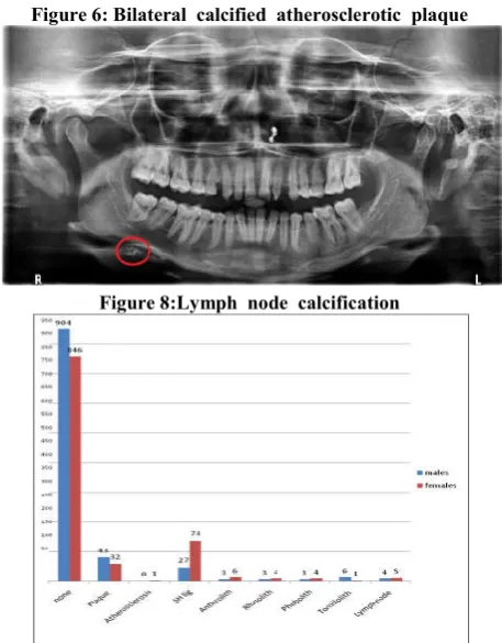
Calcified atherosclerotic plaque: They are identified as
vertical linear distribution sharply defined from surrounding soft tissues, present either superior or inferior to greater cornu of hyoid bone and adjacent to cervical vertebrae C3, C4, or the
intervertebral space between them(White, 2014) They may be
unilateral or bilateral and are considered if present in the box
7th or 12th box. (Figure 6 shows bilateral atherosclerotic
plaques).
Arteriosclerosis: They occurs due to loss of elastic fibres
followed by the deposition of calcium within the medial coat of the blood vessel. It is identified as calcification if it appeared as a parallel pair or thin, radiopaque lines which are
described as pipe stem or tram track appearance (White, 2014)
This is confirmed if present in 8th or 11th box.
Phleboliths: They are idiopathic calcifications which are
intravascular thrombi. They appear as multiple concentric radiopaque and radiolucent rings, with homogenously radiopaque internal structure giving a bull’s eye or target
appearance (White, 2014) They are confirmed if present in 8th
or 11th box (Figure 7).
Lymph node calcification: These are dystrophic calcifications
caused by the chronically inflamed lymph nodes by various diseases. They have a lobulated appearance similar to outer
shape of cauliflower.(1) It is considered if present in 8th or 11th
box which usually represents calcification of submandibular lymph node in that area.(Figure 8 shows calcification of submandibular lymph node).
RESULTS
A total of 1750 OPG's were shortlisted randomly from the period of June 2015 to June 2016. The details of OPG i.e. age, gender was blinded to the primary investigator before handing over the data to the investigator. The OPG's were evaluated for
calcifications and were tabulated and the data collected was
entered in Microsoft Excel Sheet. This was submitted to two maxillofacial radiologist who had prior experience of hard and soft tissue calcifications. These investigators excluded some of the radiographs which had poor contrast and disparity in clarity. Out of 1750 radiographs the number of radiographs with evidence of calcification was 210( 21.0%). Data analysis was done using SPSS software (v 22.0, IBM). After examination it was found that 210 radiographs showed evidence of calcification. Out of these radiographs, 131 were radiographs of females and 79 of males.(Graph 1).Age ranging from 35 to 80 years were examined. Mean age of participants with calcification was 49.01 and without calcification was 46.74(Graph 2).
In the present study radiographs showing ossified stylohyoid ligament were majority in number with 9.9% followed by calcified atherosclerotic plaque with 7.1% where as study
conducted by Vengalath J et al carotid artery calcifications
were highest in number. Antroliths and lymph node calcifications were 0.9% ,rhinoliths and tonsilloliths were 0.7% and least among all were phleboliths with 0.6% (Graph 3). In 101 radiographs of stylohyoid ligament ossification, 74 were females and 27 were males and also in 75 radiographs of carotid artery calcification, 43 were males and 32 were females. The estimation of presence of calcification with comparison of findings between males and females was analyzed using Chi-square test(p value = 0.01) and considered to be statistically significant.
DISCUSSION
Figure 2:Ossified stylohyoid ligament Figure 2:Ossified stylohyoid ligament
Figure 3:Bilateral tonsilloliths Figure 4:Rhinolith and Ossified stylohyoid ligament
Figure 5:Antrolith Figure 6: Bilateral calcified atherosclerotic plaque
Figure 7: Phleboliths Figure 8:Lymph node calcification
[image:3.595.327.549.353.464.2] [image:3.595.323.552.476.769.2]Graph 3. Age range of patients with and without calcifications
When superimposed over the bone, determination of whether the calcification is located in either the bone or the soft tissue is a complex task (Icoz, 2019; Rosana da Silva Berticelli,
2018)Among the lesions to be considered for the differential
diagnosis are the carotid artery, lymph nodes, tonsillolith, oropharyngeal tissues, and stylohyoid ligament calcifications (Nasseh, 2016). Calcification which is formed within dead and degenerative tissue is referred as dystrophic. Calcification which results from the deposition of calcium in normal tissues ,with normal calcium serologic rates it is referred as idiopathic. Calcification which occurs by the precipitation of minerals inside normal tissues with high calcium serologic rates it is referred as metastatic (White, 2014; Icoz, 2019). In this study, the highest incidence was observed for stylohyoid ossification followed by atherosclerotic plaque, least was phleboliths. No images compatible with sialoliths and arteriosclerosis were found on the radiographs. In most of previous studies conducted so far carotid artery calcifications were found highest in number but in our study, the highest found was stylohyoid ossification (9.9%) and carotid atherosclerotic plaque was 7.1%.
The ossified styloid chain is an excessive or abnormal calcification component including elongation of the styloid process and calcification of the stylohyoid ligament (Rosana da
Silva Berticelli, 2018).The average normal length of styloid
process in adult ranges from 20 to 30 mm and more than this length is considered as elongation (Garay, 2014) It was first described by Marchetti Pietro (1652) (Fusco, 2012) and took the name of Dr. Watt Eagle (1937) to become known as
Eagle’s syndrome (Moon et al., 2014). Ossification of
stylohyoid ligament usually extend from the base of the skull and most of the time occurs bilaterally. Usually ossification of stylohyoid ligament does not cause any symptoms only a few patients may have symptoms. A symptomatic ossified stylohyoid ligament referred to as Eagles Syndrome & panoramic radiography showing a length of 3cm or more is sufficient to confirm the diagnosis. Symptoms related to this ossified ligament are termed Eagles Syndrome which has two sub subtypes: (a)classic Eagles syndrome resulting from cranial nerve impingement and (b) carotid artery syndrome
resulting from impingement on carotid vessels(White, 2014)T
Kusunoki et al published a article on a case of very elongated
styloid process of 8cm in length with frequent throat pain for
10 years (Kusunoki et al., 2016). Also in the study conducted
by K Machado de et al it showed prevalence of alterations in
the morphology of the styloid process in patients with TMD (Andrade, 2012) However for patients with persistence
symptoms , the treatment will be stylohyoidectomy (Weteid, 2015). Also Galal Omami conducted a study to investigate the prevalence and pattern of calcification of stylohyoid complex in Libyan population in which he found out of 3162 images, styloid process elongation were seen in 541 (17.2%) and also were seen more in females (Galal, 2018). The reason for the majority to be seen in females is not been found. In a study
conducted by Ivonne et al tonsilloliths were found highest in
number ,also in the study conducted by Icoz D et al in year
2019 they found tonsillolith highest in number with (2.5%) followed by carotid artery calcification(2%) ,sialoloth (1%) & calcified lymph node(0.6%). In our study tonsilloliths were of 0.7% (Icoz, 2019). In many studies conducted , carotid artery calcifications were more in number. In the study conducted by
Vengalath et al also the highest incidence was observed for
carotid artery calcifications and out of 132 radiographs ,90 were radiographs of females and 42 of males (Vengalath, 2014) where as in our study out of 75 radiographs, 43 radiographs were of males and 32 were of females. In year
2018, a study conducted by Ibrahim Nasseh et al, among 500
patients, carotid artery calcification were found in 34 cases (6.8%) among them 23 females (8.18%) & 11 males (5.02%) (Nasseh, 2018) Causes of atheromatous plaque might be hypertension, diabetic mellitus, improper exercise, obesity, high stress, inadequate blood supply. In one study done by
Constantine et al. in the year 2019, carotid ultrasound was
carried out in all patients of carotid artery calcifications. In this study prevalence of carotid stenosis on ultrasound was 15.4% in those with carotid artery calcification and 5.8% for those without carotid artery calcification on OPG. The most common cause of death worldwide is coronary heart disease, cancer and stroke. The dentist could be the first person to detect impending signs of atherosclerosis.
Phleboliths are almost always associated to hemangiomas. Therefore, the existence of a hemangioma can lead a professional to research the presence of such calcifications. Regarding numbers, phleboliths are multiple. They do not have a specific location. In our study, we had only one case of phleboliths (0.1%). It is important to point out that the radiographic findings of phleboliths in soft tissues in the region of the head and neck are evidence of the presence of vascular
lesions.(4)Also we found 0.9% of lymph node calcification.
Calcification in lymph nodes occurs due to chronically inflamed nodes because of various diseases (White, 2014). Hence the dentists careful evaluation can make the OPG which is an imaging tool an economical tool for timely advise on the patient's medical health.
Conclusion
Multiple soft tissue calcifications can be noted in head and neck area as a incidental findings on panoramic radiography . Majority of the calcifications in our study were stylohyoid ligament ossification followed by atherosclerotic plaque . These calcifications may make the patients think of their health and can take advise from physician . Hence the dentist be first person in saving patients life.
REFERENCES
Andrade, KM., Rodrigues, CA., Watanabe, PC., Mazzetto, MO. 2012. Styloid process elongation and calcification in
subjects with tmd: clinical and radiographic aspects. Braz.
Dent. J, vol.23 no.4
Fusco, DJ., Asteraki, S., Spetzler, RF. 2012. Eagle’s
syndrome: Embryology, anatomy, and clinical
management. Acta Neurochir (Wien). 154(7):1119-26. Galal, Omami. 2018. Calcification of stylohyoid complex in
Libyans. Saudi Dental Journal,30:151-154.
Garay, I., Netto, H D., Olate. S. 2014. Soft tissue calcified in mandibular angle area observed by means of panoramic
radiography. Int J Clin Exp Med., 7(1):51-56.
Icoz, D., Akgunlu, F. 2019. Prevalence of detected soft tissue
calcifications on digital panoramic radiographs.SRM J Res
Dent Sci., 10: 21-5.
Kusunoki, T., Homma, H., Kidokoro, Y., Yanai, A., Fujimaki, M., Ikeda, K. 2016. A case of a very elongated styloid process 8 cm in length with frequent throat pain for 10 years. Clin Pract, 6(1):820.
Moon, CS., Lee, BS., Kwon, YD., Choi, BJ., Lee, JW., Lee
HW. 2014. Eagle’s syndrome A case report. J Korean
Assoc Oral Maxillofac Surg, 40(1):43-7.
Nasseh, I., Aoun, G. 2018. Carotid artery calcification. A
digital panoramic -Based Study. MDPI Journal, 6-15.
Nasseh, I., Sokhn, S., Noujeim, M., Aoun, G. 2016. Considerations in detecting soft tissue calcifications on
panoramic radiography. J Int Oral Health, 8(6):742-746.
Rosana da Silva Berticelli, et al. 2018. Calcifications in Soft
Tissues Observed in Panoramic Radiograph. EC Dental Science.17.11; 2012-2018.
Vengalath, J., Puttabuddi, JH., Rajkumar, B., Shivakumar, GC. 2014. Prevalence of soft tissue calcifications on digital panoramic radiographs: A retrospective study. J Indian Acad Oral Med Radiol ,26:385-9
Weteid, AS., Miloro, M. 2015. Transoral endoscopic-assisted styloidectomy: How should Eagle syndrome be managed
surgically? Int J Oral Maxillofac Surg., 4(9):1181-7.
White, SC., Pharaoh, ML. 2014. Soft tissue calcification and ossification. Oral Radiology: Principles and Interpretation.
First South Asia Edition, pp 552-65.