Received 10 May 2016 Accepted 22 May 2016
Edited by W. T. A. Harrison, University of Aberdeen, Scotland
Keywords:crystal structure; powder diffraction; density functional theory; citrate; sodium; poly-morphs.
CCDC references:1481347; 1481346; 1481345
Supporting information:this article has supporting information at journals.iucr.org/e
A second polymorph of sodium dihydrogen citrate,
NaH
2C
6H
5O
7: structure solution from powder
diffraction data and DFT comparison
Alagappa Rammohanaand James A. Kadukb*
a
Atlantic International University, Honolulu, HI, USA, andbIllinois Institute of Technology, Chicago, IL, USA. *Correspondence e-mail: [email protected]
The crystal structure of a second polymorph of sodium dihydrogen citrate, Na+H2C6H5O7 , has been solved and refined using laboratory X-ray powder
diffraction data, and optimized using density functional techniques. The powder pattern of the commercial sample used in this study did not match that corresponding to the known crystal structure [Gluskeret al.(1965).Acta Cryst. 19, 561–572; refcode NAHCIT]. In this polymorph, the [NaO7] coordination
polyhedra form edge-sharing chains propagating along the a axis, while in NAHCIT the octahedral [NaO6] groups form edge-sharing pairs bridged by two
hydroxy groups. The most notable difference is that in this polymorph one of the terminal carboxyl groups is deprotonated, while in NAHCIT the central carboxylate group is deprotonated, as is more typical.
1. Chemical context
In the course of a systematic study of the crystal structures of Group 1 (alkali metal) citrate salts to better understand the anion’s conformational flexibility, deprotonation mode, coor-dination tendencies, and hydrogen bonding, we have deter-mined several new crystal structures. Most of the new structures were solved using powder diffraction data (laboratory and/or synchrotron), but single crystals were used where available. The general trends and conclusions about the 16 new compounds and 12 previously characterized structures are being reported separately (Rammohan & Kaduk, 2016a). Three of the new structures – NaKHC6H5O7, NaK2C6H5O7,
and Na3C6H5O7– have been published recently (Rammohan
& Kaduk, 2016b,c,d).
2. Structural commentary
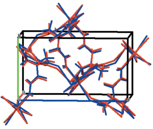
The asymmetric unit of the title compound is shown in Fig. 1. The root-mean-square deviation of the non-hydrogen atoms in the Rietveld-refined and DFT-optimized structures is 0.148 A˚ . The maximum deviation is 0.318 A˚ , at the sodium ion. The good agreement between the two structures (Fig. 2) is strong evidence that the experimental structure is correct (van de Streek & Neumann, 2014). This discussion uses the DFT-optimized structure. All of the bond lengths, bond angles, and most torsion angles fall within the normal ranges indicated by aMercury Mogulgeometry check (Macraeet al., 2008). Only the C2—C3—C4—C5 torsion angle is flagged as unusual. It lies in the tail of a minority gauche population of similar torsion angles. The citrate anion occurs in the gauche,trans
-conformation, which is one of the two low-energy conforma-tions of an isolated citrate ion. The central carboxylate group and the hydroxy group occur in the normal planar arrange-ment. The citrate chelates to one Na19 ion through the central carboxyl O9 atom and the hydroxy group O13, and to a second Na19 ion through the terminal carboxyl atom O12 and the hydroxy group O13. The Na+ion is seven-coordinate (penta-gonal–bipyramidal), and has a bond-valence sum of 1.12.
3. Supramolecular features
In this polymorph, the [NaO7] coordination polyhedra (Fig. 3)
form edge-sharing chains propagating along theaaxis, while in NAHCIT (Gluskeret al., 1965), the octahedral [NaO6] units
form edge-sharing pairs bridged by two hydroxy groups. The conformations of the citrate ions in the two structures are similar. The root-mean-square displacement of the non-hydrogen atoms is 0.11 A˚ . The conformations of the hydroxy groups differ, reflecting differences in coordination and hydrogen bonding. The most notable difference is that in this polymorph, one of the terminal carboxyl groups is deproto-nated, while in NAHCIT the central carboxylate group is deprotonated, as is more typical.
research communications
Acta Cryst.(2016). E72, 854–857 Rammohan and Kaduk Na+C
6H7O7
855
Figure 1
[image:2.610.45.296.71.246.2]The asymmetric unit, showing the atom numbering. The atoms are represented by 50% probability spheroids.
Figure 2
Comparison of the refined and optimized structures of sodium dihydrogen citrate. The refined structure is in red, and the DFT-optimized structure is in blue.
Table 1
Hydrogen-bond geometry (A˚ ,).
D—H A D—H H A D A D—H A
O7—H20 O11 1.01 1.61 2.627 176
O10—H21 O12 1.04 1.46 2.498 175
O13—H16 O8 0.97 2.50 3.033 114
[image:2.610.310.567.468.723.2]C2—H15 O8 1.09 2.50 3.166 119
Figure 3
[image:2.610.45.298.490.703.2]In this form, the hydrogen bonds occur in layers in theab
plane, while in NAHCIT the hydrogen bonds form double-ladder chains along thecaxis. The hydrogen bonds in this form contribute about 4.3 kcal mol 1 more to the lattice energy than those in NAHCIT, and seem to include a C—H O hydrogen bond (Table 1). Comparison of the DFT energies of the two polymorphs shows that this polymorph is 3.24 kcal mol 1 higher in energy than NAHCIT. Presumably it was crystallized at a higher temperature than NAHCIT, which was crystallized at 343 K.
4. Database survey
Details of the comprehensive literature search for citrate structures are presented in Rammohan & Kaduk (2016a). The crystal structure of sodium dihydrogen citrate is reported in Gluskeret al.(1965), and the powder pattern calculated from this structure is PDF entry 02-063-5032. The observed powder pattern matched PDF entry 00-016-1182 (de Wolffet al., 1966) A reduced cell search of the cell of the observed polymorph in the Cambridge Structural Database (Groom et al., 2016) (increasing the default tolerance from 1.5 to 2.0%) yielded 223 hits, but limiting the chemistry to C, H, Na, and O only resulted in no hits. The powder pattern is now contained in the the Powder Diffraction File (ICDD, 2015) as entry 00-063-1340.
5. Synthesis and crystallization
The sample was purchased from Sigma–Aldrich (lot #BCBC0142). Before measuring the powder pattern, a portion
of the sample was ground in a Spex 8000 mixer/mill and blended with a NIST SRM 640b silicon internal standard.
6. Refinement details
The powder pattern was indexed using DICVOL06 (Loue¨r & Boultif, 2007). The background andK2peaks were removed
using Jade (MDI, 2012), and Powder4 (Dragoe, 2001) was used to convert the data into an XYE file. The 10–52.22 portion of the pattern was processed inDASH 3.2(Davidet al., 2006), which suggestedP212121as the most probable space
group. Citrate and Na fragments were used to solve the structure in this space group usingDASH.
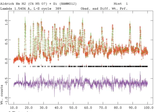
The powder pattern (Fig. 4) was indexed using Jade 9.5
(MDI, 2012). Pseudo-Voigt profile coefficients were as para-meterized in Thompsonet al.(1987) with profile coefficients for Simpson’s rule integration of the Pseudo-Voigt function according to Howard (1982). The asymmetry correction of Fingeret al.(1994) was applied and microstrain broadening by Stephens (1999).
The structure was refined by the Rietveld method using
GSAS/EXPGUI(Larson & Von Dreele, 2004: Toby, 2001). All C—C and C—O bond lengths were restrained, as were all bond angles. The hydrogen atoms were included at fixed positions, which were recalculated during the course of the refinement usingMaterials Studio(Dassault Systemes, 2014). TheUisovalues of the atoms in the central and outer portions
of the citrate were constrained to be equal, and theUisovalues
[image:3.610.46.554.93.333.2]of the hydrogen atoms were constrained to be 1.3those of the atoms to which they are attached.
Table 2
Experimental details.
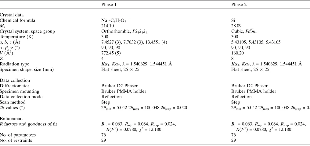
Phase 1 Phase 2
Crystal data
Chemical formula Na+C6H7O7 Si
Mr 214.10 28.09
Crystal system, space group Orthorhombic,P212121 Cubic,Fd3m
Temperature (K) 300 300
a,b,c(A˚ ) 7.4527 (3), 7.7032 (3), 13.4551 (4) 5.43105, 5.43105, 5.43105
,,(
) 90, 90, 90 90, 90, 90
V(A˚3) 772.45 (5) 160.20
Z 4 8
Radiation type K1,K2,= 1.540629, 1.544451 A˚ K1,K2,= 1.540629, 1.544451 A˚
Specimen shape, size (mm) Flat sheet, 2525 Flat sheet, 2525
Data collection
Diffractometer Bruker D2 Phaser Bruker D2 Phaser
Specimen mounting Bruker PMMA holder Bruker PMMA holder
Data collection mode Reflection Reflection
Scan method Step Step
2values (
) 2min= 5.042 2max= 100.048 2step= 0.020 2min= 5.042 2max= 100.048 2step= 0.020
Refinement
Rfactors and goodness of fit Rp= 0.063,Rwp= 0.084,Rexp= 0.024,
R(F2) = 0.0780,2 = 12.180
Rp= 0.063,Rwp= 0.084,Rexp= 0.024,
R(F2) = 0.0780,2 = 12.180
No. of parameters 76 76
No. of restraints 29 29
The Bravais–Friedel–Donnay–Harker (Bravais, 1866; Friedel, 1907; Donnay & Harker, 1937) morphology suggests that we might expect a blocky morphology for this phase. A 4th-order spherical harmonic texture model was included in the refinement. The texture index was 1.374, indicating that preferred orientation was significant for this rotated-flat-plate specimen.
7. DFT calculations
Crystal data, data collection and structure refinement details are summarized in Table 2. After the Rietveld refinement, a density functional geometry optimization (fixed experimental unit cell) was carried out using CRYSTAL09 (Dovesiet al., 2005). The basis sets for the C, H, and O atoms were those of Gattiet al.(1994), and the basis set for Na was that of Dovesi
et al.(1991). The calculation used 8k-points and the B3LYP functional, and took about 60 h on a 2.4 GHz PC. The Uiso
from the Rietveld were assigned to the optimized fractional coordinates.
References
Bravais, A. (1866). In Etudes Cristallographiques. Paris: Gauthier Villars.
Bruker (2009).DIFFRAC. Measurement. Bruker AXS Inc., Madison, Wisconsin, USA.
Crystal Impact (2015). DIAMOND. Crystal Impact GbR, Bonn, Germany. http://www.crystalimpact.com/diamond.
Dassault Systemes (2014). Materials Studio. BIOVIA, San Diego, CA, USA.
David, W. I. F., Shankland, K., van de Streek, J., Pidcock, E., Motherwell, W. D. S. & Cole, J. C. (2006).J. Appl. Cryst.39, 910– 915.
Donnay, J. D. H. & Harker, D. (1937).Am. Mineral.22, 446–467. Dovesi, R., Orlando, R., Civalleri, B., Roetti, C., Saunders, V. R. &
Zicovich-Wilson, C. M. (2005).Z. Kristallogr.220, 571–573. Dovesi, R., Roetti, C., Freyria-Fava, C., Prencipe, M. & Saunders,
V. R. (1991).Chem. Phys.156, 11–19. Dragoe, N. (2001).J. Appl. Cryst.34, 535.
Finger, L. W., Cox, D. E. & Jephcoat, A. P. (1994).J. Appl. Cryst.27, 892–900.
Friedel, G. (1907).Bull. Soc. Fr. Mineral.30, 326–455.
Gatti, C., Saunders, V. R. & Roetti, C. (1994).J. Chem. Phys.101, 10686–10696.
Glusker, J. P., Van Der Helm, D., Love, W. E., Dornberg, M., Minkin, J. A., Johnson, C. K. & Patterson, A. L. (1965).Acta Cryst.19, 561– 572.
Groom, C. R., Bruno, I. J., Lightfoot, M. P. & Ward, S. C. (2016).Acta Cryst.B72, 171–179.
Howard, C. J. (1982).J. Appl. Cryst.15, 615–620.
ICDD (2015).PDF-4+ 2015 and PDF-4 Organics 2016 (Databases), edited by S. Kabekkodu. International Centre for Diffraction Data, Newtown Square, PA, USA.
Larson, A. C. & Von Dreele, R. B. (2004). General Structure Analysis System (GSAS). Report LAUR, 86–784 Los Alamos National Laboratory, New Mexico, USA.
Loue¨r, D. & Boultif, A. (2007).Z. Kristallogr. Suppl.2007, 191–196. Macrae, C. F., Bruno, I. J., Chisholm, J. A., Edgington, P. R., McCabe, P., Pidcock, E., Rodriguez-Monge, L., Taylor, R., van de Streek, J. & Wood, P. A. (2008).J. Appl. Cryst.41, 466–470.
MDI (2012).JADE. Materials Data Inc., Livermore, CA, USA. Rammohan, A. & Kaduk, J. A. (2016a). Acta Cryst. B. Submitted. Rammohan, A. & Kaduk, J. A. (2016b).Acta Cryst.E72, 170–173. Rammohan, A. & Kaduk, J. A. (2016c).Acta Cryst.E72, 403–406. Rammohan, A. & Kaduk, J. A. (2016d).Acta Cryst.E72, 793–796. Stephens, P. W. (1999).J. Appl. Cryst.32, 281–289.
Streek, J. van de & Neumann, M. A. (2014).Acta Cryst.B70, 1020– 1032.
Thompson, P., Cox, D. E. & Hastings, J. B. (1987).J. Appl. Cryst.20, 79–83.
Toby, B. H. (2001).J. Appl. Cryst.34, 210–213. Westrip, S. P. (2010).J. Appl. Cryst.43, 920–925.
Wolff, P. de (1966). ICDD Grant-in-Aid, PFD entry 00-016-1182.
research communications
Acta Cryst.(2016). E72, 854–857 Rammohan and Kaduk Na+C
[image:4.610.46.298.68.251.2]6H7O7
857
Figure 4
sup-1
Acta Cryst. (2016). E72, 854-857
supporting information
Acta Cryst. (2016). E72, 854-857 [https://doi.org/10.1107/S2056989016008343]
A second polymorph of sodium dihydrogen citrate, NaH
2C
6H
5O
7: structure
solution from powder diffraction data and DFT comparison
Alagappa Rammohan and James A. Kaduk
Computing details
(RAMM012A_phase_1) Sodium dihydrogen citrate
Crystal data
Na+·C 6H7O7−
Mr = 214.10
Orthorhombic, P212121 Hall symbol: P 2ac 2ab
a = 7.4527 (3) Å
b = 7.7032 (3) Å
c = 13.4551 (4) Å
V = 772.45 (5) Å3
Z = 4
Dx = 1.841 Mg m−3
T = 300 K
Fractional atomic coordinates and isotropic or equivalent isotropic displacement parameters (Å2)
x y z Uiso*/Ueq
Na1 0.8787 (12) −0.2363 (10) −0.0501 (5) 0.035 (2)*
C2 0.870 (2) 0.1658 (19) 0.0733 (10) 0.0258 (15)*
C3 0.769 (2) 0.282 (2) 0.1478 (9) 0.019 (3)*
C4 0.804 (2) 0.4793 (18) 0.1383 (8) 0.019 (3)*
C5 0.723 (2) 0.564 (2) 0.2299 (10) 0.019 (3)*
C6 0.528 (3) 0.509 (2) 0.2460 (9) 0.0258 (15)*
C7 1.006 (3) 0.528 (2) 0.1354 (9) 0.0258 (15)*
O8 0.8117 (18) 0.0107 (14) 0.0697 (7) 0.0258 (15)*
O9 1.0081 (19) 0.2131 (15) 0.0280 (6) 0.0258 (15)*
O10 0.5110 (16) 0.4578 (14) 0.3329 (6) 0.0258 (15)*
O11 0.418 (2) 0.5412 (14) 0.1838 (7) 0.0258 (15)*
O12 1.062 (2) 0.6521 (14) 0.0763 (6) 0.0258 (15)*
O13 1.081 (2) 0.4474 (15) 0.2023 (7) 0.0258 (15)*
O14 0.7172 (16) 0.5561 (13) 0.0514 (6) 0.0258 (15)*
H15 0.74818 0.24337 0.22455 0.025 (4)*
H16 0.61496 0.2687 0.13343 0.025 (4)*
H17 0.69716 0.46671 0.01345 0.034 (2)*
H18 0.70618 0.69511 0.23029 0.025 (4)*
H19 0.76628 0.53492 0.29702 0.025 (4)*
H20 0.7027 −0.0047 0.0991 0.039*
supporting information
sup-2
Acta Cryst. (2016). E72, 854-857
Geometric parameters (Å, º)
Na1—O8 2.543 (12) C7—O12 1.314 (13)
Na1—O10i 2.463 (13) C7—O13 1.227 (13)
Na1—O11ii 2.363 (13) O8—Na1 2.543 (12)
Na1—O12iii 2.344 (13) O8—C2 1.271 (14)
Na1—O12iv 2.475 (16) O8—H20 0.912 (13)
Na1—O14iii 2.423 (13) O9—C2 1.253 (14)
Na1—O14ii 2.879 (13) O10—Na1v 2.463 (13)
C2—C3 1.540 (10) O10—C6 1.240 (12)
C2—O8 1.271 (14) O11—Na1iv 2.363 (13)
C2—O9 1.253 (14) O11—C6 1.198 (14)
C3—C2 1.540 (10) O11—H21 0.998 (15)
C3—C4 1.546 (10) O12—Na1vi 2.344 (13)
C3—H15 1.087 (12) O12—Na1ii 2.475 (16)
C3—H16 1.167 (18) O12—C7 1.314 (13)
C4—C3 1.546 (10) O13—C7 1.227 (13)
C4—C5 1.519 (10) O14—Na1vi 2.423 (13)
C4—C7 1.549 (10) O14—Na1iv 2.879 (13)
C4—O14 1.461 (9) O14—C4 1.461 (9)
C5—C4 1.519 (10) O14—H17 0.871 (10)
C5—C6 1.528 (10) H15—C3 1.087 (12)
C5—H18 1.022 (16) H16—C3 1.167 (18)
C5—H19 0.984 (15) H17—O14 0.871 (10)
C6—C5 1.528 (10) H18—C5 1.022 (16)
C6—O10 1.240 (12) H19—C5 0.984 (15)
C6—O11 1.198 (14) H20—O8 0.912 (13)
C7—C4 1.549 (10) H21—O11 0.998 (15)
O8—Na1—O10i 171.5 (5) C3—C4—C7 113.8 (12)
O8—Na1—O11ii 91.8 (4) C3—C4—O14 112.9 (11)
O8—Na1—O12iii 85.9 (5) C5—C4—C7 107.7 (13)
O8—Na1—O12iv 73.0 (5) C5—C4—O14 107.5 (12)
O8—Na1—O14iii 92.2 (4) C7—C4—O14 108.2 (12)
O8—Na1—H21ii 94.0 (4) C4—C5—C6 112.1 (16)
O10i—Na1—O11ii 85.0 (4) C5—C6—O10 108.5 (13)
O10i—Na1—O12iii 90.9 (4) C5—C6—O11 119.6 (15)
O10i—Na1—O12iv 114.1 (4) O10—C6—O11 131.0 (19)
O10i—Na1—O14iii 94.0 (4) C4—C7—O12 120.0 (15)
O11ii—Na1—O12iii 135.6 (6) C4—C7—O13 107.6 (13)
O11ii—Na1—O12iv 81.0 (5) O12—C7—O13 131.8 (17)
O11ii—Na1—O14iii 155.6 (5) Na1—O8—C2 131.4 (11)
O12iii—Na1—O12iv 139.0 (5) Na1v—O10—C6 142.0 (14)
O12iii—Na1—O14iii 68.8 (4) Na1iv—O11—C6 138.6 (12)
O12iv—Na1—O14iii 77.2 (4) Na1vi—O12—Na1ii 110.8 (4)
C3—C2—O8 114.0 (13) Na1vi—O12—C7 121.4 (12)
C3—C2—O9 123.3 (15) Na1ii—O12—C7 125.6 (12)
sup-3
Acta Cryst. (2016). E72, 854-857
C2—C3—C4 115.8 (13) Na1iv—H21—O11 78.3 (6)
C3—C4—C5 106.5 (13)
Symmetry codes: (i) −x+3/2, −y, z−1/2; (ii) x+1/2, −y+1/2, −z; (iii) x, y−1, z; (iv) x−1/2, −y+1/2, −z; (v) −x+3/2, −y, z+1/2; (vi) x, y+1, z.
(RAMM012A_phase_2)
Crystal data
Si
Mr = 28.09
Cubic, Fd3m
Hall symbol: -F 4vw 2vw
a = 5.43105 Å
V = 160.20 Å3
Z = 8
T = 300 K
Fractional atomic coordinates and isotropic or equivalent isotropic displacement parameters (Å2)
x y z Uiso*/Ueq
Si1 0.125 0.125 0.125 0.0304 (5)*
Geometric parameters (Å, º)
Si1—Si1i 2.3517 Si1—Si1iii 2.3517
Si1—Si1ii 2.3517 Si1—Si1iv 2.3517
Si1i—Si1—Si1ii 109.4712 Si1ii—Si1—Si1iii 109.4712
Si1i—Si1—Si1iii 109.4712 Si1ii—Si1—Si1iv 109.4712
Si1i—Si1—Si1iv 109.4712 Si1iii—Si1—Si1iv 109.4712
Symmetry codes: (i) x+1/4, y+1/4, −z; (ii) −z, x+1/4, y+1/4; (iii) y+1/4, −z, x+1/4; (iv) −x, −y, −z.
(ramm012a_DFT)
Crystal data
C6H7NaO7
Mr = 214.10
Orthorhombic, P212121
a = 7.4527 Å
b = 7.7032 Å
c = 13.4551 Å
V = 772.45 Å3
Z = 4
None; DFT calculation radiation
T = 300 K
Data collection
h = →
k = →
l = →
Fractional atomic coordinates and isotropic or equivalent isotropic displacement parameters (Å2)
x y z Uiso*/Ueq
C1 0.86930 0.18689 0.07976 0.02580*
C2 0.76864 0.31082 0.14718 0.01910*
C3 0.80887 0.50489 0.13526 0.01910*
C4 0.72870 0.60356 0.22535 0.01910*
C5 0.54276 0.54256 0.25725 0.02580*
C6 1.01121 0.54559 0.13579 0.02580*
supporting information
sup-4
Acta Cryst. (2016). E72, 854-857
O8 1.01065 0.22287 0.03877 0.02580*
O9 1.07983 0.64886 0.07729 0.02580*
O10 1.09698 0.46975 0.20891 0.02580*
O11 0.51997 0.49361 0.34617 0.02580*
O12 0.42047 0.54898 0.19138 0.02580*
O13 0.73153 0.57264 0.04623 0.02580*
H14 0.79934 0.27249 0.22351 0.02500*
H15 0.62504 0.29365 0.13548 0.02500*
H16 0.75707 0.49090 −0.00712 0.02500*
H17 0.71973 0.74075 0.20546 0.02500*
H18 0.81890 0.59174 0.28862 0.03350*
Na19 0.91363 −0.18930 −0.03840 0.03460*
H20 0.67516 0.02054 0.10415 0.03900*
H21 0.23334 0.49718 0.20429 0.03900*
Bond lengths (Å)
C1—C2 1.516 C4—H17 1.092
C1—O7 1.334 C4—H18 1.089
C1—O8 1.221 C5—O11 1.266
C2—C3 1.533 C5—O12 1.272
C2—H14 1.093 C6—O9 1.230
C2—H15 1.090 C6—O10 1.311
C3—C4 1.550 O7—H20 1.014
C3—C6 1.540 O10—H21i 1.040
C3—O13 1.428 O13—H16 0.974
C4—C5 1.525 H21—O10ii 1.040
Symmetry codes: (i) x+1, y, z; (ii) x−1, y, z.
Hydrogen-bond geometry (Å, º)
D—H···A D—H H···A D···A D—H···A
O7—H20···O11 1.01 1.61 2.627 176
O10—H21···O12 1.04 1.46 2.498 175
O13—H16···O8 0.97 2.50 3.033 114