Intracardiac Patches in Children With
Congenital Heart Disease
Jennifer S. Nelson, MD, MS, Amer Heider, MD, Ming-Sing Si, MD, and
Richard G. Ohye, MD
Department of Surgery, Division of Cardiothoracic Surgery, University of North Carolina School of Medicine, Chapel Hill, North Carolina; and Departments of Pathology and Cardiac Surgery, University of Michigan Medical School, Ann Arbor, Michigan
Background. Animal data demonstrate that intracardiac patches of decellularized porcine small intestine sub-mucosa (CorMatrix; CorMatrix Cardiovascular, Inc, Atlanta, GA) become repopulated with native cells, sug-gesting the possibility of a substrate for regenerative tissue in humans. We report the only prospective series to date of explanted CorMatrix patches placed in infants with congenital heart disease.
Methods. CorMatrix patches were implanted as the hemi-Fontan baffle in patients during their second stage of palliation. The patch material was explanted as part of the standard Fontan operation. Specimens were analyzed with the use of hematoxylin and eosin, Movat pentach-rome, and trichrome stains.
Results. Of the 12 implantations, 10 specimens were explanted. Two patients did not undergo Fontan
because of unfavorable hemodynamics. Acellular material, chronic inflammation, fibrosis, and foreign body giant cell reaction was seen in all explanted patches. Also noted in explanted specimens was calcification (n [ 2), elastic fibers (n [ 2), and eosinophils (n [ 2). No explanted CorMatrix material showed evidence of ingrowth of native cells or trans-formation into cardiac tissue at a median of 21 months after implantation.
Conclusions. Although the CorMatrix remained pliable and most did not exhibit calcification, these intracardiac patches did not show evidence of native heart tissue ingrowth at a median of 21 months in vivo.
(Ann Thorac Surg 2016;102:1329–35) Ó2016 by The Society of Thoracic Surgeons
T
he ability to replace damaged or absent heart struc-tures with the patient’s own tissue remains one of the ultimate, but as of yet unachieved, goals of congenital heart surgeons. Regenerated native tissue, whether culti-vated outside the body and implanted or developed over time on an implanted biological scaffold, would have many advantages. These advantages include durability and the potential for growth, important in the pediatric population. One such scaffold, porcine small intestinal submucosa extracellular matrix (SIS ECM), has shown promise in regenerating native tissue. The ECM is implanted and by a process of ingrowth of native cells may be theoretically transformed into native tissue. CorMatrix (CorMatrix Cardiovascular, Inc, Atlanta, GA) is a commercially avail-able ECM product that is approved by the Food and Drug Administration for use as a pericardial replacement and as an intracardiac patch.Animal studies have shown remodeling of CorMatrix patches into myocardial tissue with contractile properties
[1]. However, recent retrospective human studies have had disappointing results[2–4]. Of all available studies to examine explanted CorMatrix patches used for intracar-diac use in congenital heart disease, none have shown native tissue ingrowth[2–5]. Prior studies are limited by their retrospective nature, heterogeneity of implant sites, and short times in vivo [2–4]. To our knowledge, the current study represents the first prospective histologic study of CorMatrix for intracardiac use in children with congenital heart disease.
The purpose of this study was to describe the histo-pathologic process of a prospective series of explanted CorMatrix patches used as a hemi-Fontan baffle in chil-dren undergoing routine single-ventricle palliation.
Material and Methods
The University of Michigan Medical School Institutional Review Board approved this study. Informed, written consent was obtained from all patients by their parents or legal representative.
Study Population
The medical records for consecutive patients scheduled for stage 2 palliation for a single-ventricle lesion between June 1, 2012, and February 1, 2013. at C.S. Mott Children’s
Accepted for publication March 22, 2016.
Presented at the Fifty-second Annual Meeting of The Society of Thoracic Surgeons, Phoenix, AZ, Jan 23–27, 2016.
Address correspondence to Dr Nelson, Division of Cardiothoracic Sur-gery, 3041 Burnett-Womack Bldg, University of North Carolina School of Medicine, Chapel Hill, NC 27514; email:[email protected].
Ó2016 by The Society of Thoracic Surgeons 0003-4975/$36.00
Published by Elsevier http://dx.doi.org/10.1016/j.athoracsur.2016.03.086
CO
NGENITAL
H
Hospital were reviewed. Patients with anatomy amenable to hemi-Fontan procedure (HFP) were approached for parental consent. Enrollment was closed after the recruitment of 12 patients.
Operative Details
On the basis of institutional preference, a modified HFP is performed for stage 2 palliation whenever anatomically possible. Our technique is described elsewhere[6]. A variety of patch materials are available for routine use for intra-cardiac baffle creation, including polytetrafluoroethylene, bovine pericardium, pulmonary homograft, autologous pericardium, and decellularized porcine small intestine submucosa (CorMatrix; CorMatrix Cardiovascular Inc).
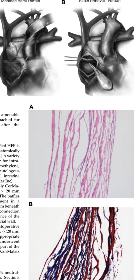
In this study, all patients received circular 4-ply CorMa-trix patches that measured approximately 20 20 mm implanted as the standard hemi-Fontan baffle. The baffles were sutured with polypropylene monofilament in a running fashion completely within the right atrium beneath the superior vena caval-pulmonary artery connection
(Figs 1A, 1B). Therefore, the entire circumference of the
CorMatrix patch was in contact with the right atrial wall. After operation, patients received routine postoperative care. In general, the baffles were exposed to low (<20 mm Hg) pressures while in vivo. When clinically appropriate according to institutional criteria, patients underwent completion lateral-tunnel Fontan. As a routine part of the Fontan operation, the previously implanted CorMatrix patches were excised (Fig 1B).
Histologic Examination
Explanted CorMatrix patches werefixed in 10% neutral-buffered formalin and embedded in paraffin. Sections 5-
m
m thick were stained with hematoxylin and eosin, Masson’s trichrome, and Movat pentachrome stains. The histologic examination was evaluated for evidence of native tissue ingrowth, specifically any tissue resemblingFig 1. (A) Hemi-Fontan patch baffle location within the right atrium. The patch separates the superior vena caval/ pulmonary artery junction from the body of the right atrium. (B) Patch removal at Fontan operation. (Reprinted with permission from Elsevier from Bove EL, Mosca RS. Surgical repair of the hypo-plastic left heart syndrome. Prog Pediatr Cardiol 1996;5:23–35.)
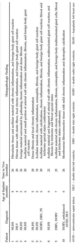
Fig 2. (A) Control; collagen and ghost outlines of nuclei are shown. (Hematoxylin and eosin;10.) (B) Control; connective tissue with collagen stained blue. (Trichrome;10.)
CO
NG
EN
IT
A
L
HEA
R
right atrial tissue. Specimens were photographed, and the histopathologic results were recorded. An unused CorMatrix patch was analyzed as a control after being prepared according to the manufacturer’s instructions
(Figs 2A,2B).
Results
CorMatrix hemi-Fontan patches were implanted in 12 infants, ranging in age from 4 to 8 months (Table 1). Time spent in vivo ranged from 18 to 26 months (median, 21 months). Two patients did not undergo completion Fontan because of unfavorable hemodynamics.
Fibrosis, acellular material, chronic inflammation, and foreign body giant cell reaction were seen in all explanted specimens (Table 2; Figs 3A, 3B). Disorga-nized elastic fibers (2 specimens), dystrophic calcifi ca-tion (2 specimens), and eosinophils (2 specimens) were less frequentfindings (Figures 4–6). No explanted Cor-Matrix material showed evidence of ingrowth of native cells or transformation into cardiac tissue at a median of 21 months after implantation.
Comment
The potential for regeneration of native cardiac tissue in humans with CorMatrix remains unclear. To our Table
2. Histopathologic Variables (n ¼ 10) Pati ent Di agnosis Ag e a t Implant (months ) Tim e in Vivo (mon ths) Histopa tholog ic Find ing 1 H L H S 8 26 Fibro elastic tissue and acellula r m a terial with chro nic in fl am matio n, and foreig n body giant cell reaction 2 DILV 4 2 0 Fibro us tiss ue with elastic fi bers, focal chro nic in fl am matio n, and fo reign body giant cell reac tion 3 H L H S 5 26 Fore ign ma terial surro unded b y palisadin g chro nic in fl amma tion and dens e fi bros is 4 H L H S 6 20 Acellu lar mate rial and small port ion of arter ial wall with chro nic in fl am mation , fi brosis, and fo reign body giant cell reaction 5 H L H S 5 20 De nse fi bros is and focal chro nic in fl amma tion 6 H L H S 6 18 Acellu lar mate rial, chro nic in fl amma tion, eosino phils , fi b rosis, and foreig n b ody giant cell reaction 7 DOR V, HRV, PS 6 2 0 Acellu lar mate rial with calci fi catio ns, chro nic in fl ammatio n, fi bros is, and foreign body gia nt cel l reac tion; Mo vat and trichrome spe cial stains co n fi rma tory 8 H L H S 6 22 Acellu lar mate rial and fragme nt of art erial wa ll with chron ic in fl amma tion, multin ucleated giant cell reaction; and fi brosi s b y trichro me and Mo vat spe cial stains 9 DIRV, DOR V 7 25 Acellu lar materia l with chro nic in fl amma tion, fi bros is, eosinop hils, and foreign body multinu cleated gian t cells; Movat and trichro me stains co n fi rma tory 10 DOR V, unbalan ced AV SD, H L V 6 2 6 Membr anous fi broconn ective tiss ue with mild chro nic in fl amma tion and dystro phic calci fi ca tion AVSD ¼ atrioventricular septal defect; DILV ¼ double inlet left ventricle; DIRV ¼ double inlet right ventricle; DORV ¼ double outlet right ventricle; HLHS ¼ hypoplastic left heart syn-drome; HLV ¼ hypoplastic left ventricle; HRV ¼ hypoplastic right ventricle; PS ¼ pulmonary stenosis.
Table 1. Clinical Characteristics of Study Population (n¼12)
Characteristic Value*
Male sex 9 (75)
Diagnosis
HLHS 7 (58)
DILV 1 (8)
DORV, HRV, PS 1 (8)
Unbalanced AVSD, HLV, DORV, heterotaxy, dextrocardia, interrupted IVC
1 (8)
DIRV, DORV 1 (8)
DORV, unbalanced AVSD, HLV 1 (8)
Operation at implant
HFP 10 (83)
HFP/Kawashima 2 (17)
Operation at explant (n¼10)
Fontan 10 (100)
Patch not explanted 2 (17)
Median age at implant, months 6
Median age at explant, months 27
Median time in vivo, months 21
* Values may not sum to expected total because of rounding.
Data are n (%) unless otherwise noted.
AVSD ¼ atrioventricular septal defect; DILV¼ double inlet left ventricle; DIRV¼double inlet right ventricle; DORV¼double outlet right ventricle; HFP¼hemi-Fontan procedure; HLHS¼ hypoplastic left heart syndrome; HLV ¼ hypoplastic left ventricle; HRV¼hypoplastic right ventricle; IVC¼inferior vena cava; PS¼pulmonary stenosis.
CO
NGENITAL
H
knowledge, this study represents the first prospective evaluation of explanted intracardiac CorMatrix patches in children with congenital heart disease. In 100% of explanted patches, chronic inflammation and scar were identified with no evidence of native tissue ingrowth or regeneration. However, patches performed well in the HFP baffle application and remained pliable and largely without calcification.
CorMatrix ECM has been marketed as an intracardiac patch material that can generate new healthy tissue because it is completely resorbed over time [7]. The proposed mechanism is that the SIS ECM elicits a regenerative pathway that is immune-mediated and provides a scaffold for native tissue ingrowth[1].
Early animal studies of SIS products were encour-aging[1, 8–12]. Badylak and colleagues[1]used circular patches created from sheets of ECM derived from
porcine small intestinal submucosa or porcine urinary bladder matrix to repair full-thickness defects of the right ventricular anterior walls of six pigs. They re-ported complete replacement of the ECM with a mixture of tissue types, including cartilage and contractile myocardial tissue[1].
In general, experimental animal models with SIS ECM for cardiac use have involved suturing the patch material to an area of induced full-thickness injury [1]. In our study, as would be the case in a typical atrial septal defect closure in humans, patches are sewn to uninjured tissue with an intact endothelium. The process of creating a cardiac defect in animal models initiates an inflammatory pathway that could potentially contribute to a difference infindings. Future studies to investigate the role of su-turing the patch to an edge of cut myocardium, as opposed to an intact endothelialized surface such as the right atrial wall, may be useful.
Fig 3. (A) Low-power magnification with hematoxylin and eosin (H&E) stain showing section of the CorMatrix patch with a band of chronic inflammation and aggregates of histiocytes. (B) High-power magnification with H&E stain showing a section of the CorMatrix patch. This photomicrograph highlights the foreign body multinucleated giant cell reaction.
Fig 4. Trichrome stain at low-power magnification highlighting the acellular CorMatrix and the associatedfibrosis.
Fig 5. Movat pentachrome stain at low-power magnification with disorganized elasticfibers (stained black).
CO
NG
EN
IT
A
L
HEA
R
Most specimens in this study demonstrated a robust foreign body giant cell reaction to the CorMatrix patch, similar to the findings of Woo and colleagues [2] and Rosario and colleagues [3]. The appearance of the aggregate of histiocytes (Figs 3A, 3B) is similar to what would be expected with arteritis within the media of a large vessel wall. Although elasticfibers were seen in two cases on pentachrome staining, they were disorganized, not well defined, and did not form a distinct layer (Fig 5). In addition, the background of mucin-containing ground substance, staining light blue on pentachrome stain, has an appearance similar to a fibrinous exudate. These
findings do not support the theory that the CorMatrix was in the process of forming an organized, elastic, or muscular tissue layer such as atrial wall.
Badylak and colleagues [13]suggested that the forces present in the mechanical environment might affect the ability of SIS ECM products to remodel. Because our patches were exposed to a low-pressure system, it is possible that the lack of mechanical loading contributed to a microenvironment that was not conducive to remodeling in the time frame studied.
In the current study, the median time patches spent in vivo (21 months) is greater than previously published reports, and all were removed electively[4, 14, 15]. In general, time in vivo in prior reports has been short, and most patches were removed for failure[4, 5, 14–16]. Additional strengths of this study are the homogeneity of
the patient population, the standardization of the im-plantation technique, and the consistent site of implan-tation within the right atrium. Prior studies of explanted CorMatrix represent a mix of patient ages and implanta-tion sites [2–4, 15, 17]. Although CorMatrix is only approved as a pericardial replacement or for intracardiac repair, sites of implantation from prior reports of explanted CorMatrix vary widely from the right ventric-ular outflow tract, the mitral valve, pulmonary artery, pulmonary valve, and aortic arch[2–4, 14, 15, 18, 19].
As has been demonstrated, CorMatrix may be a clinically suitable patch material for the closure of septal defects and reconstruction of the great vessels[15, 19]. Note that, in some studies, the tissues were not examined for transformation, only for clinical efficacy
[16, 18]. Although some investigators have noted
suboptimal functional performance of CorMatrix when used for pulmonary artery wall reconstruction, for example, most studies have found that CorMatrix patches remained pliable and did not exhibit calcifi -cation at mid-term follow-up, much like our experience
[3, 15, 18–20]. In the current study, CorMatrix performed
well as a HFP baffle, but there is no evidence in our study that CorMatrix demonstrates regenerative prop-erties in humans.
References
1. Badylak S, Obermiller J, Geddes L, Matheny R. Extracellular
matrix for myocardial repair. Heart Surg Forum 2003;6:
E20–6.
2. Woo JS, Fishbein MC, Reemtsen B. Histologic examination of
decellularized porcine intestinal submucosa extracellular matrix (CorMatrix) in pediatric congenital heart surgery.
Cardiovasc Pathol 2016;25:12–7.
3. Rosario-Quinones F, Magid MS, Yau J, Pawale A,
Nguyen K. Tissue reaction to porcine intestinal Sub-mucosa (CorMatrix) implants in pediatric cardiac patients: a single-center experience. Ann Thorac Surg 2015;99:
1373–7.
4. Zaidi AH, Nathan M, Emani S, et al. Preliminary experience
with porcine intestinal submucosa (CorMatrix) for valve reconstruction in congenital heart disease: histologic evalu-ation of explanted valves. J Thorac Cardiovasc Surg 2014;148;
2216–4, 2225.e1.
5. McConnell PI, Hibino N. Something to consider: porcine
intestinal submucosa as a biologic scaffold, not a simple
patch. J Thorac Cardiovasc Surg 2014;148:1767–9.
6. Hirsch-Romano JC, Bove EL, Si MS, Ohye RG. Modified
hemi-Fontan procedure. Oper Tech Thorac Cardiovasc Surg
2013;18:117–23.
7. CorMatrix [product brochure]. Atlanta, GA: CorMatrix Car-diovascular, Inc; 2010.
8. Fallon A, Goodchild T, Wang R, Matheny RG. Remodeling of
extracellular matrix patch used for carotid artery repair.
J Surg Res 2012;175:e25–34.
9. Badylak SF. The extracellular matrix as a biologic scaffold
material. Biomaterials 2007;28:3587–93.
10. Kochupura PV, Azeloglu EU, Kelly DJ, et al.
Tissue-engineered myocardial patch derived from extracellular matrix provides regional mechanical function. Circulation
2005;112(9 Suppl):I144–9.
11. Robinson KA, Li J, Mathison M, et al. Extracellular matrix
scaffold for cardiac repair. Circulation 2005;112(9 Suppl):
I135–43.
12. Padalino MA, Castellani C, Dedja A, et al. Extracellular
matrix graft for vascular reconstructive surgery: evidence of Fig 6. (A) Low-power and (B) high-power hematoxylin and eosin
stain showing membranousfibroconnective tissue with mild chronic inflammation and dystrophic calcification (arrow).
CO
NGENITAL
H
autologous regeneration of the neoaorta in a murine model.
Eur J Cardiothorac Surg 2012;42:e128–35.
13. Badylak SF, Freytes DO, Gilbert TW. Extracellular matrix as a
biological scaffold material: structure and function. Acta
Biomater 2009;5:1–13.
14. Scholl FG, Boucek MM, Chan KC, Valdes-Cruz L,
Perryman R. Preliminary experience with cardiac recon-struction using decellularized porcine extracellular matrix scaffold: human applications in congenital heart disease.
World J Pediatr Congenit Heart Surg 2010;1:132–6.
15. Witt RG, Raff G, Van Gundy J, Rodgers-Ohlau M, Si MS.
Short-term experience of porcine small intestinal submucosa patches in paediatric cardiovascular surgery. Eur J
Car-diothorac Surg 2013;44:72–6.
16. Quarti A, Nardone S, Colaneri M, Santoro G, Pozzi M.
Pre-liminary experience in the use of an extracellular matrix to
repair congenital heart diseases. Interact Cardiovasc Thorac
Surg 2011;13:569–72.
17. Luk AA, Rao V, Cusimano RJ, David TE, Butany J. CorMatrix
extracellular matrix used for valve repair in the adult: is there de novo valvular tissue seen? Ann Thorac Surg 2015;99:
2205–7.
18. Gerdisch MW, Shea RJ, Barron MD. Clinical experience with
CorMatrix extracellular matrix in the surgical treatment of
mitral valve disease. J Thorac Cardiovasc Surg 2014;148:1370–8.
19. Padalino MA, Quarti A, Angeli E, et al. Early and mid-term
clinical experience with extracellular matrix scaffold for congenital cardiac and vascular reconstructive surgery: a multicentric Italian study. Interact Cardiovasc Thorac Surg
2015;21:40–9; discussion 49.
20. Sanders SP, Padera RF Jr. Reply to the editor. J Thorac
Car-diovasc Surg 2014;148:1769–70.
DISCUSSION
DR ALI DODGE-KHATAMI(Jackson, MS): That was a beautiful presentation. Congratulations on your results. As you mentioned, a lot of the things that have been published on CorMatrix are kind of retrospective and have almost the feel of the believers and the nonbelievers of CorMatrix; some saying that it is great stuff, and others saying that it is just as good as any other patch material.
Based on the study that you did, which is very controlled, the patches are in a low-pressure system. That may somehow in-fluence your results. To the believers of CorMatrix, do you think that part of the success would be the fact that they are implanting CorMatrix as part of aortic valve repairs or conduit substitutes? Do you think pressure or volume can play a role as to how CorMatrix behaves?
DR NELSON:In other words, do I think that it would be more successful in native tissue regeneration in a higher pressure system?
I think there is a lot of evidence that looks at explanted valve repair specimens which does not show native tissue remodeling either. For the low-pressure example, in the dog studies that were referenced in the CorMatrix product brochure, there were ASD patches placed in 2 dogs and right ventricular free wall patches in 2 other dogs. When explanted, by that report, they were completely indistinguishable from otherwise normal car-diac tissue.
The high-pressure example was looked at by Zaidi in 2014, and those aortic and mitral valve specimens were basically the results we have shown here. So no, I do not think so.
DR ANDREW LODGE(Durham, NC): I have always been con-cerned that implanting this material as a patch where it is not sewn to the cut edge could affect the ability for native tissue to replace the patch. So thefirst question I have is: Do you think that the fact that this patch was sewn to an endothelialized sur-face could have had an impact on what you saw?
And the second is: Do you have any experience explanting patches that are sewn to a cut surface, such as a ventriculotomy or an arterial wall, that have shown different results?
DR NELSON: I will answer your second question first. No experience in the evaluation of explanted patches that were sewn to a cut edge.
To answer yourfirst question, I think that it is possible. And in those studies I mentioned in dogs, those ASDs had been created for that model; therefore, there was a cut edge. Whereas in our example, you are right, with the perpendicular attachment of the
patch to the right atrial wall it is an otherwise undisturbed, uncut endothelialized surface. So I suppose that is a possible explana-tion, although I reserve my enthusiasm.
DR MUHAMMAD MUMTAZ (San Antonio, TX): I have two questions. One is that in your histopathologic studies did you see any evidence of neovascularization of this tissue?
DR NELSON:We did not.
DR MUMTAZ:Did you look for that?
DR NELSON:We did.
DR MUMTAZ:And the second question is, which I’m assuming
the answer would be yes, is that at the time when you were putting this patch, at the time of hemi-Fontan, let’s suppose that patch size was X, then by the time you took it out the child had grown and so the actual diameter of the patch is now larger than when it was placed. Did you look at that? And this may not represent growth, I understand that, but just in terms of plain geometry of the patch, the size that was put in and the size that was explanted, was there a difference?
DR NELSON: There was a difference. The explanted patches were slightly smaller. They appeared whitish on the gross in-spection, and they did appear slightly contracted.
DR MUMTAZ:So that is interesting. My own observation is that they are not smaller but they have actually grown to the size of whatever structure they are sutured to. Whereas if it was actually smaller, would you expect the hemi-Fontan to develop a gradient? I’m a little surprised by your answer.
DR NELSON:I would not expect a gradient in the hemi-Fontan pathway just by nature of theflow patterns and the position of the baffle which just really creates a barrier between the superior venocaval-pulmonary artery junction and the body of the right atrium. So contraction of this patch in that location would not cause obstruction toflow there. I would also say that the degree of contraction was almost imperceptible, but we were looking really hard at that. And the difference in the size of the right atrium was not of clinical significance.
DR CONSTANTINE MAVROUDIS (Orlando, FL): This was a delightful presentation with excellent data. The fact that you only have a few patients, 10 or 12, should not matter at all. I think you
CO
NG
EN
IT
A
L
HEA
R
presented your data in a forthright manner, and let the science speak for itself.
However, I have a question on what you are doing now, because that might be telling to the audience. I think there is a bias either for or against these kinds of modified pericardial patches. So the question for you is, what are you using now?
DR NELSON:We have returned to using PTFE for this patch in this position.
DR JOHN E. MAYER (Boston, MA): I would say I am not surprised by yourfindings. I think the question about whether
the tissue is going to shrink or stretch, or I hesitate to use the word, grow, is clearly going to depend on the hemodynamic forces to which the material is subjected. I think the important thing is that you have demonstrated quite clearly this is nonliving tissue. Nonliving tissue that is continuouslyflexed or stretched is going to stretch out and get bigger. If it is not subjected to those forces, then it is likely to shrink as a part of the inflammatory response to this foreign body. I think the results that you show are exactly predictable if viewed from that perspective.
DR NELSON:Thank you for your comments.
CO
NGENITAL
H