University of Pennsylvania
ScholarlyCommons
Publicly Accessible Penn Dissertations
1-1-2014
Regulating Gene Expression With Light-Activated
Oligonucleotides
Julianne C. Griepenburg
University of Pennsylvania, [email protected]
Follow this and additional works at:http://repository.upenn.edu/edissertations Part of theBiochemistry Commons, and theChemistry Commons
This paper is posted at ScholarlyCommons.http://repository.upenn.edu/edissertations/1297
For more information, please [email protected].
Recommended Citation
Griepenburg, Julianne C., "Regulating Gene Expression With Light-Activated Oligonucleotides" (2014).Publicly Accessible Penn Dissertations. 1297.
Regulating Gene Expression With Light-Activated Oligonucleotides
Abstract
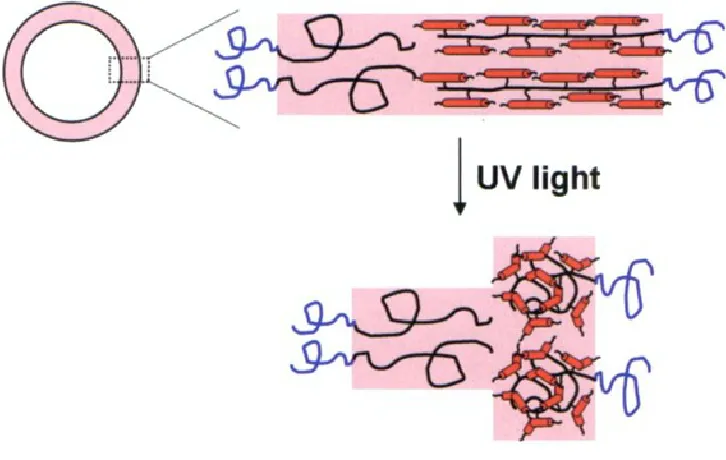
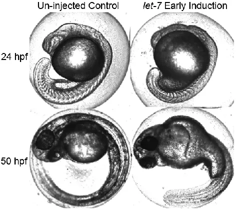
The work in this thesis identifies new photochemical approaches to gain high spatiotemporal control over molecular structure and function, for broad applications in materials and biological science. "Caged" compounds provide a method for temporarily blocking function until acted upon by an external trigger, typically near-UV light. To enable multiplexing studies, three new biomolecular caging strategies were developed that can be activated with various wavelengths of near-UV or visible light. The first method, an oligonucleotide hairpin structure incorporating one or two nitrobenzyl photolinkers, was applied to a miRNA antagomir and used to "turn off " let-7 miRNA in zebrafish embryos with 365 nm light. To achieve
bidirectional control over miRNA, a circular construct was designed for the ability to "turn on" the release of exogenous miRNA into zebrafish embryos with 365 nm light. A second oligonucleotide caging method, using a ruthenium-based photolinker (RuBEP), was designed to extend photoactivation to the visible spectrum, with additional potential for two-photon activation. RuBEP was used to cage antisense morpholinos through circularization via a Cu(I)-mediated [3+2] Huisgen cycloaddition reaction. RuBEP-caged morpholinos were photoactivated to "turn on" antisense activity and successfully knocked down zebrafish chd and ntl genes with 450 nm light, with limited background activity prior to irradiation. A third method of caging was based on encapsulation within photoresponsive nano-polymersomes. Self-assembly of nano-polymersomes was optimized to generate visible-light-responsive vesicles that incorporate a porphyrin dimer in the hydrophobic membrane. These nanovesicles were shown to encapsulate a variety of cargo, including 25mer
oligonucleotides, a small molecule fluorescent dye, and two biologically relevant metal ions, Zn2+ and Ca2+. The photoresponsiveness of the system was modulated with light wavelength, irradiation time, and the presence of dextran in the aqueous core.
Degree Type
Dissertation
Degree Name
Doctor of Philosophy (PhD)
Graduate Group
Chemistry
First Advisor
Ivan J. Dmochowski
Keywords
Light-activated, Oligonucleotides, Photo-activated, Polymersomes, Ruthenium, Zebrafish
Subject Categories
Biochemistry | Chemistry
REGULATING GENE EXPRESSION WITH LIGHT-ACTIVATED OLIGONUCLEOTIDES
Julianne C. Griepenburg
A DISSERTATION in
Chemistry
Presented to the Faculties of the University of Pennsylvania in
Partial Fulfillment of the Requirements for the Degree of Doctor of Philosophy
2014
Supervisor of Dissertation
________________________
Dr. Ivan J. Dmochowski
Associate Professor of Chemistry
Graduate Group Chairperson
________________________
Dr. Gary A. Molander
Hirschmann-Makineni Professor of Chemistry
Dissertation Committee
Dr. David W. Christianson, Roy and Diana Vagelos Professor of Chemistry and Chemical Biology Dr. Barry S. Cooperman, Professor of Chemistry
ii
ACKNOWLEDGMENT
I would first like to thank my advisor, Dr. Ivan Dmochowski, for his continued guidance throughout my graduate career. I truly appreciate the advice and support he has provided. The opportunities I have had in the Dmochowski Lab have helped me grow both scientifically and professionally. I would also like to thank my committee members, Dr. David Christianson, Dr. Barry Cooperman, and Dr. Tobias Baumgart. I have truly appreciated their insight, knowledge, and constructive criticism which has helped to guide me in my research.
iii
I would also like to thank all of the friends that I have made over the years, starting with the great group of friends from my first year of graduate school, Ariane Perez-Gavilan, Genette "G" McGrew, Josh Stecher, and Najat Khan. I wouldn't have survived the early years of graduate school without them. We spent so many days and nights studying, but also had a ton of fun, exploring Philadelphia together and cooking our weekly dinners. I know we will continue to stay in touch as we all move forward with our careers. Another wonderful friend that I was so lucky to meet in graduate school is Anne Wagner. Anne was my go-to for stress relief, whether it was going to lunch, or taking a quick coffee break and talking about dogs! Anne has a very special talent of making everyone around her smile! I'd also like to thank Nimil Sood as both a collaborator and friend. Nimil and I worked so well together on a very difficult project and have become great friends in the process. I will miss our happy hours after long days of experiments! Finally, I'd like to give much credit to my best friend since middle school, Ellen McColl, for always being there for me either for support, advice, or a good laugh!
iv ABSTRACT
REGULATING GENE EXPRESSION WITH LIGHT-ACTIVATED
OLIGONUCLEOTIDES
Julianne C. Griepenburg
Ivan J. Dmochowski
The work in this thesis identifies new photochemical approaches to gain high
spatiotemporal control over molecular structure and function, for broad applications in
materials and biological science. "Caged" compounds provide a method for temporarily
blocking function until acted upon by an external trigger, typically near-UV light. To
enable multiplexing studies, three new biomolecular caging strategies were developed
that can be activated with various wavelengths of near-UV or visible light. The first
method, an oligonucleotide hairpin structure incorporating one or two nitrobenzyl
photolinkers, was applied to a miRNA antagomir and used to “turn off” let-7 miRNA in
zebrafish embryos with 365 nm light. To achieve bidirectional control over miRNA, a
circular construct was designed for the ability to “turn on” the release of exogenous
miRNA into zebrafish embryos with 365 nm light. A second oligonucleotide caging
method, using a ruthenium-based photolinker (RuBEP), was designed to extend
photoactivation to the visible spectrum, with additional potential for two-photon
activation. RuBEP was used to cage antisense morpholinos through circularization via a
Cu(I)-mediated [3+2] Huisgen cycloaddition reaction. RuBEP-caged morpholinos were
v
chd and ntl genes with 450 nm light, with limited background activity prior to irradiation.
A third method of caging was based on encapsulation within photoresponsive
nano-polymersomes. Self-assembly of nano-polymersomes was optimized to generate
visible-light-responsive vesicles that incorporate a porphyrin dimer in the hydrophobic
membrane. These nanovesicles were shown to encapsulate a variety of cargo, including
25mer oligonucleotides, a small molecule fluorescent dye, and two biologically relevant
metal ions, Zn2+ and Ca2+. The photoresponsiveness of the system was modulated with
vi
TABLE OF CONTENTS
ABSTRACT ... ii
LIST OF ILLUSTRATIONS ... xii
CHAPTER 1: INTRODUCTION TO LIGHT-ACTIVATION ... 1
I. "Uncaging" with light ... 2
II. Common caging groups ... 4
III. Light-activated oligonucleotides ... 6
A. Caged hairpin oligonucleotides ... 8
B. Circular caged oligonucleotides ... 10
C. Other designs of caged oligonucleotides ... 11
III. Moving away from ultraviolet light activation ... 20
A. One-photon vs. two-photon activation ... 20
B. Two-photon photolinkers ... 21
C. [Ru(bpy)2XY]n+ ligand dissociation with visible and two-photon light ... 21
D. [Ru(bpy)2(X)2]2+ as a photolinker ... 23
IV. Light-activated polymersomes... 29
vii
B. Light-activated polymersomes ... 30
V. Research Aims ... 37
VI. References... 38
CHAPTER 2: CAGED OLIGONUCLEOTIDES FOR BIDIRECTIONAL PHOTOMODULATION OF LET-7 IN ZEBRAFISH EMBRYOS ... 45
I. Introduction ... 46
II. Experimental procedures ... 49
A. Synthesis, purification, and characterization of light-activated miRNA antagomirs: CHANT1 and CHANT2... 49
B. Synthesis and purification of CIRClet7 ... 50
C. Characterization ... 52
D. In vivo studies ... 53
E. Materials ... 54
III. Results and discussion ... 59
A. Photoactivatible miRNA antagomir: CHANT1 ... 59
B. Photoactivatible miRNA antagomir: CHANT2 ... 61
C. Caged circular miRNA, CIRClet7 ... 72
viii
V. References ... 79
CHAPTER 3: RUTHENIUM-CAGED ANTISENSE MORPHOLINOS FOR REGULATING GENE EXPRESSION IN ZEBRAFISH EMBRYOS... 83
I. Introduction ... 84
II. Experimental procedures ... 87
A. Synthesis of [Ru(bpy)2(3-ethynylpyridine)2](PF6)2 (RuBEP) ... 87
B. Circularization procedure for DNA and morpholino ... 88
C. HPLC purification for N3-DNA, Ru-DNA, N3-DNA-N3, and Ru-cDNA ... 89
D. Gel-shift assay and PAGE analysis ... 90
E. Light Sources ... 90
F. Molecular beacon hybridization assay ... 91
G. Zebrafish microinjection experimental details ... 91
H. Materials ... 92
I. Instrumentation ... 93
III. Results and Discussion ... 101
A. RuBEP ... 101
B. Ru-cDNA ... 103
C. Ru-cMO ... 116
ix
V. References ... 132
CHAPTER 4: CAGING METAL IONS WITH LIGHT-RESPONSIVE NANO-POLYMERSOMES ... 135
I. Introduction ... 136
II. Experimental procedures ... 139
A. Self-assembly of micron-scale polymersomes ... 139
B. Self-assembly of nanoscale polymersomes ... 139
C. Encapsulation and purification ... 140
D. Dynamic light scattering measurements... 141
E. Cryo-TEM measurements ... 142
F. Cargo release from polymersomes ... 143
G. Detection of cargo release from nano-polymersomes ... 143
H. Microinjection into zebrafish embryos... 145
I. Porphyrin dimer (PZn2) wavelength shift determination ... 146
J. Materials ... 147
III. Results and discussion ... 148
A. Thin film self-assembly vs. Direct injection ... 148
B. Effect of mixing, aqueous-to-organic ratio, and polymer concentration on self-assembly of nanoscale polymersomes... 149
x
D. Encapsulation and release of oligonucleotides ... 163
E. FITC loading and release from nanovesicles ... 171
F. Loading and releasing metal ions with photoresponsive nano-polymersomes ... 173
G. Nano-polymersomes in vivo ... 180
IV. Conclusion: ... 185
V. References ... 186
CHAPTER 5: CONCLUSIONS AND FUTURE DIRECTIONS ... 189
I. Conclusions ... 190
II. Future directions ... 192
III. References: ... 193
APPENDIX A. CRYSTAL STRUCTURE DETERMINATION OF RU(BPY)2 (3-ETHYNYLPYRIDINE)2(PF6)2... 194
I. Methods ... 194
xi
LIST OF TABLES
Table 2-1. Gradient for HPLC purification of CHANT1 or CHANT2 after cleavage from
solid support. ... 50
Table 2-2. Gradients for HPLC purification of CIRClet7 A) after solid-phase synthesis and B) after circularization. ... 52
Table 2-3. Masses determined for CHANT1 and CHANT2 by MALDI-TOF MS. ... 53
Table 3-1. Stoichiometries for DNA and MO circularization reactions ... 89
Table 3-2. Gradient used for Ru-DNA and Ru-cDNA HPLC purification ... 91
Table 3-3. Relative fluorescence intensities for molecular beacon targeting ntl-DNA .. 115
Table 3-4. Phenotypic scoring for Ru-cMO-chd in vivo... 129
Table 3-5. Phenotypic scoring for Ru-cMO-ntl in vivo ... 129
Table 3-6. Ru-cMO MALDI data. ... 129
Table 3-7. Oligonucleotide sequences, 5′ to 3′ ... 130
Table A1-1. Summary of structure determination of [RuBEP](PF6)2 ... 198
Table A1-2. Refined positional parameters for [RuBEP](PF6)2 ... 199
Table A1-3. Positional parameters for hydrogens in [RuBEP](PF6)2 ... 202
Table A1-4. Refined thermal parameters (U's) for [RuBEP](PF6)2 ... 205
Table A1-5. Bond distances in [RuBEP](PF6)2, Å ... 208
xii
LIST OF ILLUSTRATIONS
Figure 1-1. Structure of o-nitrobenzyl and derivatives ... 13
Figure 1-2. Structure of MCM ester (coumarin) and derivative, brominated
7-hydroxycoumarin-4-ylmethyl ester (Bhc). ... 14
Figure 1-3. Azobenzene structure ... 15
Figure 1-4. Cartoon representation of A) caged backbone, or photocleavable bases and B)
caged nucleobases. ... 16
Figure 1-5. Structure of antisense morpholino ... 17
Figure 1-6. Cartoon representation of A) caged antisense hairpin and B) circular antisense
oligonucleotide. ... 18
Figure 1-7. Cartoon representation of A) RNA bandage or Photomorph and B)
Photomorph "off to on" antisense oligonucleotide. ... 19
Figure 1-8. General structure of [Ru(bpy)2XY]n+ complexes ... 26
Figure 1-9. Jablonski diagram showing mechanism of photo-triggered ligand dissociation
... 27
Scheme 1-1. General scheme for single ligand exchange of [Ru(bpy)2XY]n+ in H2O ... 28
Figure 1-11. General schematic of a polymersome. ... 34
Figure 1-12. Cartoon of a PEG-b-PBD/PEG-b-PMAzo444 polymersome and response to
UV light. ... 35
Figure 1-13. Nitrobenzyl-linked light-responsive polymersome. ... 36
xiii
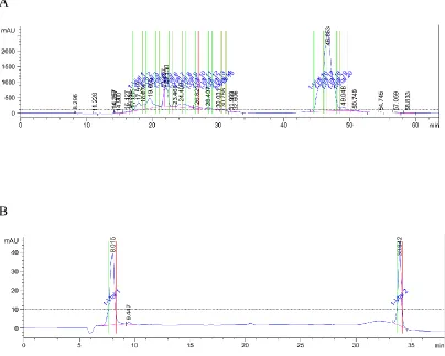
Figure 2-2. HPLC trace for CHANT1. ... 56
Figure 2-3. HPLC traces for purification of CIRClet7. ... 57
Figure 2-4. Masses determined for A) CHANT1 and B) CHANT2 by MALDI-TOF MS. ... 58
Figure 2-5. Structures of caged hairpin antagomirs CHANT1 and CHANT2. ... 66
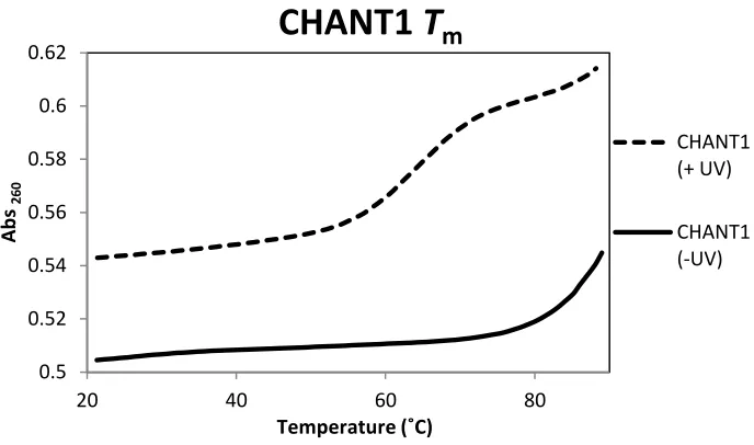
Figure 2-6. Melting temperature data for CHANT 1. ... 67
Figure 2-7. CHANT1 in vivo experiments. ... 68
Figure 2-8. Melting temperature data for CHANT2 ... 69
Figure 2-9. CHANT2 in vivo experiments. ... 70
Figure 2-10. CHANT1 vs. CHANT2 in vivo efficiency. ... 71
Figure 2-11. Synthesis and photocleavage of circular caged miRNA (CIRClet7) ... 74
Figure 2-12. Gel electrophoresis of CIRClet7 crude reaction mixture and CIRClet7 after HPLC purification. ... 75
Figure 2-13. CIRClet7in vivo experiments. ... 76
Figure 2-14. Comparison of control, RNA-induced phenotype, and 2′-OMe RNA-induced phenotype. ... 77
Scheme 3-1. RuBEP conjugation and irradiation ... 95
Scheme 3-2. Synthetic scheme of RuBEP ... 96
Figure 3-1. UV/Vis monitoring product formation... 97
Figure 3-2. Structure of bis-azido morpholino ... 98
xiv
Figure 3-4. HPLC traces for N3-DNA-N3 ... 100
Figure 3-5. Change in UV-Vis spectrum of RuBEP upon 450-nm focal irradiation. ... 107
Figure 3-6. Electrospray mass spectrometry of [RuBEP])PF6)2 +/- light ... 108
Figure 3-7. 1H NMR, pre- and post-photolysis of RuBEP in D2O. ... 109
Figure 3-8. Quantuim yield determination of RuBEP. ... 110
Figure 3-9. Time-course gel of DNA circularization... 111
Figure 3-10. Mono-azide DNA click reaction ... 112
Figure 3-11. 20%, 7 M urea PAGE analysis of Ru-cDNA after HPLC purification ... 113
Figure 3-12. Calibration curve for molecular beacon targeting ntl ... 114
Figure 3-13. Molecular beacon hybridization assay for Ru-cDNA ... 115
Figure 3-14. chd-MO knockdown phenotype ... 121
Figure 3-15. ntl-MO knockdown phenotype ... 122
Figure 3-16. Gel-shift assay showing Ru-cMO formation and photolysis ... 123
Figure 3-17. ntl-MO circularization ... 124
Figure 3-18. Time course gel-shift for Ru-cMO-ntl ... 125
Figure 3-19. Molecular beacon assay showing Ru-cMO-chd caging ... 126
Figure 3-20. in vivo testing of Ru-cMO-chd ... 127
Figure 3-21. Ru-cMO-ntl in vivo data... 128
Figure 4-1. Abs260 measurement of Amicon flow-through for vesicle purification ... 147
Figure 4-2. Effect of stirring vs. sonicating during nanovesicle self-assembly ... 152
xv
Figure 4-4. Effect of vortex time on nano-vesicle morphology... 154
Figure 4-5. Effect of organic to aqueous ratio on nano-vesicle size ... 155
Figure 4-6. Effect of organic to aqueous ratio on vesicle morphology ... 156
Figure 4-7. Effect of polymer concentration on nanovesicle size ... 157
Figure 4-8. DLS and cryo-TEM showing final conditions for nanovesicle self-assembly ... 158
Figure 4-9. Structure of meso-to-meso ethyne-bridged (porphinato)zinc(II) dimer (PZn2) ... 161
Figure 4-10. Membrane deformation detected by PZn2 emission blue-shift. ... 162
Figure 4-11. Encapsulation of fluorescein-labeled MO in micron vesicles... 166
Figure 4-12. Dual encapsulation of Alexa 488-DNA and PZn2 in micron vesicles ... 167
Figure 4-13. Unloaded micron vesicles ... 168
Figure 4-14. Fluorescence intensity of fl-MO as a function of irradiation time ... 169
Figure 4-15. Release curve for fl-MO loaded nanovesicles ... 170
Figure 4-16. Release curve of FITC-loaded polymersomes ... 175
Figure 4-17. FITC release from nano-vesicles without PZn2 or dextran ... 176
Figure 4-18. Morphological change in polymersomes after light irradiation. ... 177
Figure 4-19: Hydrodynamic diameter of metal ion-loaded nanovesicles determined by DLS. ... 178
Figure 4-20. Release curves of metal ion-loaded polymersomes. ... 179
xvi
Figure 4-22. Cell uptake of nano-polymersomes ... 183
Figure 4-23. PZn2 wavelength shift in vivo ... 184
1
Chapter 1
2
I. "Uncaging" with light
The understanding of complex biological systems is advanced by tools that can
help manipulate structure, function, and or localization of molecules with high spatial and
temporal resolution. One strategy is to put an active compound under the control of a
conditional trigger. This concept of blocking a compound's biological activity until acted
on by an internal or external stimulus was termed "caging" in 1978 by Kaplan et. al. with
the photolytic release of adenosine 5'-triphosphate.1 Light-activation dates back to 1943,
however, with the example of azobenzene modified amino acids.2 Although the term
"caged" is now widely used in biochemistry, it is somewhat of a misnomer because most
caging strategies involve the use of one or more small photoactive moieties rather than
true molecular confinement.
Light in the UV to near IR (NIR) window is a commonly used external stimulus
for the activation of caged molecules as it can be very easily manipulated both temporally
and spatially. Spatial control depends on the irradiation source and activation wavelength,
but generally, confocal laser scanning microscopy (CLSM) and two-photon microscopy
provide excellent and well established spatial resolution with the ability to irradiate and
image simultaneously. Two-photon microscopy achieves high spatial resolution, with the
ability to control light in three dimensions down to the sub-cellular level and femtoliter
volumes.3,4 Although UV light has been commonly used for uncaging due to the broad
availability of UV-active caging groups5, uncaging with longer wavelengths of light has
3
as scattering has a λ-4 dependence on wavelength. Visible light is far less damaging to
cells than UV light6, and has the potential to penetrate biological systems up to 1 cm.7
This makes visible light activation feasible for small biological model systems and cells.
However, when transitioning to larger biological systems, significantly higher depth
penetration is necessary. The NIR window (700 - 900 nm) has a very low absorption
coefficient in biological tissue, and combined with low levels of scattering, NIR light has
the potential to penetrate tissue to depths of several centimeters.7 Above 900 nm, this is
hindered by strong water absorption.
It is important to expand the currently available caging toolkit to include a
broader range of biologically active molecules that can be controlled light. Specifically,
there is a need to be able to cage molecules ranging from small metal ions to large
proteins and plasmids. Additionally, it is of equal importance to expand the wavelengths
that can be used for uncaging. Several examples exist in the literature for caging moieties
that can be activated with UV light, but fewer exist for longer wavelength light in the
visible and near-IR region. Ideally, the future will hold a vast library of caged
biomolecules that can be activated with high spatiotemporal resolution at different
wavelengths of light spanning the UV, visible and near-IR spectrum.
This introduction will focus on literature precedent for three types of caging
relevant to the work presented in this thesis: 1) The commonly used UV-active moieties
and their applications in caging short oligonucleotides, 2) Visible and two-photon light
4
II. Common caging groups
Three photoresponsive caging groups that have been commonly used for
biological applications are nitrobenzyl, coumarin, and azobenzene moieties, shown in
Figures 1-1, 1-2, and 1-3. These caging groups and their derivatives have received
attention for their ability to be incorporated site-specifically into biomolecules to control
structure and function with UV light.
The most commonly used caging group8, and the caging group used most
frequently in our laboratory is the nitrobenzyl group, shown in Figure 1-1. The
ortho-nitrobenzyl has been widely used as a synthetic protecting group since initial reports in
1970.9,10 Ortho-nitrobenzyl is photoactive at 365 nm, but the wavelength can be tuned by
adding substituents. Common derivatives of 2-nitrobenzyl are the 4,5-dimethoxy analog
(DMNB), 1-(2-nitrophenyl)ethyl (NPE) and 4,5-dimethoxy analog (DMNPE) and
α-carboxy analog (CNB) which range in activation wavelength from 260 nm to 365 nm.11
These analogs provide benefits and drawbacks, for example, CNB has increased
hydrophilicity due to the carboxy groups, but activates at shorter wavelengths (maximum
at 260 nm).12 DMNPE and DMNB activate at longer wavelengths (maximum at 355
nm), but photolysis rates and quantum yields are typically much lower than for CNB.
Recently, styryl-2-nitrobenzyl (SNB) moieties were presented to extend conjugation and
red-shift activation wavelengths to 370 nm, allowing tail-end activation up to 450 nm.13
The nitrobenzyl moiety has become so widely used for photocontrol over
5
solid-phase synthesis,14,15 and is now commercially available through Glen Research
(Sterling, VA).
Initial literature on coumarin protecting groups dates back to 1984 with the report
on 7-methoxycoumarin-4-ylmethyl (MCM) esters by Givens et al.16 Irradiation of
coumarin results in the release of the protected carboxylic acid and formation of the
corresponding hydroxymethyl coumarin. Typical MCM esters have activation
wavelengths in the UV (340 - 360 nm) range, but the spectral range of irradiation can be
altered with the addition of substituents, which can also change the hydrophilicity and
quantum yield of photolysis. Brominated 7-hydroxycoumarin-4-ylmethyl esters (Bhc) is a
coumarin derivative reported by Furuta et al. that has been modified to push the
activation wavelength to 365 nm. Additionally, the extended conjugation and high
extinction coefficient for π-π* transitions makes Bhc two-photon active with a cross
section of 1 GM.17 This two-photon active coumarin has been applied to the design and
synthesis of a caged glutamate and used to resolve three-dimensional maps of neuron
glutamate sensitivity in intact mouse brain slices.17 Structures of coumarin (MCM ester)
and derivative Bhc are shown in Figure 1-2. Recently, coumarin derivatives have been
synthesized for photoactivation with visible light. Variations of
(coumarin-4-yl)methoxycarbonyl (CMOC) have been shown to light-activate in the visible region, and
by adding carboxylate substituents, these chromophores can be made more water
6
Azobenzene is another popular choice as a photoactive moiety that has a different
light-activated mechanism than other caging groups. Azobenzene undergoes trans-cis
isomerization upon irradiation with UV light, resulting in a structural change instead of
photocleavage. Unlike the previously presented groups, nitrobenzyl and coumarin,
azobenzene has the ability to "photoswitch" and reverse back to trans, initiated either
thermally or upon irradiation with visible light. Many derivatives have been made to
adjust the wavelength, quantum yield, and to prevent the thermally activated
isomerization back to the trans configuration to have better control over the system with
light.20 Several examples in the literature have used azobenzene, from the reversible
photocontrol of oligonucleotide duplex formation,21-23 to the incorporation of azobenzene
derivatives in diblock copolymers for materials science applications.24-26 The reversible
isomerization of azobenzene is shown in Figure 1-3.
III. Light-activated oligonucleotides
Caging a wide variety of small and large biologically active molecules has been a
popular area of research and has had much success in recent years. Examples of caging
can be found for molecules including metal ions,27 peptides,28 oligonucleotides,29
proteins,30 and plasmids.31,32 This review will focus on literature pertaining to caging
short oligonucleotides that control gene expression, primarily antisense oligonucleotides.
Over the past two decades, several methods for controlling oligonucleotide
7
their complexity and large size, and thus, many different methods have been explored.
Initial efforts towards light-activated oligonucleotides were made in 1995 by
Ordoukhanian et. al., with the design of photoresponsive DNA "building blocks" which
could be site-specifically incorporated into a short oligonucleotide synthesis, and
destabilize hybridization upon photolysis with 355 nm light.33 Another example of using
photoresponsive nucleobases to induce DNA strand breaks was presented in 2002 by
Dussy et. al.34 More commonly, strategies have been developed that involve the use of
caging groups on the nucleosides to disrupt Watson-Crick base pairing and destabilize
duplex formation until irradiation.35,36 This strategy has been used widely for a variety of
applications including siRNA,37-39 peptide nucleic acids,40,41 caged fluorescent
oligonucleotides,42 antisense oligonucleotides,43-45 and miRNA.43,46 A cartoon
representation of these designs is represented in Figure 1-4 A-B.
Although caged nucleobases have proven effective towards light-activating
hybridization based functions of oligonucleotides, drawbacks of this approach include the
need for unique nucleobase monomers, which makes it difficult to design a generalizable
method for caging. Another drawback to this method is that typically multiple caging
groups are necessary to effectively disrupt hybridization which increases the light dose
necessary for uncaging. This can be especially problematic since many of these designs
use UV-active moieties, and high doses of UV irradiation can be damaging in biological
systems. For these reasons, our lab and others have moved towards caged hairpin and
8
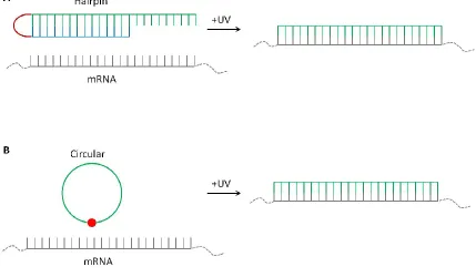
A. Caged hairpin oligonucleotides
Caged hairpins consist of a biologically active strand and a shorter
complementary strand, covalently attached with one or more photocleavable linkers.
Through hybridization of a shorter blocking strand, the biologically active strand is
rendered inactive. Covalent attachment of the two complementary sequences achieves a
higher effective concentration, resulting in a higher thermal stability for the duplex.
While covalently held in the hairpin structure, the biologically active strand is unable to
bind to its target. Upon irradiation with UV light, the photolinker is cleaved which
destabilizes the duplex. This results in duplex dissociation, and more favorable
hybridization to the full length target. A schematic representation of a caged hairpin is
shown in Figure 1-6A.
Our lab has designed and synthesized various caged hairpins, for RNase
H-mediated mRNA digestion, antisense oligonucleotides, and most recently, for harvesting
mRNA from single cells.47 Additional examples for caged hairpins have been presented
by the Chen lab.48-50 One of the first examples published by Tang and Dmochowski
reported a caged antisense DNA hairpin.51 This hairpin blocked function of a 20-mer
DNA when covalently attached to a blocking strand via a nitrobenzyl-based
heterobifunctional photocleavable linker. Upon photocleavage with 365 nm light,
antisense DNA was revealed to bind to target mRNA and recruit RNase H for mRNA
degradation. This design was applied to target c-myb, a hematopoietic transcription factor
9
antisense negatively charged peptide nucleic acid.53 This caged antisense hairpin was
used to photomodulate gene expression in zebrafish embryos for two early developmental
genes, chordin and bozozok.
Additional efforts have been made towards caging antisense morpholinos, which
are currently the gold standard in achieving gene knockdown in many model
developmental organisms, including sea urchin, ascidian, zebrafish, frog, chick, and
mouse.54 Morpholinos (Figure 1-5) have been demonstrated to have high nuclease
resistance due to their highly modified backbone. Initial reports of caging morpholinos by
Shestopalov et al. involved a hairpin structure, where the morpholino, inhibitor sequence
and dimethoxynitrobenzyl moiety were linked through a Cu(I)-catalyzed Huisgen
1,3-dipolar cycloaddition.48 Additional features of this design included 3′ fluorescein for
construct visualization in vivo. Successful photomodulation of zebrafish notail was
achieved with this design. Following up on this design, Shestopalov et al. reported an
additional caged morpholino with a simplified synthesis.49,50 A DMNB-based
bifunctional linker was used to conjugate the antisense morpholino and its
complementary inhibitor strand in three steps, starting with commercially available
morpholinos. Generalizable methods pertaining to inhibitor placement and length
optimization were also explored. Additionally, a bromohydroxyquinoline (BHQ)-based
10
B. Circular caged oligonucleotides
More recently, caging of short oligonucleotides has been achieved through
circularization.55-59 By covalently attaching the 5′ and 3′ ends of the oligonucleotide with
a photocleavable linker so that it forms a circular structure, the oligonucleotide is
structurally restricted from hybridizing to a complementary target. A cartoon
representation of this design is shown in Figure 1-6. This design has many benefits that
arise from the lack of a blocking sequence. Primarily, the circularization scheme is
sequence independent. Additionally, there is no risk of the blocking strand having
biological activity and off-target effects after photolysis and dissociation. As there is only
one photocleavable linker in most circular designs, this significantly lowers the light dose
necessary for uncaging.
Initial reports of circular oligonucleotides were by Richards et al. where a
photolabile circular DNAzyme was enzymatically synthesized using T4 ligase and
photomodulation of RNA digestion was achieved.58 Tang et al. presented the first
example of photomodulating RNA digestion by RNase H through the synthesis of
light-activated circular DNA antisense oligonucleotides.57 The first example of a caged circular
morpholino was reported by Yamazoe et al.,55 shortly followed by Wang et al. who also
caged a 25-mer morpholino by linking the two ends in a circular structure with a
nitrobenzyl photocleavable moiety.56 These caged cMOs were successfully used to
11
Yamazoe et al. compared background activity between circular and hairpin structures
and found significantly less background activity in vivo with circular constructs.
C. Other designs of caged oligonucleotides
Another method for caging oligonucletides reported by the Dmochowski lab is an
"RNA bandage" design that provides a method of blocking mRNA translation and
restoring it upon irradiation with UV light.60 These constructs perform opposite of the
previously described caged hairpins and circular caged oligonucleotides, as they can
regulate gene expression from "off to on" as opposed to "on to off". A schematic
representation is shown in Figure 1-7A. Bandages were designed and synthesized using
2′-OMe RNA antisense oligonucleotides and linked by a nitrobenzyl-based
photocleavable moiety that activates with 365 nm irradiation. These RNA bandages were
demonstrated to be successful at photomodulating in vitro translation.
A similar design strategy was implemented by Tallafuss et al. to cage an antisense
morpholino.61 In this design, two shorter blocking sequences were linked with a
nitrobenzyl photocleavable moiety and hybridized to a 25-mer target morpholino for
turning genes from "off to on" with light (Figure 1-7A). Additionally, this design can be
used for "on to off" gene photomodulation by linking two shorter MO sequences together
with a nitrobenzyl moiety to form a full length active morpholino that is able to bind to
12
are liberated, resulting in dissociation from the mRNA. This technology is commercially
13
14
15
Figure 1-3. Azobenzene structure
16
Figure 1-4. Cartoon representation of A) caged backbone, or photocleavable bases and B) caged nucleobases.
17
18
Figure 1-6. Cartoon representation of A) caged antisense hairpin and B) circular antisense oligonucleotide.
19
Figure 1-7. Cartoon representation of A) RNA bandage or Photomorph and B) Photomorph "off to on" antisense oligonucleotide.
20
III. Moving away from ultraviolet light activation
A. One-photon vs. two-photon activation
The primary goal of caged compounds is the ability to control biological
processes with light with high spatiotemporal resolution. Although much progress has
been made towards this goal through the use of 1-photon active moieties such as the UV
active o-nitrobenzyl group and derivatives, significant improvements can be achieved
with the implementation of two-photon photolinkers. The use of 2-photon activation in
caged compounds can provide even greater spatial resolution in three dimensions through
the addition of depth control.62
Two-photon photolysis replaces the absorption of one photon with two
longer-wavelength, lower-energy photons, typically in the NIR region, of equivalent total
energy. The simultaneous absorption of these photons is governed by the light intensity
and the two-photon absorption cross section, measured in GM (Göppert-Mayer). A GM is
equal to 10-50cm4 s photon-1, therefore, confining the uncaging event to a very small
region of focus with negligible out of focus irradiation.63 This allows for uncaging in
very small regions of interest and volumes as small as 1 femtoliter.4 Additional benefits
of NIR light used for two-photon uncaging is significantly less scattering than shorter
21
B. Two-photon photolinkers
Nitrobenzyl and coumarin derivatives have been shown to be two-photon active,
however their two-photon absorption cross sections are very low, in the 0.01 - 0.1 GM
range.65 Although NIR light is significantly less damaging than UV light, high laser
powers required to activate compounds with such low two-photon cross sections result in
heating and toxicity due to water strongly absorbing 700-900 nm light.66 For this reason,
it is necessary to develop two-photon caging groups with significantly higher
cross-sections (3-30 GM).63
There are few applications that have been demonstrated for two-photon caged
biomolecules, likely due to the limited availability of two-photon caging groups. Some
examples of two-photon caged compounds using coumarin derivatives are caged calcium,
azid-1,67 and caged glutamate.68 Although successful two-photon uncaging has been
achieved with these caged compounds,69,70 cross-sections were measured to be below the
3 - 30 GM target level, therefore requiring high levels of laser power.4
C. [Ru(bpy)2XY]n+ ligand dissociation with visible and two-photon light Octahedral Ru(II) polypyridyl complexes have been shown to be a promising
method of caging molecules for release with visible and two-photon excitation.
Specifically, complexes of the type [Ru(bpy)2XY]n+, a Ru(II) center with two bipyridines
and two monodentate ligands (Figure 1-8), undergo photochemistry resulting in the
22
were published by Dwyer et al.,72 with in-depth photochemical studies followed two
decades later.73 Photochemical reactions typically follow equation (1) and sometimes also
equation (2), where X and Y are monodentate ligands, and S is solvent.
A general mechanism of photorelease is demonstrated through the Jablonski
diagram in Figure 1-9. Upon irradiation, absorption into the MLCT (typically 400-500
nm) populates the single 1MLCT band. Intersystem crossing (ISC) occurs, populating the
triplet 3MLCT state. This process occurs very rapidly, on the order of 40 fs measured for
Ru(bpy)32+.74 Decay can occur through multiple pathways, non-radiative (nr), radiative
(rad), and in the case of [Ru(bpy)2XY]n+, cross-over to the ligand field (3LF) state where
ligand dissociation occurs.75 Population of the ligand field state is thermally activated,
and directly correlates with the energy gap between 3MLCT and 3LF. At a given
temperature, a higher energy 1MLCT results in a higher energy 3MLCT band and thus, a
smaller gap between 3MLCT and 3LF. This results in a blue-shifted MLCT ground-state
absorption band and therefore, a higher yield of photosubstitution (i.e., a higher quantum
yield). In contrast, a larger gap between the 3MLCT and 3LF will result in lower quantum
yields of photosubstitution.75
h
23
D. [Ru(bpy)2(X)2]2+ as a photolinker
The tunable photophysical properties of [Ru(bpy)2XY]n+ make these complexes
very attractive for caging applications. The MLCT in the visible region is beneficial for
use in biological systems, as visible light is far less damaging to cells, and also penetrates
tissue samples deeper due to the longer wavelength.71 Additionally, as shown in Scheme
1-1, the mechanism of uncaging is through a ligand-exchange process instead of a
photo-cleavage process which allows for very clean and efficient whole-molecule uncaging.
Many examples of whole-molecule caging with these ruthenium complexes have been
presented in the literature.
The first example of ruthenium caging of a biologically relevant molecule was
presented by Zayat et. al. in 2003 with the caging of 4-aminopyridine.76 In this example,
4-aminopyridine (4-AP), a neurocompound that blocks K+ channels,77 was cooordinated
directly to Ru(bpy)2Cl2 through the amine. The resulting water-soluble compound,
[Ru(bpy)2(4-AP)2]Cl2 had a MLCT centered at 489 nm and underwent successful
uncaging, as confirmed by a free 4-AP ligand seen by 1H NMR spectroscopy.
Experiments using [Ru(bpy)2(4-AP)2]Cl2 showed successful uncaging and neuronal
stimulation in a leech ganglion, with no toxicity observed. Additional compounds were
designed by the Etchenique lab using similar strategies, including a cholinergic agonist
nicotine, [Ru(bpy)2(Nic)2]2+,78 and a caged γ-aminobutyric acid (GABA).79 These caged
24
The octahedral ruthenium center on [Ru(bpy)2XY]n+ provides flexibility in
coordination chemistry. The choice of ligand depends primarily on the desired
photophysical characteristics, such as wavelength and quantum yield, providing excellent
potential for designing photolinkers. Selecting ligands that are much weaker σ-donors
will result in an electronically depleted Ru(II) center, thus, shifting the activation to
higher energy (shorter wavelengths). This blue-shifting results in a significantly higher
quantum yield.80 Another consideration is whether or not one or both monodentate
ligands will dissociate. This also depends on the electronics of the ligands. For example,
if X and Y are different ligands, substitution will occur at the ligand that is a weaker
σ-donor. In cases where the monodentate ligands are identical, the substitution will proceed
on one ligand to form the mono-aquo product, unless the ligand is a weaker σ-donor than
the solvent, in which case both ligands will be exchanged.71
Another benefit of using [Ru(bpy)2XY]n+ compounds as photolinkers is their
demonstrated two-photon activation. Salierno et al. presented a caged glutamate,
Ru(bpy)2(PMe3)(Glu), which could be efficiently uncaged with one-photon (450 nm) or
two-photon (800 nm) light.81 The two-photon cross section for this caged glutamate was
determined to be 0.14 GM. Additional examples of two-photon activation of
[Ru(bpy)2XY]n+ compounds include the two-photon uncaging of a caged dopamine,
[Ru(bpy)2(PMe3)(Dopa)](PF6)2 which has an even higher two-photon cross-section of
0.24 GM.82 These two-photon cross sections are significantly higher than what has been
25
potential for tuning the Ru ligands to achieve the desired 3-30 GM target. Compounds of
the type [Ru(bpy)2XY]n+ are promising for developing a library of photoactive moieties,
26
27
28
29
IV. Light-activated polymersomes
A. Introduction to polymersomes
Polymersomes are a class of synthetic vesicles that self-assemble from
amphiphilic diblock copolymers.83 Polymersomes have attracted much attention since
their initial discovery by Hammer, Discher, and Eisenberg a little over a decade ago84,85
due to their robustness, tunability, ability to mimic biological membranes and the ability
to encapsulate a broad variety of molecules. Polymersomes are composed of two
components, a large hollow aqueous core, and a thick hydrophobic membrane which
separates the aqueous core from the outer medium. These two structural components
provide versatility in encapsulation possibilities, from hydrophobic drugs (ie, paclitaxel,
doxorubicin, quantum dots) encapsulated in the membrane, to small molecules and large
biomolecules (i.e., siRNA, DNA, plasmids, proteins, enzymes) encapsulated in the
aqueous core.
Polymersomes have many benefits over liposomes as carrier systems, namely,
they are fully synthetic which provides the possibility to tune many characteristics such
as membrane thickness, vesicle size, and vesicle composition. Since polymersomes are
comprised of high molecular weight polymers as opposed to small phospholipids like
liposomes, membrane thickness typically spans from 3-30 nm in comparison to 3-5 nm.83
These high molecular weights also offer benefits such as decreased membrane
30
shown to have significantly longer circulation times in vivo than unmodified liposomes.86
Circulation time of liposomes is typically on the order of hours,87 but can be increased by
functionalizing the outer surface with poly(ethylene glycol) (PEG), known as "stealth
liposomes".88 In contrast, polymersomes have intrinsically long circulation times due to
their composition.
Polymersomes can generally be prepared by two different methods,
phase-inversion, and polymer rehydration. The phase-inversion technique involves dissolving
the diblock copolymer in an organic solvent, followed by hydration with aqueous solvent.
Typically, this process yields fairly uniform nanovesicles that can be tuned through
polymer concentration and organic-to-water ratio.83 This process results in self-assembly
by increasing the interfacial tension between the hydrophobic polymer blocks and the
hydration solution. The polymer rehydration method involves dissolving the diblock
copolymer in organic solvent and forming a thin film by evaporation of the organic
solvent. Self-assembly is promoted by hydration with water. With the polymer
rehydration technique, vesicles are formed as the water permeates the polymer film
through defects, causing the polymer to lift from the surface. Polymer rehydration
typically yields larger vesicles that can be made smaller and more uniform through
extrusion.89
B. Light-activated polymersomes
Much attention has been dedicated to designing "smart" polymersomes, or
31
internal or external trigger such as degradation by hydrolysis, temperature, pH, magnetic
fields, or light.90 This review will focus on examples of polymersomes that undergo
membrane disruptions in response to light. Light-responsive polymersomes are an
attractive method of controlled release because light can be easily externally manipulated
for release of cargo with high spatial and temporal resolution.
One such system by Mabrouk et al. used an azobenzene group incorporated
within the diblock copolymer to promote rapid membrane disruptions in response to UV
light.91 Asymmetric micron-sized polymersomes were assembled through two block
copolymers, an inert copolymer, polyethyleneglycol-polybutadiene (PEG-b-PBD), and a
liquid crystal based copolymer, PEG-b-PMAazo444 (PAzo). The liquid crystal block
contained an azobenzene group which underwent a trans-to-cis configurational transition
in response to UV light. The isomerization caused a conformational change in the block
from a rod to a coil, which induced an area difference between the two polymer
monolayers sufficient to trigger membrane rupture.
Another example of a light-responsive micron-sized polymersome system was
presented by Robbins et al.92 This system was the first example of a photoactive
polymersome formed by incorporating a protein in the aqueous interior and a
meso-to-meso ethyne-bridged bis[(porphinato)zinc] (PZn2) chromophore in the membrane.
Micron-size vesicles were self-assembled using a polyethylene oxide-polybutadiene
(PEO30-PBD46, denoted OB29) diblock copolymer. Two different proteins, horse spleen
32
core. Incorporation of the PZn2 chromophore into the vesicle's hydrophobic membrane
allowed for light absorption of near-UV to near-IR wavelengths. Irradiation with 488,
543, and 633 nm induced irreversible membrane deformities ranging from "budding" to
complete rupture. It was hypothesized that the protein associated with the inner
membrane, causing asymmetrical membrane deformation upon PZn2 energy dissipation
as heat, which ultimately resulted in membrane rupture. A small molecule, biocytin, was
encapsulated within the core and 25-50% release was demonstrated upon light exposure.
The work by Robbins et al.92 was further explored by Kamat et al.93 and
subsequently resulted in a general method for producing photoresponsive micron-sized
vesicles with chromophore PZn2 in the hydrophobic membrane. Following up on the
work by Robbins et al., it was hypothesized that any luminal solute that associates with
the inner leaflet of the membrane can induce polymersome rupture when combined with
PZn2 in the membrane. Dextran was investigated as a luminal encapsulant due to its
biocompatibility and aqueous solubility. It was shown that inclusion of dextran could
induce membrane instability upon irradiation. Studies were performed varying the size of
the dextran and molecular weight of the polymer, and it was shown that
photoresponsiveness increased with molecular weight and dextran concentration.
Additional work by Kamat et al. explored this system for use as a membrane stress
sensor.94 cIt was demonstrated that PZnn fluorophores underwent significant red emission
33
To date, only one example exists of a photoresponsive nanoscale polymersome. It
is important to develop light-responsive vesicles on the nano-scale for biological
applications, as micron vesicles are not sized appropriately for in vivo experiments.
Mammalian cells, such as HeLa cells, are typically 10-20 µM in diameter, therefore it is
necessary to develop much smaller carrier systems for these applications. Additionally,
passive uptake of polymer-based nanoparticles by red blood cells has been shown to
occur for particles less than 200 nm.95
Cabane et al. presented a photocleavable amphiphilic diblock copolymer,
poly(methyl caprolactone)-ONB-poly(acrylic acid) (PMCL-ONB-PAA) that
self-assembled into micelles and 150 nm polymersomes.96 qcThis block copolymer contained
an o-nitrobenzyl moiety as a photocleavable linker between the hydrophilic and
hydrophobic polymer chains. UV-irradiation induced a successful cleavage of the diblock
copolymer chains, both in THF and aqueous solution, as well as in self-assembled
vesicles. This work was further extended to probe the ability of this system to encapsulate
and release two small molecules, as well as a large biomolecule, green fluorescent
protein.97 Irradiation with 365 nm light induced polymersome disintegration within
minutes. Upon irradiation, cargo was released and polymersome morphology was shown
by cryo-TEM to transition from vesicles to micelles. This nanoscale system provides a
promising route to deliver cargo in vivo with UV light, and can be tuned for delivery
34
Figure 1-11. General schematic of a polymersome.
35
Figure 1-12. Cartoon of a PEG-b-PBD/PEG-b-PMAzo444 polymersome and response to UV light.
Under UV illumination, isomerization of the azobenzene induces a conformational change of the diblock copolymer, resulting in vesicle rupture.
36
Figure 1-13. Nitrobenzyl-linked light-responsive polymersome.
Chemical structure of the poly(methyl caprolactone)-ONB-poly(acrylic acid) diblock copolymer and degradation products upon UV irradiation are shown.
37
V. Research Aims
Work presented in this thesis demonstrates three unique designs for biomolecular
caging, all responsive to different wavelengths of light. The first two designs involve the
site-specific incorporation of a caging moiety, and the third design involves encapsulation
within a light-activated polymersome nanocarrier. Specifically, Chapter 2 presents a
caged antagomir for the regulation of let-7 miRNA, in zebrafish embryos. This caged
antagomir provides the ability to block the activity of a miRNA in vivo upon
photoactivation with 365 nm light. Additionally, Chapter 2 presents a caged miRNA,
providing a method of introducing an exogenous miRNA into zebrafish embryos and
activating it upon irradiation. Used together, these two constructs provide the ability for
bidirectional control of a miRNA. Chapter 3 focuses on the design, synthesis, and
application of a ruthenium-based visible-light photolinker, RuBEP. Two antisense
morpholinos were circularized with RuBEP through click-chemistry, and activated in
zebrafish embryos with 450-nm light. Chapter 3 demonstrates an encapsulation-based
approach for caging through the use of photo-responsive nano-polymersomes. These
nano-polymersomes were used to encapsulate oligonucleotides, small molecules (FITC),
38
VI. References
(1) Kaplan, J. H.; Forbush, B.; Hoffman, J. F. Biochemistry1978, 17, 1929-1935. (2) Karrer, P.; Keller, P.; Szönyi, G. Helv. Chim. Acta1943, 26, 38-50.
(3) Denk, W.; Strickler, J. H.; Webb, W. W. Science1990, 248, 73-76.
(4) Brown, E. B.; Shear, J. B.; Adams, S. R.; Tsien, R. Y.; Webb, W. W. Biophys. J.
1999, 76, 489-499.
(5) Mayer, G.; Heckel, A. Angew. Chem., Int. Ed. Engl.2006, 45, 4900-4921.
(6) Sinha, R. P.; Hader, D.-P. Photochem. Photobiol. Sci.2002, 1, 225-236.
(7) Weissleder, R. Nat. Biotech.2001, 19, 316-317.
(8) Shao, Q.; Xing, B. Chem. Soc. Rev.2010, 39, 2835-2846.
(9) Barltrop, J. A.; Plant, P. J.; Schofield, P. Chem. Commun. (London) 1966, 822-823.
(10) Patchornik, A.; Amit, B.; Woodward, R. B. J. Am. Chem. Soc. 1970, 92, 6333-6335.
(11) Corrie, J. E. T.; Furuta, T.; Givens, R.; Yousef, A. L.; Goeldner, M. In Dynamic Studies in Biology; Wiley-VCH Verlag GmbH & Co. KGaA: 2005, p 1-94.
(12) Schaper, K.; Madani Mobarekeh, S. A.; Doro, P.; Maydt, D. Photochem. Photobiol.2010, 86, 1247-1254.
(13) Bao, C.; Jin, M.; Li, B.; Xu, Y.; Jin, J.; Zhu, L. Org. Biomolec. Chem.2012, 10, 5238-5244.
(14) Olejnik, J.; Krzymanska-Olejnik, E.; Rothschild, K. J. Nucleic Acids Res. 1996,
39
(15) Olejnik, J.; Krzymanska-Olejnik, E.; Rothschild, K. J. Nucleic Acids Res. 1998,
26, 3572-3576.
(16) Givens, R. S.; Matuszewski, B.; Athey, P. S.; Stoner, M. R. J. Am. Chem. Soc.1990, 112, 6016-6021.
(17) Furuta, T.; Wang, S. S.-H.; Dantzker, J. L.; Dore, T. M.; Bybee, W. J.; Callaway, E. M.; Denk, W.; Tsien, R. Y. Proc. Natl. Acad. Sci. U. S. A. 1999, 96, 1193-1200.
(18) Kotzur, N.; Briand, B.; Beyermann, M.; Hagen, V. J. Am. Chem. Soc. 2009, 131, 16927-16931.
(19) Yamazoe, S.; Liu, Q.; McQuade, L. E.; Deiters, A.; Chen, J. K. Angew. Chem., Int. Ed. Engl.2014, n/a-n/a.
(20) Bandara, H. M. D.; Burdette, S. C. Chem. Soc. Rev.2012, 41, 1809-1825.
(21) Asanuma, H.; Liang, X.; Yoshida, T.; Komiyama, M. ChemBioChem2001, 2, 39-44.
(22) Asanuma, H.; Matsunaga, D.; Liu, M.; Liang, X.; Jhao, J.; Komiyama, M. Nucleic Acids Res.2003, 117-118.
(23) Liang, X.; Yoshida, T.; Asanuma, H.; Komiyama, M. Nucleic Acids Symp. Ser.
2000, 277-278.
(24) del Barrio, J.; Oriol, L.; Sanchez, C.; Serrano, J. L.; Di Cicco, A.; Keller, P.; Li, M. H. J. Am. Chem. Soc.2010, 132, 3762-3769.
(25) Mabrouk, E.; Cuvelier, D.; Brochard-Wyart, F.; Nassoy, P.; Li, M.-H. Proc. Natl. Acad. Sci. U. S. A.2009, 106, 7294-7298.
(26) Li, M.-H.; Auroy, P.; Keller, P. Liq. Cryst.2000, 27, 1497-1502. (27) Ellis-Davies, G. C. R. Chem. Rev.2008, 108, 1603-1613.
40
(29) Tang, X.; Dmochowski, I. J. Molecular BioSyst.2007, 3, 100-110.
(30) Riggsbee, C. W.; Deiters, A. Trends Biotechnol., 28, 468-475.
(31) Monroe, W. T.; McQuain, M. M.; Chang, M. S.; Alexander, J. S.; Haselton, F. R.
J. Biol. Chem.1999, 274, 20895-20900.
(32) Ando, H.; Furuta, T.; Tsien, R. Y.; Okamoto, H. Nat. Genet.2001, 28, 317-325.
(33) Ordoukhanian, P.; Taylor, J.-S. J. Am. Chem. Soc.1995, 117, 9570-9571. (34) Dussy, A.; Meyer, C.; Quennet, E.; Bickle, T. A.; Giese, B.; Marx, A.
ChemBioChem2002, 3, 54-60.
(35) Heckel, A. In Curr. Protoc. Nucleic Acid Chem.; John Wiley & Sons, Inc.: 2001.
(36) Buff, M.; Mack, T.; Heckel, A. CHIMIA2009, 63, 261-264. (37) Mikat, V.; Heckel, A. RNA2007, 13, 2341-2347.
(38) Govan, J. M.; Young, D. D.; Lusic, H.; Liu, Q.; Lively, M. O.; Deiters, A. Nucleic Acids Res.2013, 41, 10518-10528.
(39) Wu, L.; Pei, F.; Zhang, J.; Wu, J.; Feng, M.; Wang, Y.; Jin, H.; Zhang, L.; Tang, X. Chemistry2014.
(40) Guha, S.; Graf, J.; Goricke, B.; Diederichsen, U. J. Pept. Sci.2013, 19, 415-422. (41) Watanabe, T.; Hoshida, T.; Sakyo, J.; Kishi, M.; Tanabe, S.; Matsuura, J.;
Akiyama, S.; Nakata, M.; Tanabe, Y.; Suzuki, A. Z.; Watanabe, S.; Furuta, T.
Org. Biomol. Chem.2014, 12, 5089-5093.
(42) Tang, X.; Dmochowski, I. J. Org. Lett.2005, 7, 279-282.
41
(44) Deiters, A.; Garner, R. A.; Lusic, H.; Govan, J. M.; Dush, M.; Nascone-Yoder, N. M.; Yoder, J. A. J. Am. Chem. Soc.2010, 132, 15644-15650.
(45) Young, D. D.; Lusic, H.; Lively, M. O.; Yoder, J. A.; Deiters, A. ChemBioChem
2008, 9, 2937-2940.
(46) Connelly, C.; Deiters, A. In Cancer Cell Signaling; Robles-Flores, M., Ed.; Springer New York: 2014; Vol. 1165, p 99-114.
(47) Lovatt, D.; Ruble, B. K.; Lee, J.; Dueck, H.; Kim, T. K.; Fisher, S.; Francis, C.; Spaethling, J. M.; Wolf, J. A.; Grady, M. S.; Ulyanova, A. V.; Yeldell, S. B.; Griepenburg, J. C.; Buckley, P. T.; Kim, J.; Sul, J.-Y.; Dmochowski, I. J.; Eberwine, J. Nat. Methods2014, 11, 190-196.
(48) Shestopalov, I. A.; Sinha, S.; Chen, J. K. Nat. Chem. Biol.2007, 3, 650-651.
(49) Ouyang, X.; Shestopalov, I. A.; Sinha, S.; Zheng, G.; Pitt, C. L.; Li, W. H.; Olson, A. J.; Chen, J. K. J. Am. Chem. Soc.2009, 131, 13255-13269.
(50) Shestopalov, I. A.; Chen, J. K. Methods Cell Biol.2011, 104, 151-172.
(51) Tang, X.; Dmochowski, I. J. Angew. Chem., Int. Ed. Engl.2006, 45, 3523-3526.
(52) Tang, X.; Swaminathan, J.; Gewirtz, A. M.; Dmochowski, I. J. Nucleic Acids Res.
2008, 36, 559-569.
(53) Tang, X.; Maegawa, S.; Weinberg, E. S.; Dmochowski, I. J. J. Am. Chem. Soc.2007, 129, 11000-11001.
(54) Heasman, J. Dev. Biol.2002, 243, 209-214.
(55) Yamazoe, S.; Shestopalov, I. A.; Provost, E.; Leach, S. D.; Chen, J. K. Angew. Chem., Int. Ed. Engl.2012, 51, 6908-6911.
(56) Wang, Y.; Wu, L.; Wang, P.; Lv, C.; Yang, Z.; Tang, X. Nucleic Acids Res.2012,
40, 11155-11162.
42
(58) Richards, J. L.; Seward, G. K.; Wang, Y. H.; Dmochowski, I. J. ChemBioChem
2010, 11, 320-324.
(59) Griepenburg, J. C.; Ruble, B. K.; Dmochowski, I. J. Bioorg. Med. Chem. 2013,
21, 6198-6204.
(60) Richards, J. L.; Tang, X.; Turetsky, A.; Dmochowski, I. J. Bioorg. Med. Chem. Lett.2008, 18, 6255-6258.
(61) Tallafuss, A.; Gibson, D.; Morcos, P.; Li, Y.; Seredick, S.; Eisen, J.; Washbourne, P. Development2012, 139, 1691-1699.
(62) Denk, W.; Strickler, J.; Webb, W. Science1990, 248, 73-76.
(63) Pelliccioli, A. P.; Wirz, J. Photochem. Photobiol. Sci.2002, 1, 441-458. (64) Fomina, N.; McFearin, C. L.; Sermsakdi, M.; Morachis, J. M.; Almutairi, A.
Macromolecules2011, 44, 8590-8597.
(65) Aujard, I.; Benbrahim, C.; Gouget, M.; Ruel, O.; Baudin, J. B.; Neveu, P.; Jullien, L. Chemistry2006, 12, 6865-6879.
(66) Tsai, C.-L.; Chen, J.-C.; Wang, W.-J. Med. Biol. Eng.2001, 21, 7-14.
(67) Adams, S. R.; Lev-Ram, V.; Tsien, R. Y. Chem. Biol.1997, 4, 867-878.
(68) Pettit, D. L.; Wang, S. S.; Gee, K. R.; Augustine, G. J. Neuron1997, 19, 465-471.
(69) Matsuzaki, M.; Ellis-Davies, G. C.; Kasai, H. J. Neurophysiol. 2008, 99, 1535-1544.
(70) Konishi, M.; Yamashita, T.; Nakayama, S.; Kokubun, S. Jpn. J. Physiol. 2001,
51, 127-132.
(71) Zayat, L.; Filevich, O.; Baraldo, L. M.; Etchenique, R. Philos. Trans. A Math Phys. Eng. Sci.2013, 371, 20120330.
43
(73) Pinnick, D. V.; Durham, B. Inorg. Chem.1984, 23, 1440-1445.
(74) McCusker, J. K. Acc. Chem. Res.2003, 36, 876-887.
(75) Garner, R. N.; Joyce, L. E.; Turro, C. Inorg. Chem.2011, 50, 4384-4391.
(76) Zayat, L.; Calero, C.; Alborés, P.; Baraldo, L.; Etchenique, R. J. Am. Chem. Soc.2003, 125, 882-883.
(77) Muller, M.; Dierkes, P. W.; Schlue, W. R. Brain Res.1999, 826, 63-73.
(78) Filevich, O.; Salierno, M.; Etchenique, R. J. Inorg. Biochem. 2010, 104, 1248-1251.
(79) Filevich, O.; Etchenique, R. Photochem. Photobiol. Sci.2013, 12, 1565-1570.
(80) Campagna, S.; Puntoriero, F.; Nastasi, F.; Bergamini, G.; Balzani, V. In
Photochemistry and Photophysics of Coordination Compounds I; Balzani, V., Campagna, S., Eds.; Springer Berlin Heidelberg: 2007; Vol. 280, p 117-214.
(81) Salierno, M.; Marceca, E.; Peterka, D. S.; Yuste, R.; Etchenique, R. J. Inorg. Biochem.2010, 104, 418-422.
(82) Araya, R.; Andino-Pavlovsky, V.; Yuste, R.; Etchenique, R. ACS Chem. Neurosci.2013, 4, 1163-1167.
(83) Meng, F.; Zhong, Z. J. Phys. Chem. Lett.2011, 2, 1533-1539.
(84) Discher, B. M.; Won, Y.-Y.; Ege, D. S.; Lee, J. C.-M.; Bates, F. S.; Discher, D. E.; Hammer, D. A. Science1999, 284, 1143-1146.
(85) Discher, D. E.; Eisenberg, A. Science2002, 297, 967-973.
(86) Photos, P. J.; Bacakova, L.; Discher, B.; Bates, F. S.; Discher, D. E. J. Controlled Release2003, 90, 323-334.
44
(88) Immordino, M. L.; Dosio, F.; Cattel, L. Int. J. Nanomed.2006, 1, 297-315.
(89) Lee, J. S.; Feijen, J. J. Controlled Release2012, 161, 473-483.
(90) De Oliveira, H.; Thevenot, J.; Lecommandoux, S. Wiley Interdiscip. Rev.: Nanomed. Nanobiotechnol.2012, 4, 525-546.
(91) Tong, X.; Wang, G.; Soldera, A.; Zhao, Y. J. Phys. Chem. B 2005, 109, 20281-20287.
(92) Robbins, G. P.; Jimbo, M.; Swift, J.; Therien, M. J.; Hammer, D. A.; Dmochowski, I. J. J. Am. Chem. Soc.2009, 131, 3872-3874.
(93) Kamat, N. P.; Robbins, G. P.; Rawson, J.; Therien, M. J.; Dmochowski, I. J.; Hammer, D. A. Adv. Funct. Mater.2010, 20, 2588-2596.
(94) Kamat, N. P.; Liao, Z.; Moses, L. E.; Rawson, J.; Therien, M. J.; Dmochowski, I. J.; Hammer, D. A. Proc. Natl. Acad. Sci. U. S. A.2011.
(95) Shang, L.; Nienhaus, K.; Nienhaus, G. J Nanobiotechnology2014, 12, 5.
(96) Cabane, E.; Malinova, V.; Meier, W. Macromol. Chem. Phys. 2010, 211, 1847-1856.
(97) Cabane, E.; Malinova, V.; Menon, S.; Palivan, C. G.; Meier, W. Soft Matter2011,
45
Chapter 2
Caged oligonucleotides for bidirectional photomodulation of
let-7
in
zebrafish embryos
46
I. Introduction
MicroRNA (miRNA) is a large class of non-coding RNA that interferes with
post-transcriptional gene expression through binding to the 3′ UTR of mRNA. Since the
discovery of the first miRNA in 1993, lin-4, it has been found that miRNAs are abundant
short RNAs that have important roles in normal developmental and cellular processes, as
well as in human disease.1,2
Unlike short interfering RNA (siRNA), a single miRNA can interfere with
multiple gene targets, as binding does not require perfect complementarity except in the
5′ seed region.3,4 This makes elucidation of miRNA function particularly challenging as a
single miRNA can have multiple roles that vary with cellular location and timing.5
Loss-of-function miRNA studies are useful in determining miRNA pathways,6 but fall short in
identifying multiple miRNA functions with spatial and temporal resolution.
Loss-of-function studies of miRNA are most commonly performed using
anti-miRNA oligonucleotides, also known as antagomirs.7 These oligonucleotides are
perfectly complementary to the biologically active sequence of the miRNA and sterically
block function by outcompeting the target mRNA. Various oligonucleotide backbone and
ribose modifications have been previously studied to improve the efficacy of antagomirs,
such as phosphorothioation, 2′-F RNA, 2′-OMe RNA, locked nucleic acid (LNA), as well
as morpholinos which are commonly used in antisense applications.4 Reverse


![Figure 1-8. General structure of [Ru(bpy)2XY]n+ complexes](https://thumb-us.123doks.com/thumbv2/123dok_us/9366544.1470384/44.612.240.403.70.237/figure-general-structure-of-ru-bpy-xy-complexes.webp)