INTERNATIONAL JOURNAL OF PHARMACEUTICAL, CHEMICAL AND BIOLOGICAL SCIENCES
Available online at
www.ijpcbs.com
STUDIES ON THE SYNTHESIS AND CHARACTERIZATION OF THE
TRANSITION METAL COMPLEXES OF NOVEL MANNICH BASE
A. Balakrishnan
1and A. Sankar
2*
1Department of Chemistry, Govt Arts College,
Dharmapuri, Tamilnadu, India.
2Department of Chemistry,Kandaswami Kandar’s College,
P.Velur, Namakkal, Tamilnadu, India.
1.INTRODUCTION
During last two decades, there has been increased research activity in the field of organometallic complexes particularly derived from Mannich bases. Mannich reaction is a three component condensation in which a compound containing an active hydrogen ( substrate) is reacted withformaldehyde and a secondary amine.It is a prototype of carbon-carbon bond forming reaction involves the addition
resonance stabilized nucleophile to
iminiumions.During the course of the reaction, three compounds condense with concomitant release of water to produce a new base, called Mannich base in which the active hydrogen in
the substrate is replaced with an
aminomethylgroup.The formation of both C-C bond and C-N bond makes this reaction a extremely useful synthetic procedure.Literature survey reveals that some Mannich bases possess broad spectrum biological activities which include Antineoplastic1, Antibacterial 2,3,
Antifungal 4,5,Anti HIV 6,7, Anti cancer8,9,and
Antimalarial 10-14.In the present work, we report
the synthesis, characterization, antibacterial studiesof a new Mannich base and its transition metal complexes.
2.EXPERIMENTAL
2.1 MATERIALS AND METHODS
Reagents such as salicylaldehyde, acetone, formaldehyde and piperidine were of Merck products and were used as such. The melting point of all the synthesized compounds was determined in open capillaries and is uncorrected. The UV-Vis spectra were recorded in DMSO solvent on Shimadzu UV mini-1240 spectrophotometer, IR spectra were recorded
on Agilent Resolutions FT-IR
spectrophotometer using KBr pellets and 1H
NMR spectra were recorded with Bruker AMX400 NMR spectrophotometer using DMSO solvent.
2.2 Synthesis of the ligand
2.2.1. Synthesis of Salicylideneacetone This compound was synthesised in the following manner. A mixture of salicylaldehyde and acetone in1:1 molar ratio was prepared. It was added to a solution of NaOH (10 gm in 100 ml water and 80 ml ethanol) with a constant stirring, during which a red coloured precipitate was formed. The precipitate was filtered and washed with cold water to eliminate unreacted NaOH and was dried at room temperature upon filter paper. It was recrystallised from hot rectified sprit. The sample was dried in vacuo over fused calcium chloride and then analysed. The reaction is given in Scheme.1.
Research Article
ABSTRACT
In this study, we report the synthesis of a novel Mannich base derived from Salicylidene acetone and its transition metal complexes . Here Salicylidene acetone was used as a precursor and treated with formaldehyde and piperidine. The so formed base is complexed with transition metals. The structure of the synthesized compounds are confirmed by UV,IR, and 1H NMR spectroscopic techniques.The antibacterial activity of the ligand and the complexes were examined and found that the metal complexes showed good activity than the free ligand.
2.2.2 Synthesis of Salicylidineactenomethylpiperidine ( SAMP )
0.1 mol of Piperidine(8.5mL) and 0.1 mol of Formaldehyde (3 mL) are dissolved in 50mL of ethanol and taken in a 100mL RB flask. The contents of the flask are stirred well in ice bath using magnetic stirrer for about 2 hrs. Then 0.1 mol of the Salicylideneacetone is added gradually with constant stirring to the reaction mixture kept in icebath and the stirring was continued for about 1hr.Then it is kept in refrigerator for overnight.Then the contents are refluxed for about 4hrs.After that it is kept in refrigerator again.Next day, the solvent was
recovered from the mixture by
distillation.Mannich base separates out. It is filtered and washed with hot water, recrystallised in alcohol and dried in air-oven at 60°C.The yield is found out to be about 72%.Reaction is given in scheme.2.
2.3 Synthesis of complexes
Hot ethanolic solution of the ligand ( 1 equivalent ) was slowly mixed withhot ethanolic solution of metal chloride ( 1 equivalent) under reflux condition with constant stirring.The mixture was refluxed for 1-2 hours and after that it was cooled and kept in refrigerator for few hours. The colored solid complexes were separated out in each case.It was filtered ,washed with 50% alcohol and finally dried.
3. RESULTS AND DISCUSSION 3.1 1H NMR Spectra
The 1H NMR spectra (Fig.1) of the Mannich
base under study exhibit a multiplet at 7.2-7.4ppm for the hydrogens of the aromatic rings. The appearance of peaks at 3.5 & 2.5 ppm indicates the methylene hydrogens attached with the phenolic ring& methylene hydrogens of the piperidine ring respectivelyand the aromatic –OH is appears at 10ppm.Further, the formation of the ligand is ascertained by the disappearance of a signal corresponding to the – NH proton of secondary amine as it was eliminated in the Mannich reaction.
3.2 IR Spectra
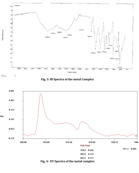
The important observation is the presence of an intense band at ~ 1670 cm-1 which is due to νC=O carbonyl group ( Fig.2).The most notable change in the IR spectra is the disappearance of the – NH stretching vibration and appearance of an intense band at ~ 1211 cm-1 due to νC-N-C stretching. The absence of band at 3300 cm-1 due to amino -NH disappears implying its condensation after deprotonation. In all the complexes, (Fig.3) band due to νC=O and νC-N
indicating the nitrogen and carbonyl oxygen are involved in coordination with metal ions.The new bands at 730 cm-1 corresponding to M-O bond.The presence of coordinated water molecules is determined by the presence of bands around 3391cm-1 and a band at 833 cm-1 is assignable to –OH stretching mode of vibration.The presence of phenolic –OH group in all the complexes in between 3443- 3360 cm-1 indicates that which was not involved in coordination.
3.3 UV-Visible spectra
The UV-Visible spectra of the complexes(Fig.4) were recorded in the range of 200-1100 nm. The UV spectrum mostly showed two intense maxima bands around 47540 cm-1 and 29890 cm-1 which belong to the π →π* and n →π* transitions respectively.The Cu (II) complex under present study exhibit a broad band in the region 26700cm-1 due to transition between 2Eg
→ 2 T2g which indicated the octahedral
geometry.The Ni (II) complex showed broad signals at 26255 cm-1 and 28540 cm-1which is assigned to 3 A2g → 3 T1gand 3 A2g → 3
T2gtransitions respectively which further
confirms its octahedral geometry. The position of bands observed for Co (II) complex also shows it is also having the octahedral geometry.
3.4 Suggested structure of the complexes
Based on the foregoing results we suggest the following structure for the complexes synthesized using the Mannich base ligand.
OH
C
H CH C O CH2 CH2 N M Cl Cl
H
2O
H
2O
Where M – Cu(II), Co(II) or Ni(II)
3.5 Antimicrobial activity
that the chelating tends to make the ligand to act as more potent bactericidal agents,thus destroying more bacteria than the free ligand.
4.CONCLUSION
It may be concluded that the newly prepared igand behaves as a bidentate chelating agent thro’ the N and O donor sites and the
spectroscopic data is in support of our expected structure. The antimicrobial property of the complexes were better than that of the free ligand observed.This may be attributed to the permeation of metal complexes through the cell membranes is much feasible due to coordinated bond than the free ligand.
OH
CHO
H3C C
O
CH3
+
OH
CH HC C
O
CH3
NaOH
Scheme . 1:
OH
C
H CH C
O
CH3
OH
C
H CH C
O
CH2 CH2 N
+ HCHO + HN
Fig. 1: NMR spectra of the ligand
Fig. 3: IR Spectra of the metal Complex
Fig. 4: UV Spectra of the metal complex
Table 1: Physical data of the ligand and the complexes
Compund Yield (%) Colour Mp (°C)
SAMP Ligand 78 Colorless 253
SAMP-Co 75 Pale pink 255
SAMP-Ni 72 Pale green 232
Table 2: IR Spectral data of the ligand and the complexes
Compound IR Stetching Frequency ( in cm-1)
-C=O -CNC M-N M-O
SAMP Ligand 1670 1211 ---- ----
SAMP-Co 1655 1206 883 810
SAMP-Ni 1654 1207 882 808
SAMP-Cu 1655 1207 883 808
Table 3: Antibacterial activity
S. No. Bacteria Standard Antibiotic
Disk(Ciprofloxacin)
Zone of inhibition mm in diameter (10μg/disc)
SAMP-L SAMP-Co SAMP-Ni SAMP-Cu
1 Staphylococcus aureus 21 10 15 13 17
2 Bascillussubtilis 16 12 17 15 19
3 Escherichia coli 26 09 19 12 16
4 Pseudomonas aeruginosa 18 08 13 14 17
5.REFERENCES
1. Ivanova Y, Momekov G, Petrov O, Karaivanova M and Kalcheva V. Eur J Med Chem. 2007;42: 1382-1387. 2. Sridhar SK, Saravanan M and Ramesh A.
Eur J Med Chem. 2001;36:615-625. 3. Joshi S, Khosla N, Khare D and Sharda R.
Bioorg Med ChemLett. 2005;15:221-226.
4. Chipeleme A, Gut J, Rosenthal PJ and
Chibale K. Bioorg Med Chem.
2007;15:273-282.
5. Ravichandran V, Mohan S and Suresh Kumar K. ArkivocNewslett. 2007;14:51-57.
6. Pandeya SN, Sriram D, Nath G and De Clercq E. IL Farmaco. 1999;54:624-628. 7. Surendra N, Pandeyaa, Dhamrajan
Srirama, Gopal Nathb and Erik De Clercq. Eur J Med Chem. 2000;35:249-255.
8. Perumal Yogeeswari, Dharmarajan Sriram, Ramkumar Kavya and Sonali
Tiwari. Biomed Pharmacother.
2005;59:501-510.
9. Shivarama Holla B, Veerendra B, Shivananda MK and Boja Poojary. Eur J Med Chem. 2003;38: 759-767.
10. InciGul H, Vepsalainen J, Gul M, Erciyas E and Hanninen O. Pharmaceutica Acta Helvetiae. 2000; 74:393-398.
11. Mohamed Ashraf Ali and Mohammad Shaharyar. Bioorg Med Chem Lett. 2007;17:3314-3316.
12. Sridhar SK, Pandeya SN, Stables JP and
Ramesh A. Eur J Pharm Sci.
2002;16:129-132.
13. Maria Grazia Ferlin, Gianfranco Chiarelotto, Francesca Antonucci, Laura Caparrotta, Guglielmina Froldi. Eur J Med Chem. 2002;37:427-434.