Light Microscopy
Peter TC So,
Massachusetts Institute of Technology, Cambridge, Massachusetts, USA Two-photon fluorescence microscopy allows three-dimensional imaging of biological specimensin vivo. Compared with confocal microscopy, it offers the advantages of deeper tissue penetration and less photodamage but has the disadvantage of slightly lower resolution.Nature of Two-photon Absorption
The invention of two-photon fluorescence light micro-scopy by Denk, Webb and co-workers (Denket al., 1990) revolutionized three-dimensional (3D)in vivoimaging of cells and tissues. In 1931, the theoretical basis of two-photon excitation was established by Maria Go¨ppert-Mayer, and this photophysical effect was verified experi-mentally by Kaiser and Garret in 1963. Two-photon excitation is a fluorescence process in which a fluorophore (a molecule that fluoresces) is excited by the simultaneous absorption of two photons (Figure 1). The familiar one-photon fluorescence process involves exciting a fluoro-phore from the electronic ground state to an excited state by a single photon. This process typically requires photonsin the ultraviolet or blue/green spectral range. However, the same excitation process can be generated by the simultaneous absorption of two less energetic photons (typically in the infrared spectral range) under sufficiently intense laser illumination. This nonlinear process can occur if the sum of the energies of the two photons is greater than the energy gap between the molecule’s ground and excited states. Since this process depends on the simulta-neous absorption of two infrared photons, the probability of two-photon absorption by a fluorescent molecule is a quadratic function of the excitation radiance. Under sufficiently intense excitation, three-photon and higher-photon excitation is also possible and deep UV microscopy based on these processes has been developed.
Article Contents . Nature of Two-photon Absorption . Design of a Two-photon Microscope . Fluorophores for Two-photon Microscopy . Comparison of Conventional, Confocal and
Two-photon Light Microscopy Methods
. Photobleaching and Photodamage in Two-photon Microscopy
. Application Trends in Two-photon Microscopy
(a) (b) Electronic ground state One-photon excitation Excitation photon Emission photon Fluorescence emission Excitation photons Two-photon excitation Fluorescence emission Emission photon Vibrational relaxation Vibrational states First electronic excited state
Figure 1 Jablonski diagram of one-photon (a) and two-photon (b) excitation, which occurs as fluorophores are excited from the ground state to the first electronic states. One-photon excitation occurs through the absorption of a single photon. Two-photon excitation occurs through the absorption of two lower-energy photons via short-lived intermediate states. After either excitation process, the fluorophore relaxes to the lowest energy level of the first excited electronic states via vibrational processes. The subsequent fluorescence emission process for both relaxation modes is the same.
Design of a Two-photon Microscope
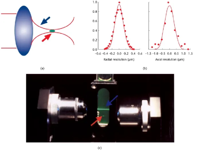
Although the possibility of nonlinear excitation was recognized in 1978 (Sheppard and Kompfner, 1978), two-photon microscopy was not demonstrated until 1990 (Denket al., 1990). The investigators noted that the two-photon process can be exploited to implement microscopy imaging in three dimensions. Depth discrimination is one of the most important properties of two-photon micro-scopes equipped with high numerical aperture objectives. For a spatially uniform specimen, fluorescence signals are generated equally from eachz-section above and below the focal plane for one-photon excitation. In contrast, over 80% of the total fluorescence signal can be confined to a region 1mm thick about the focal point using two-photonexcitation. This depth discrimination results from the quadratic dependence of the fluorescence probability on the spatial distribution of the excitation radiance. Appreci-able two-photon fluorescence occurs only at the micro-scope focal volume, where the photon density is high; negligible fluorescence is excited outside of this volume (Figure 2a,b). The typical two-photon excitation point spread function has a full width at half-maximum of 0.3mm in the radial direction and 0.9mm in the axial direction when 960 nm excitation light is focused by a 1.25 NA objective (Figure 2c).
Two-photon excitation efficiency is maximized when laser light is focused to a diffraction-limited volume.
Figure 3shows a typical two-photon microscope design. A critical component in a two-photon microscope is its light
Figure 2 A schematic representation of the localization of two-photon excitation. (a) Infrared light (blue arrow) is focused by an objective lens and fluorescence (red arrow) occurs only at the focal volume. (b) A detailed excitation profile of the two-photon excitation volume. The full width at half-maximum of the excitation profile is 0.3mm along the radial direction (left) and is 0.9mm along the longitudinal direction (right) at a laser wavelength of 960 nm. (c) A demonstration of the localization of two-photon excitation volume. Fluorescein solution is excited by one-photon excitation (blue arrow) via a 0.1 numerical aperture objective; fluorescence excitation is observed throughout the path of the laser beam. For two-photon excitation using a second objective with the same numerical aperture (red arrow), fluorescence excitation occurs only from a 3D localized spot.
source; a high-radiance light source on the order of 1010– 1012W cm22 is required for efficient excitation. This radiance level can be achieved by focusing light from a 1 W continuous-wave laser to a 1029cm2 diffraction-limited focal volume. Two-photon microscopy based on continuous-wave lasers has been demonstrated. However, the high average laser power is a concern for pigmented biological samples with appreciable one-photon absorp-tion. High repetition rate (100 MHz), ultrafast (femtose-cond or picose(femtose-cond pulse widths) lasers, such as titanium– sapphire and Nd:YLF lasers, are the most widely used light sources. The higher peak power and the lower duty cycle of these lasers minimize average power deposition in the specimen while maximizing two-photon excitation effi-ciency.
Figure 3shows laser excitation directed into the micro-scope via an epiluminescence light path. The excitation light is reflected by a dichroic mirror to the microscope objective and is focused in the specimen. Two-photon induced fluorescence is generated at the diffraction-limited volume. Images are constructed by raster scanning the fluorescent volume in three dimensions using a galvan-ometer-drivenx–yscanner and a piezo-objectivez-driver. The emission signal is collected by the same objective and transmitted through the dichroic mirror along the emission path. An additional barrier filter is needed to further attenuate the scattered excitation light. High-sensitivity
detection electronics, such as single-photon counting circuitry, are used to ensure maximal detection efficiency and signal dynamic range.
Fluorophores for Two-photon
Microscopy
A fluorophore that is one-photon active at wavelengthl can often be excited by two photons of twice the wavelength (2l). However, one should recognize that one-photon and two-photon excitation are fundamentally different quantum-mechanical processes and have very different selection rules. A fluorophore’s two-photon excitation spectrum scaled to half the wavelength is typically not equivalent to its one-photon excitation spectrum. However, a fluorophore’s emission spectrum, in the absence of ground-state heterogeneity, is indepen-dent of the excitation mechanism, since the molecule relaxes to the same excited state through vibrational mechanisms before emission.
Since the two-photon spectra of many molecules can be significantly different from their scaled one-photon equivalent, it is important to characterize the spectral properties of common fluorophores under two-photon
Specimen Photon sensor Dichroic mirror Objective lens Piezoelectric translator Ti-Sapphire laser Galvanometer-driven x–y mirror
Figure 3 A schematic drawing of typical components in a two-photon microscope. This system typically consists of a high-peak-power pulsed laser, a high-throughput scanning microscope and high-sensitivity detection circuitry.
excitation. The two-photon excitation spectra of over 20 fluorophores have been measured (Xuet al., 1996).
Further, fluorophores designed for one-photon excita-tion are not necessarily optimized for good two-photon absorption characteristics. Enhanced two-photon fluor-ophores with cross-sections two orders of magnitude
higher are found in p-conjugated molecules with large
quadrupole transition moment upon excitation (Albota
et al., 1998). Similar success has been achieved in designing photolabile caged groups for two-photon photolysis experiments (Furutaet al., 1999).
Comparison of Conventional, Confocal
and Two-photon Light Microscopy
Methods
Conventional light microscopy is an important tool, but its ability to resolve microscopic structures in optically thick specimens is limited because the image at the focal plane is blurred by out-of-focus noise. The invention of confocal microscopy in the 1960s and two-photon microscopy in the 1990s has started to address 3D imaging needs. Confocal microscopy is a technique very similar to two-photon microscopy. Confocal microscopy achieves 3D resolution using a set of conjugate apertures, one for illumination and one for detection of the scattered or fluorescent light. These conjugate pinholes, functioning as spatial filters, ensure that the microscope will illuminate and detect light from the same volume within the specimen.
Although two-photon and confocal microscopies are very similar, two-photon microscopy has a number of advantages. First, two-photon excitation wavelengths are typically about twice the one-photon excitation wave-lengths. This wide separation between excitation and emission spectrum ensures that the excitation light and the Raman scattering can be rejected while filtering out a minimum of fluorescence photons. Second, two-photon microscopy is particularly suited for imaging in optically thick specimens. Near-infrared radiation used in two-photon excitation has orders of magnitude less absorption in biological specimens than UV or blue-green light. The attenuation of excitation light from scattering is also reduced, as the scattering cross-section decreases with increasing wavelength. Third, confocal microscopy uses the emission pinhole aperture to reject out-of-focus light. Inside thick specimens, scattering of the fluorescent photons is inevitable. The resultant path deviation causes a significant loss of these photons at the confocal pinhole. Two-photon microscopy requires no pinhole aperture and minimizes signal loss.
While two-photon microscopy has a number of unique advantages when compared with the confocal approach, it has lower spatial resolution. The resolution of a
micro-scope system scales inversely with the wavelength of light used. For a given fluorophore, two-photon excitation requires the use of excitation at twice the one-photon wavelength, resulting in approximately half the resolution. Furthermore, confocal microscopes can generate images based on specimen refractive index variation in addition to fluorophore distribution.
Photobleaching and Photodamage in
Two-photon Microscopy
Compared with confocal microscopy operating in the UV or blue-green excitation wavelengths, two-photon micro-scopy minimizes photobleaching and photodamage. Un-like in confocal microscopy, which illuminates the specimen with a double inverted cone of light, photo-bleaching and photodamage are limited to a sub-femtolitre volume for two-photon microscopy. This difference is critical; confocal microscopy obtains 3D resolution by limiting the observation volume, whereas two-photon microscopy limits the excitation volume. Photodamage in a confocal microscope occurs when any region of the inverted cone of light intersects with the specimen. Two-photon photodamage is limited to a sub-femtolitre volume at the focal point. The reduction in the photodamage volume results in a dramatic increase in viability of biological specimens.
At the focal volume where photochemical interactions occur, two-photon microscopy can still cause considerable photodamage. Three major mechanisms of two-photon photodamage have been recognized. (1) Photodamage can be caused by two-photon or higher-photon excitation of endogenous and exogenous fluorophores similar to that of ultraviolet irradiation. These fluorophores act as photo-sensitizers in photooxidative processes. Photoactivation of these fluorophores results in the formation of reactive oxygen species that trigger the subsequent biochemical damage cascade in cells (Koniget al., 1996). (2) Single-photon absorption of the high-power infrared radiation can produce thermal damage. This effect is particularly pronounced in pigmented specimens. (3) Photodamage may also be caused by mechanisms, such as dielectric breakdown, resulting from the intense electromagnetic field of the femtosecond laser pulses.
The unique potential of two-photon imaging for noninvasive biological studies is well demonstrated. Two-photon microscopy has been used successfully to study the development ofCaenorhabditis elegansembryos. This is an application for which traditional confocal microscopy failed because photodamage resulted in developmental arrest of the organism (Mohleret al., 1998).
Application Trends in Two-photon
Microscopy
Two-photon microscopy is expected to have an impact in areas such as physiology, neurobiology, embryology and tissue engineering, for which imaging of highly scattering tissue is required. Highly opaque tissues such as human skin have been visualized with cellular detail (Masterset al., 1997). Clinically, two-photon microscopy may find an application in noninvasive optical biopsy, for which high-speed imaging is required. This need has been addressed by
video rate two-photon microscopy (Bewersdorf et al.,
1998). In cell biology, the most promising applications are those that rely on two-photon excitation to produce localized chemical reactions, such as in 3D resolved uncaging and photobleaching recovery studies (Denk
et al., 1994).
References
Albota M, Beljonne D, Bredas JL et al. (1998) Design of organic molecules with large two-photon absorption cross sections.Science 281: 1653–1656.
Bewersdorf J, Rainer P and Hell SW (1998) Multifocal multiphoton microscopy.Optics Letters23: 665–667.
Denk W, Strickler JH and Webb WW (1990) Two-photon laser scanning fluorescence microscopy.Science248: 73–76.
Denk W, Delaney KR, Gelperin A et al. (1994) Anatomical and functional imaging of neurons using 2-photon laser scanning microscopy.Journal of Neuroscience Methods54: 151–162. Furuta T, Wang SS, Dantzker JL et al. (1999) Brominated
7-hydroxycoumarin-4-ylmethyls: photolabile protecting groups with biologically useful cross-sections for two photon photolysis. Proceed-ing of the National Academy of Sciences of the USA96: 1193–1200. Konig K, So PTC, Mantulin WW, Tromberg BJ and Gratton E (1996)
Two-photon excited lifetime imaging of autofluorescence in cells during UVA and NIR photostress. Journal of Microscopy 183: 197–204.
Masters BR, So PTC and Gratton E (1997) Multiphoton excitation fluorescence microscopy and spectroscopy ofin vivo human skin. Biophysical Journal72: 2405–2412.
Mohler WA, Simske JS, Williams-Masson EM, Hardin JD and White JG (1998) Dynamics and ultrastructure of developmental cell fusions in theCaenorhabditis eleganshypodermis.Current Biology8: 1087–1090.
Sheppard CJR and Kompfner R (1978) Resonant scanning optical microscope.Applied Optics17: 2879–2882.
Xu C, Zipfel W, Shear JB, Williams RM and Webb WW (1996) Multiphoton fluorescence excitation: new spectral windows for biological nonlinear microscopy.Proceedings of the National Academy of Sciences of the USA93: 10763–10768.
Further Reading
Berland KM, So PT and Gratton E (1995) Two-photon fluorescence correlation spectroscopy: method and application to the intracellular environment.Biophysical Journal68: 694–701.
Brakenhoff GJ, Squier J, Norris Tet al. (1996) Real-time two-photon confocal microscopy using a femtosecond, amplified Ti:sapphire system.Journal of Microscopy181: 253–259.
Centonze VE and White JG (1998) Multiphoton excitation provides optical sections from deeper within scattering specimens than confocal imaging.Biophysical Journal75: 2015–2024.
Goeppert-Mayer M (1931) Uber Elementarakte mit zwei Quanten-sprungen.Annals of Physics (Leipzig)5: 273–294.
Lakowicz JR (1997) Nonlinear and two-photon-induced fluorescence. In: Lakowicz JR (ed.)Topics in Fluorescence Spectroscopy. New York: Plenum Press.
Lakowicz JR, Gryczynski I, Malak H et al. (1997) Time-resolved fluorescence spectroscopy and imaging of DNA labeled with DAPI and Hoechst 33342 using three-photon excitation.Biophysical Journal 72: 567–578.
Pawley JB (1995)Handbook of Confocal Microscopy. New York: Plenum Press.
Piston DW, Masters BR and Webb WW (1995) Three-dimensionally resolved NAD(P)H cellular metabolic redox imaging of thein situ cornea with two-photon excitation laser scanning microscopy.Journal of Microscopy178: 20–27.
Piston DW, Summers RG, Knobel SM and Morrill JB (1998) Characterization of involution during sea urchin gastrulation using two-photon excited photorelease and confocal microscopy. Micro-scopy and Microanalysis4: 404–414.
Shear JB, Xu C and Webb WW (1997) Multiphoton-excited visible emission by serotonin solutions.Photochemistry and Photobiology65: 931–936.