http://cjm.asm.md
http://dx.doi.org/10.19261/cjm.2019.636
© Chemistry Journal of Moldova
EFFECT OF TEMPERATURE ON GREEN SYNTHESIS OF SILVER
NANOPARTICLES USING VITEX AGNUS-CASTUS EXTRACT
Oksana Stavinskaya
a, Iryna Laguta
a*, Tetiana Fesenko
a, Marina Krumova
baChuiko Institute of Surface Chemistry of National Academy of Sciences of Ukraine, 17, General Naumov str., Kiev 03164, Ukraine
bKonstanz Universität, 10, Universitätsstraße, Konstanz 78464, Germany *e-mail: [email protected]
Abstract. The Vitex agnus-castus leaves extract has been used for green synthesis of silver nanoparticles. The effect of the temperature on both silver ions reduction and silver nanoparticles formation was investigated. It was found that fast reduction of silver ions occurs even at 40C, while effective synthesis of silver nanoparticles requires elevated temperatures of 60-80C. It is concluded that the extract contains both strong reducing and effective stabilizing agents that provide rapid reduction of silver ions and hinder the fast particles growth, respectively.
Keywords: nanoparticle, silver, green synthesis, Vitex agnus-castus extract.
Received: 09 September 2019/ Revised final: 03 November 2019/ Accepted: 08 November 2019
Introduction
Many plant extracts are known to contain significant amounts of reducing agents and are widely used in cosmetic, pharmaceutical and food industries. Recently, the possibility to use plant extracts in the processes of green chemistry, in particular, in the synthesis of metal nanoparticles (NPs), has attracted a great attention [1]. The advantages of green synthesis of metal nanoparticles as compared to traditional chemical methods are the mild synthesis conditions, low cost, simplicity, the absence of toxic reagents and by-products. Metal cations, in the green processes, may be reduced by the extract components such as flavonoids, phenolic acids, terpenoids, monosaccharides, and other compounds with reducing properties [2,3]. Many of these substances, as well as amino acids and proteins, may also form an organic coating on NPs surface and thus stabilize the colloidal particles [3,4]. Due to the absence of toxic components, such colloids may be used for biological and medical purposes [5-7].
Various plant leaves extracts are used in the green synthesis of metal NPs. For example, silver nanoparticles (AgNPs) colloids were successfully obtained using the extracts from the leaves of
Carica papaya [8], Murraya koenigii [9],
Morinda tinctoria [10], Pinus desiflora, Diopyros
kaki, Ginko biloba [11]. Stevia rebaudiana and
Magnolia kobus leaves extracts were also very
effective reactants for the synthesis of AgNPs
[5,11,12]. The literature [13] and our preliminary results suggest that the Vitex genus leaves extracts contain large amounts of bioactive compounds (phenols, terpenoids, monosaccharides, etc.), possess high reducing properties and significant activity in green synthesis of AgNPs.
The aim of this study was to continue the investigation of Vitex extract use in the synthesis of AgNPs and to examine the effect of temperature on the reduction of silver ions and on the growth of silver nanoparticles.
Experimental Materials
All reagents were obtained from commercial sources (Merck, Germany) and used without further purification.
Vitex agnus-castus leaves were used to
prepare the extract for green synthesis of AgNPs.
The active compounds were extracted using
ethanol solution (70%), the dried leaves to extractant ratio was 1 g/100 mL.
Methods
For the synthesis of AgNPs, a volume of
1 mL of Vitex extract was added to 9 mL of 1 mM AgNO3 aqueous solution, then Vitex extract +
AgNO3 reaction mixture was stirred for various
solutions maintained at 80C were diluted by 6 times.
The concentration of the remaining free Ag+ ions in the solutions was determined
according to the procedure described elsewhere [14]. A volume of 1 mL of tested solution was placed into volumetric flask, already containing 1 mL of 10-1 M EDTA disodium salt solution,
1 mL of 10-3 M 1,10-phenanthroline solution,
1 mL of 20% ammonium acetate solution and 2 mL of 10-4 M bromopyrogallol red solution. The
resulting solution was diluted to 50 mL with distilled water and the absorbance at maximum of about ~600 nm was measured against a blank, containing all of the above reagents except silver. To calculate the concentration of free Ag+ ions, a
plot of absorbance against silver concentration was preliminary obtained using the fresh prepared AgNO3 solutions of various concentrations.
UV-Vis spectra of all the solution and
reaction mixtures were recorded in the 220-800 nm wavelength range on a Perkin Elmer Lambda 35 UV-Vis double beam Spectrophotometer at 25C using a cuvette with path length of 10 mm.
For transmission electron microscopy study
(TEM), the colloidal solution was deposited on a carbon coated cupper grid and dried at ambient temperature. TEM images were obtained on a Zeiss Libra 120 instrument operated at 120 kV with resolution of 0.5 nm.
Results and discussion
Figure 1 gives the UV-Vis spectra for the
Vitex extract + AgNO3 reaction mixture stirred at
80C for 0.75-5 h. The absorbance band with the maximum at around 420 nm is due to AgNPs surface plasmon (SP) resonance, with the intensity of the band being dependent on the concentration of AgNPs in the solution. Figure 2 gives the absorbance at the maximum of the band versus time of the reaction for the mixtures maintained at temperatures of 50-80C. Figure 2 shows that, the changes in the intensity of the SP resonance band are strongly affected by the synthesis temperature. At 80C, the fast formation of AgNPs was observed, with the resonance absorption intensity being increased with the reaction time without saturation (Figures 1 and 2). For lower temperatures, a slower increase of the intensity of SP band in time was registered, showing a delayed particles growth (Figure 2, curves 1-3).
The spectra corresponding to the mixture kept at 40C did not contain a pronounced signal
in the region of ~400 nm even after 48 h of the reaction (Figure 3).
Figure 1. UV-Vis spectra of the initial Vitex extract + AgNO3 mixture (1) and of the mixtures stirred at
80C during 0.75 (2), 1.5 (3), 3 (4) and 5 h (5).
Figure 2. Absorbance at the maximum of SP resonance band in the UV-Vis spectra of the Vitex extract + AgNO3 mixtures versus time of
the reaction at 50C (1), 60C (2), 70C (3) and 80C (4).
Figure 3. UV-Vis spectra of the initial
Vitex extract + AgNO3 mixture (1),
Nevertheless, by subtracting the spectra of the initial Vitex extract + AgNO3 mixture from the
spectra of the mixture, maintained at 40C during 48 h, a very broad line with the maximum at about 400-420 nm is obtained with the full width at half maximum equivalent to ~1 eV. For small metal particles, the width of SP resonance peak is inversely proportional to the radius, thus the sizes of NPs responsible for the band registered at 40C may be estimated as smaller than 1 nm [15]. These are very small clusters/particles that may not possess sufficient stability [16]. While only slow formation of small clusters/particles occurs at 40C, the synthesis at 60-80C appears to be both more effective and more appropriate from the point of view of the colloidal properties.
The results of the UV-Vis spectroscopic study given above are in agreement with TEM data. Figures 4 and 5 show TEM images of AgNPs formed in the reaction mixtures at temperatures of 40 and 80C, respectively.
Both images reveal the presence of several sufficiently large particles/aggregates with a diameter of 10-20 nm (partially, these particles/aggregates may be formed during the colloidal solutions storage and during drying the samples before TEM observation). Beside these particles, the mixture prepared at 80C contains a large number of NPs of several nanometers in diameter (Figure 5). For the mixture prepared at 40C, the registering of small NPs was unsuccessful (Figure 4): clusters/particles with diameters less than 1 nm, which are supposed to form at 40C, may be just indistinguishable in the image.
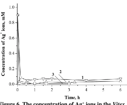
Figure 6 shows the data on the content of free Ag+ ions in the mixtures versus time of the
reaction. It was determined that no more than 10% of Ag+ ions remain in the mixture in several
minutes even at 40C (Figure 6, curve 1). Similar amounts of free Ag+ ions are registered in the
mixtures kept at temperatures of 60 and 80C (Figure 6, curves 2,3). Thus, the reduction of silver ions is only slightly affected by the time and the temperature of the reaction. Nevertheless, as it was mentioned above, the intensity of SP resonance absorption band in the spectra of the colloidal solutions and, therefore, the quantity of AgNPs in the mixtures, strongly depend on the synthesis conditions.
In general, the process of metal NPs formation includes several steps: reduction of metal precursor and formation of metal atoms (probably, coupled with the molecules of reducing agents), aggregation of such metal monomers in the dimmers and primary clusters/particles,
growth of primary clusters/particles due to their coalescence or due to diffusion and absorption of metal monomers on the particles surface [3,16]. Depending on the synthesis conditions and the properties of reducing agents, the growth of metal NPs may occur through various mechanisms. In the case of mild reducing agent, the reduction of metal precursor occurs relatively slow and the process of NPs growth may be limited by this monomer-supplying reaction.
Figure 4. TEM image of the Vitex extract + AgNO3
mixture stirred at 40C during 24 h.
Figure 5. TEM image of the Vitex extract + AgNO3
mixture stirred at 80C during 5 h.
Figure 6. The concentration of Ag+ ions in the Vitex
extract + AgNO3 mixture versus time of the
For the strong reducing agents, the reduction of metal ions and the formation of primary clusters/particles may take place almost immediately after the reagents mixing, and the following particles growth occurs mainly owing to coalescence of the clusters/particles. The kinetics of NPs growth in the coalescence process is determined by the ability of the particles to overcome the “aggregation barrier”, which originates from electrostatic repulsion of the electrical double layers formed on the charged particles surface. The additional factor hindering the particles aggregation is the presence of stabilizing agents, which absorb on the NP surface and provide their electrostatic and/or steric stabilization.
The data on the concentration of Ag+ ions
in the Vitex extract + AgNO3 mixture (Figure 6)
suggests that the extract contains strong reducing agents, which ensure the rapid formation of metal monomers and primary clusters. The process of the AgNPs growth was found to be strongly temperature-dependent, and it is relatively slow as compared to the processes described elsewhere [16,17]. This assumes the involvement of the large molecules – the components of the extracts, which appear to form the organic coating on the particles surface thus hindering the particles growth. On the one hand, the existence of such a coating requires higher temperature for AgNPs growth; on the other hand, it ensures the stability of colloidal particles. Thus, Vitex agnus-castus extract seems to be a promising agent for the synthesis of stable colloidal AgNPs at elevated temperature; the resulting NPs do not contain toxic components and may be used for medical application.
Conclusions
In this paper, the effect of temperature on green synthesis of silver nanoparticles using the extract of Vitex agnus-castus is reported; the novelty of the work consists in the simultaneous study of the processes of silver nanoparticles (AgNPs) formation and silver ions reduction.
Rapid reduction of silver ions was found to occur even at a relatively low temperature of 40C while the effective synthesis of AgNPs was registered only at 60-80C. The results obtained suggest that the extract contains both strong reducing and effective stabilizing agents. The extract can be a promising alternative in the synthesis of colloidal AgNPs, suitable for biological and medical applications.
References
1. Iravani, S. Green synthesis of metal nanoparticles using plants. Green Chemistry, 2011, 13(10), pp. 2638−2650.
DOI: 10.1039/c1gc15386b.
2. Mittal, A.K.; Chisti, Y.; Banerjee, U.C. Synthesis of metallic nanoparticles using plant extracts. Biotechnology Advances, 2013, 31(2), pp. 346–356. DOI: https://doi.org/10.1016/ j.biotechadv.2013.01.003
3. Makarov, V.V.; Love, A.J.; Sinitsyna, O.V.; Makarova, S.S.; Yaminsky, I.V.; Taliansky, M.E.; Kalinina, N.O. “Green” nanotechnologies: synthesis of metal nanoparticles using plants. Acta Naturae, 2014, 6(1), pp. 35–44.
http://actanaturae.ru/catalog/225.aspx
4. Khandel, P.; Yadaw, R.K.; Soni, D.K.; Kanwar, L.; Shahi, S.K. Biogenesis of metal nanoparticles and their pharmacological applications: present status and application prospects. Journal of Nanostructure in Chemistry, 2018, 8(3), pp. 217–254.
DOI: https://doi.org/10.1007/s40097-018-0267-4
5. Laguta, I.; Fesenko, T.; Stavinskaya, O.; Dzjuba, O.; Shpak, L. Antioxidant and antimicrobial properties of Stevia leaves extracts and silver nanoparticles colloids. Chemistry Journal of Moldova. General, Industrial and Ecological Chemistry, 2016, 11(2) pp. 46–51. DOI:
https://dx.doi.org/10.19261/cjm.2016.11(2).08
6. Roy, A.; Bulut, O.; Some, S.; Mandal, A.K.; Yilmaz, M.D. Green synthesis of silver nanoparticles: biomolecule-nanoparticle organizations targeting antimicrobial activity. RSC Advances, 2019, 9(5), pp. 2673–2702.
DOI: 10.1039/C8RA08982E
7. Vallet-Regí, M.; González, B.; Izquierdo-Barba, I. Nanomaterials as promising alternative in the infection treatment. International Journal of Molecular Sciences, 2019, 20(15), pp. 3806–3823. DOI: https://doi.org/10.3390/ijms20153806
8. Banala, R.R.; Nagati, V.B.; Karnati, P.R., Green synthesis and characterization of Carica papaya leaf extract coated silver nanoparticles through X-ray diffraction, electron microscopy and evaluation of bactericidal properties. Saudi Journal of Biological Sciences, 2015, 22(5), pp. 637–644. DOI: https://doi.org/10.1016/j.sjbs.2015.01.007 9. Christensen, L.; Vivekanandhan, S.; Misra, M.;
Mohanty, A.K. Biosynthesis of silver nanoparticles using Murraya koenigii (curry leaf): An investigation on the effect of broth concentration in reduction mechanism and particle size. Advanced Materials Letters, 2011, 2(6), pp. 429–434.
DOI: 10.5185/amlett.2011.4256
10.Ramesh, K.K.; Nattuthurai, N.; Gopinath, P.; Mariappan, T. Biosynthesis of silver nanoparticles from Morinda tinctoria leaf extract and their larvicidal activity against Aedes aegypti linnaeus 1762. Journal of Nanomedicine & Nanotechnology, 2014, 5(6), pp. 242–246.
11.Song, J.Y.; Kim, B.S. Rapid biological synthesis of silver nanoparticles using plant leaf extracts. Bioprocess and Biosystems Engineering, 2009, 32(1), pp. 79–84.
DOI: https://doi.org/10.1007/s00449-008-0224-6
12.Yilmaz, M.; Turkdemir, H.; Akif Kilic, M.; Bayram, E.; Cicek, A.; Mete, A.; Ulug, B. Biosynthesis of silver nanoparticles using leaves of Stevia rebaudiana. Materials Chemistry and Physics, 2011, 130(3), pp. 1195–1202. DOI:
https://doi.org/10.1016/j.matchemphys.2011.08.068
13.Rani, A.; Sharma, A. The genus Vitex: A review. Pharmacognosy Reviews, 2013, 7(14), pp. 188–198.
DOI: 10.4103/0973-7847.120522
14.Dagnall, R.M.; West, T.S. A selective and sensitive colour reaction for silver. Talanta, 1964, 11(11),
pp. 1533–1541. DOI: https://doi.org/10.1016/0039-9140(64)80225-3
15.Garcia, M.A. Surface plasmons in metallic nanoparticles: fundamentals and applications. Journal of Physics D: Applied Physics, 2011, 44(28), pp. 283001. DOI:
https://doi.org/10.1088/0022-3727/44/28/283001
16.Polte, J. Fundamental growth principles of colloidal metal nanoparticles – a new perspective. CrystEngComm, 2015, 17(36), pp. 6809–6830. DOI: 10.1039/c5ce01014d
17.Ahmad, N.; Sharma, S.; Alam, Md.K.; Singh, V.N.; Shamsi, S.F.; Mehta, B.R.; Fatma, A. Rapid synthesis of silver nanoparticles using dried medicinal plant of basil. Colloids and Surfaces B: Biointerfaces, 2010, 81(1), pp. 81–86.