University of New Orleans Theses and
Dissertations Dissertations and Theses
12-17-2004
Development and Characterization of Controlled Drug Delivery
Development and Characterization of Controlled Drug Delivery
Using Nanoparticles
Using Nanoparticles
Li Chen
University of New Orleans
Follow this and additional works at: https://scholarworks.uno.edu/td
Recommended Citation Recommended Citation
Chen, Li, "Development and Characterization of Controlled Drug Delivery Using Nanoparticles" (2004). University of New Orleans Theses and Dissertations. 186.
https://scholarworks.uno.edu/td/186
This Thesis is protected by copyright and/or related rights. It has been brought to you by ScholarWorks@UNO with permission from the rights-holder(s). You are free to use this Thesis in any way that is permitted by the copyright and related rights legislation that applies to your use. For other uses you need to obtain permission from the rights-holder(s) directly, unless additional rights are indicated by a Creative Commons license in the record and/or on the work itself.
NANOPARTICLES
A Thesis
Submitted to the Graduate Faculty of the
University of New Orleans
in partial fulfillment of the
requirements for the degree of
Master of Science
in
The Department of Chemistry
by
Li Chen
B.S, Zhengzhou University, China, 1993
ii
ACKNOWLEDGEMENTS
Firstly, I would like to express my greatest gratitude to my advisor, Prof. Zeev
Rosenzweig, not only for his support and invaluable advice on the research work, but also for his
encouragement and patience during the past years.
I would also like to thank Prof. Nitsa Rosenzweig for her help in cell cultures; committee
member Prof. Matthew A. Tarr and Prof. Jiye Fang for their advices and discussions on the
research work.
I would like to thank everyone in my group for their help and advices. I’ll remember the
time we spent together.
Lastly, I would like to thank my husband Jiachang Gong, my lovely twin girls Katherine
iii
LIST OF FIGURES ...v
ABSTRACT... viii
CHAPTER 1: INTRODUCTION ...1
1.1 Controlled Drug Delivery ...1
1.2 Strategy of Controlled Release ...2
1.3 Scope of Controlled Drug Delivery Systems...4
1.3.1 Liposomes and Miclelles ...4
1.3.2 Drug Conjugates ...6
1.3.3 Nanoparticles and Microparticles ...7
1.4 Fluorescence and its Applications in Controlled Drug Delivery...8
1.4.1 Fluorescence ...8
1.4.2 Applications in Drug Delivery ...10
CHAPTER 2: EXPERIMENTAL...11
2.1 Materials and Reagents ...11
2.2 Procedures and Protocls...13
2.2.1 synthesis of PLGA and Doxorubicin-loaded Nanoparticles ...13
2.2.2 Synthesis of Mesoporous Silica Nanoparticles ...13
2.2.3 Drug ananlog –tetramentylrhodamine dextran loading ...14
2.2.4 Preparation of Lipids Membrane Coated Silica Nanoparticles ...15
2.2.4 Cell Culture ...15
2.3. Instrumentation ...16
2.3.1 Spectrofluorometry ...16
2.3.2 Digital Fluorescence Imaging Microscopy ...17
2.3.3 Transmission Electron Microscopy ...18
CHAPTER 3: TUMOR TARGETED DELIVERY OF THE ANTICANCER DRUG-DOXORUBICIN USING POLYMERIC NANOPARTICLES AS CARRIERS...19
3.1. Introduction...19
3.2. Specific Experimental and Technical Details ...20
3.2.1 Characterization of Nanoparticles...20
3.2.2 Fluorescence Measurements ...20
3.2.3 Loading Studies of Doxorubicin ...21
iv
3.3.1 Charcterization of PLGA Nanoparticles...23
3.3.2. Fluorescence Studies...24
3.3.3 In vitro Release Studies ...28
3.3.4 Cellular Uptake and In vitro Cytoxicity Studies ...30
3.4 Summary and Conclusions ...34
CHAPTER 4: NOVEL SILICA NANOPARTICLES-BASED DRUG DELIVERY SYSTEM TRIGGERED BY ANTIMICROBIAL PEPTIDES ...35
4.1 Introduction ...35
4.2 Specific Experimental and Technical Details ...37
4.2.1 Characterization of Silica Nanoparticles ...37
4.2.2 Studies of TMR-Dex Release Profiles ...39
4.2.3 Effects of antimicrobial peptide cecropin-melittin ...39
4.3 Results and Discussion ...40
4.3.1 Synthesis of Mesoporous Silica Nanoparticles ...40
4.3.2 Loading Studies of TMR-Dex into Silica Nanoparticles ....41
4.3.3 Lipids Membrane Coating of Silica Nanoparticles ...42
4.3.4 The induced release of TMR-Dex by antimicrobial peptide ...45
4.4 Summary and Conclusions ...49
CHAPTER 5: SUMMARY AND CONCLUSIONS ...50
REFERENCES ...52
v
LIST OF FIGURES
Figure 1.1 Jablonski diagram illustrating the processes involved in the creation of an excited
electronic singlet state by optical absorption and subsequent emission of fluorescence and
phosphorescence ...9
Figure 2.1 Digital Fluorescence Imaging Microscopy system ...18
Fig 3.1 Transmission electron images of PLGA nanoparticles (The sample was negatively stained.)
...25
Fig. 3.2 a) Fluorescence emission spectrum of DXR in PBS solution ...26
Fig 3.2 b) Fluorescence image of DXR-loaded nanoaprticles (magnification 400x) ...26
Fig 3.3 Emission spectra of a) Released Doxorubicin from PLGA nanoparticles, b) free
Doxorubicin c) Doxorubicin encapsulated in PLGA nanoparticles. (The fluorescence
intensities are not comparable since these samples do not have the same concentration)
...27
Fig 3.4 In vitro release profile of doxorubicin from PLGA nanoparticles (pH 7.4, 37oC)
...29
vi
...29
Fig 3.6 Transmission images of MCF-7 cells treated with (a) free DXR (c) DXR-loaded PLGA
nanoaprticles. Fluorescence images of (b) free DXR (d) nanoparticles. The cells were incubated with
5µM equivalent DXR concentration. All images were taken with 400x magnification ...31
Fig 3.7 The cytocoxicity of DXR-loaded PLGA nanoparticles (mean size 110nm). The density of
blank particles is the same as that of drug-load nanoaprticles. Untreated cell culture were used as a
reference as 100% viability ...32
Fig. 4.1 Schematic representation of formation of lipids coated mesoporous silica nanoparticles drug
delivery system ...38
Fig. 4.2 TEM image and Fluorescence image of TMR-Dex loaded silica nanoparticles
...41
Fig. 4.3 Release profile of TMR-Dex loaded silica particles in 10mM HEPES pH 7.4 buffer
solutions ...44
Fig. 4.4 Release profile of TMR-Dex loaded silica DMPC-lipobeads in 10mM HEPES pH7.4
vii
Fig. 4.6 Peptide concentration-dependent release. (The measurements were taken 2 hours after
addition of cecropin-melittin peptide) ...47
Fig. 4.7 Fluorescence spectra of silica DMPC-lipobeads (Measurements were taken 2 hours after
peptides were added) ...48
Fig. 4.8 Fluorescence spectra of silica PS-lipobeads (Measurements were taken 2 hours after
viii
ABSTRACT
The objective of this project was to develop new controlled drug delivery systems using
nanomeric particles and characterize the delivery of drugs into cells in real time by digital
fluorescence imaging microscopy techniques. The project is based on the idea that it could be
possible to improve efficacy of drug molecules when encapsulated in nanometer-sized particles.
Due to their small dimensions the particles could permeate through cells and tissues and even
through the blood brain barrier.
The anti-cancer drug Doxorubicin was encapsulated into biodegradable Poly
(DL-lactide-co-glycolide) (PLGA) nanoparticles by simple nanoprecipitation method. The small size of
these particles (<200nm) could be beneficial to realize passive tumor-targeted drug delivery
through enhanced permeability and retention (EPR) effects. These drug-containing particles
showed a sustained release profile. Fluorescence images indicated that these particles can be
internalized by human breast cancer MCF-7 cells by non-specific endocytosis. The bioactivity
of the drugs was also tested against cell culture. The results indicated that DXR-loaded PLGA
nanoaprticles could be used to deliver Doxorubicin into breast cancer cells.
As the second approach, a novel silica nanoparticles-based stimuli-responsive drug
delivery system has been developed. The feasibility of these unique carriers was demonstrated by
coating the dye-loaded silica nanoaprticles with phospholipids membrane. The release was
induced by the addition of the antimicrobial peptide cecropin (1-8)-melittin (1-18). The
CHAPTER 1 INTRODUCTION
1.1 Controlled Drug Delivery
From the earliest times, people have found ways to introduce drugs into the body. This
process began with the chewing of leaves and roots of medicinal plants. Throughout the history of
medicine delivery of drugs to humans has evolved from primitive extracts and inhalants to more
reliable dosages forms, such as injections, tablets and capsules. These drug delivery systems are
expected to be further optimized to increase drug activity and reduce toxicity. For instance, one of
the most common ways of administering drugs to the body is via injection into the bloodstream. The
injected material is circulated throughout the body and thus commonly termed systemic delivery.
The drawbacks of this delivery method are that the concentration of the injected material is
extremely diluted and the material acts on most tissues of the body and may be toxic to some of
them. The problem could be solved by controlled drug delivery. In controlled drug delivery systems,
the active agent is released in a predesigned manner. Drug delivery systems can influence the
performance of a drug by manipulating its concentration, location and duration of exposure.
In the past 30 years, controlled drug delivery technology has represented one of the most
will contribute significantly to human health. These drug delivery systems offer numerous
advantages compared to conventional dosage forms:
♦ increasing the efficacy of currently used drugs
♦ providing opportunities for the use of new agents currently precluded from clinical use due
to challenges including low drug solubility and systemic toxicity
♦ reducing harmful side effects
♦ precise control of dose
♦ decreasing number of dosages
♦ improving patient compliance and convenience
1.2 Strategy of Controlled Release
There are three strategies to achieve controlled drug delivery. One method is to prepare
drug delivery systems that release drugs over extended duration. Numerous works have been
done based on biodegradable polymers [6-9]. In the conventional drug delivery, the drug
concentration in the blood rises when drug is taken, then peaks and declines. Since each drug has
a plasma level above which it is toxic and below which it is ineffective, the plasma drug
concentration in a patient at a particular time depends on compliance with the prescribed routine.
In contrast, with controlled release systems, the rate of drug release matches the rate of drug
elimination. Therefore, the drug concentration is within the therapeutic range for a longer time.
This release pattern is highly beneficial for drugs that are rapidly metabolized and eliminated
The second approach is to prepare a feedback controlled devices that release the
appropriate amount of drug in response to a therapeutic marker. In recent years, several research
groups have been developing responsive systems [10-14]. These systems can be classified as
external regulated and self-regulated systems. The external controlled devices apply external
triggers for pulsed delivery such as: magnetic, ultrasonic, thermal and electric triggers. In the
self-regulated system, the release rate is controlled by feedback information. The self-regulated
systems utilize several approaches such as pH-sensitive polymers, enzyme substrate reactions
and competitive binding, as rate-control mechanisms.
The third strategy is to control drug distribution in the body. The idea is to deliver a drug
to the precise location in the body where it will be most effective. There are two basic types of
targeting systems: passive and active. Passive targeting systems rely on non-specific interactions
such as hydrophobic or electrostatic interactions, and the body physical characteristics. The size
of drug carriers has been extensively studied for passive targeting. It was found that particles
larger than 5-7 µm in diameter usually become trapped in the lung [15] and particles smaller than
1 µm in diameter rapidly phagocytosed by the Kupffer cells of the liver [16]. When the particle
size is reduced below 100 nm, the particles can appear in the bone marrow [17]. It was also
demonstrated that drug carriers small than 200 nm can be accumulated efficiently in tumor
through enhanced permeability and retention (EPR) effect due to the abnormality of tumor tissue,
resulting in the enhanced vascular permeability compared to healthy tissues [18-20]. On the other
hand, active targeting systems utilize specific interactions, such as antigen-antibody and
ligand-receptor binding, to achieve specific targeting goals. In this approach, the therapeutic index of
drugs could be enhanced by keeping drugs away from healthy cells. The types of receptors that
receptors (tumor cells) [22], albumin receptors (cardiac and lung) [23] and growth factors
receptors [24].
1.3 Scope of Controlled Drug Delivery Systems
While these fundamental ideas of controlled drug delivery are extremely attractive,
achieving controlled drug delivery is not a simple or straightforward task. Currently, the vast
majority of the work in this area is focusing on liposomes, micelles, drug conjugates, and
particles.
1.3.1 Liposomes and Micelles
Although liposomes were initially developed as models of biological membranes, their
potential as a drug delivery system have undergone intensive investigations for over 25 years [25].
Liposomes are formed by the equilibration of natural phospholipids with excess water or aqueous
salt solution. They contain one or several (concentric) lipid bilayers, which can solubilize
hydrophobic drugs. Alternating aqueous compartments can entrap hydrophilic drugs [26-29].
Liposomes with mean diameter smaller than 100 nm selectively spread in leaky tissues (eg. solid
tumors), and exhibit target specificity with negligible adverse effects to normal tissues [30].
There are several advantages of liposomes as drug delivery carriers. Liposomes are able to
protect drugs from degradation. They are relatively easy to prepare and prevent accumulation of
elimination, increased circulation life times). When coupled with antibodies, liposomes serve as a
means to confer active targeting [31]. Some issues such as contents retention, circulation lifetime,
biodistribution and immunogenicity can be managed by the formulation of the liposomal drug
carriers [32-34]. Liposome properties such as size, surface charges, membrane rigidity and phase
transitions within the bilayer can be controlled either by selecting appropriate lipid compositions or
by changing external conditions such as temperature, acidity or the presence of specific agents
[35-39]. Liposomes have been used as carriers for many drugs with low molecular weight, peptides
[40-45] and oligonucleotides [46, 47]. However, liposomes also have limitations. One of the problems
with liposomes as drug delivery carriers is their lack of stability in biological fluids. Consequently,
drug molecules leak to normal tissues and cause undesirable side effects.
Micellar drug carriers are formed from amphiphilic block copolmers composed of
hydrophilic and hydrophobic segments. The hydrophobic segments form the inner core of the
micelle and are surrounded by an outer shell consisting of the hydrophilic segments. Micelles are
commonly of the order of 50 nm [48], which compares with the dimension of viruses, and thus may
be able to penetrate the sinusoidal and fenestrated capillaries that have pores approximately 100 nm
in size. However, micellar carriers are generally considered to be poor delivery systems because
micellar complexes are in dynamic equilibrium with free molecules in solution. They continuously
break down and reform, and they are generally unstable on dilution.
1.3.2 Drug conjugates
The basic idea is to develop drug conjugates by chemically modify a drug in order to
Thus, drugs have been attached to soluble macromolecules such as proteins, polysaccharides, and
polymers via degradable linkages [49, 50]. This process changes the drug’s size and other properties,
resulting in different pharmacokinetics and biodistribution. One active area is to conjugate
antitumor drugs to polymers. For example, doxorubicin, an antitumor agent was coupled to
N-(2-hydroxypropyl) methacrylamide copolymers, which resulted in radically altered pharmacokinetics
and reduced toxicity. The half-life of the drug in plasma and the drug levels in the tumor were
increased while the concentrations in the periphery decreased [51]. Polymers such as polyethylene
glycol (PEG), can be attached to drugs to lengthen their lifetime. PEG-asparaginase is used for
patients with leukemia [52]. Receptors such as transferrin were also used to conjugate antitumor
agents for tumor targeting. Recently, dendrimers emerged as promising drug carriers. Compared
with polymers, it is relatively easy for dendrimers to control molecular weight and functional groups.
Recent studies showed that dendrimers attenuate the toxicity of approved drugs – methotrexate and
6-mercaptopurine, suggesting that higher dosing might be attainable [53].
1.3.3 Nanoparticle and microparticles
The application of nanoparticles and microparticles as drug delivery systems has received
increasing attention. They have been widely applied in the delivery of drugs, genes, and vaccines to
specific cells and tissues of interest with potential reduction of toxicity as well as increased
therapeutic effects [54, 55]. Compared to liposomes, particles appear to offer an interesting
alternative. They possess higher stability in biological fluids and during storage. In addition, they
One of widely studied areas is polymer nanoparticles, either natural or synthetic, as drug
carriers. Polymer particles have the ability to deliver a wide range of drugs to varying areas of the
body for sustained period of time. The active agent can be released from polymeric systems by
diffusion, degradation and swelling, depending on the nature of the polymer. The most common
used polymers have been poly (lactic acid) (PLA), poly (glycolic acid) (PGA), poly (Є-caprolactone)
(PCL) and the copolymer (PLGA) of PLA and PGA, These polymers are known for their
biocompatibility and biodegradability [56]. The degradation rate and accordingly, the drug release
rate can be manipulated by varying the polymer composition. Other than these polymers, natural
hydrophilic polymers such as chitosan, sodium alginate and gelatin have also been used to prepare
drug-loaded nanoparticles. These particles have shown good association with proteins, such as
bovine serum albulmin, tetanus toxoid, diptaheria toxoid, insulin and oligonucleotides [57-59].
Porous polymeric particles are also attractive as delivery systems due to their special
structures. Langer et al developed a new type of inhalation aerosol, which is based on porous
microparticles composed of 50:50 PLGA and poly (lactic acid-co-lysine-graft-lysine) [60]. Results
revealed that inhalation of large porous insulin particles resulted in elevated systemic levels of
insulin and suppressed systemic glucose levels for 96 hours, whereas small nonporous insulin
particles had this effect for only 4 hours.
Recently, advances in nanomaterials science and biotechnology facilitated the development of
new drug delivery systems. Silica naoparticles are well known for their compatibility in biological
system [61]. Their surfaces can be easily modified with different functional groups. These features
make silica nanoparticles promising as drug carriers. Prasad et al demonstrated the potential of
ceramic-based nanoparticles as drug carriers for photodynamic therapy [62]. Several other groups
attractive features such as large surface area, tunable pore sizes and surface properties [63-66]. Their
research demonstrated that silica materials could play a significant role in developing a new
generation of controlled drug delivery systems.
1.4 Fluorescence and its Applications in Controlled Drug Delivery
1.4.1 Fluorescence
Fluorescence is the property of some molecules and atoms to absorb light at a particular
wavelength and to subsequently emit light of longer wavelength after a brief interval.
Fluorescence typically occurs from poly-aromatic hydrocarbons or heterocycles molecules that
are called fluorescent dyes or fluorophore. Fluorescence occurs when an excited molecule
returns to the electronic ground state from the excited singlet state by the emission of a photon.
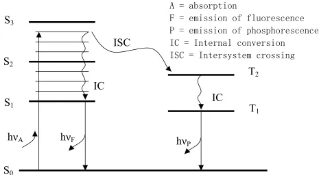
Fig. 1.1 describes radiative and nonradiative processes that occur following excitation of
a molecule. S0, S1, S2, and Sn represent the singlet state of ground, 1st, 2nd, and nth electronic
energy levels, respectively. T1, T2 represent the triplet states of 1st and 2nd electronic energy
levels. The absorption of photons typically occurs from the lowest vibration level of S0 to a
vibration state of S1 or S2. After the light was absorbed by the fluorophore, several processes take
place. Nonradiative, fast relaxation brings the molecule down to the lowest excited state S1
within 10-12 s. This process is defined as internal conversion. Fluorescence brings back the
molecule to one of the vibronic sublevels of the ground state S0. The fluorescence lifetime of S1
back to its ground state without photon emission. A third type of process presenting in organic
dyes is intersystem crossing to the first excited triplet state T1. Relaxation from this excited state
back to the ground state is defined as phosphorescence. The lifetime of this state is in the order
of microseconds to milliseconds. The entire fluorescence process is cyclical. Unless the
fluorophore is irreversibly destroyed in the excited state, the same fluorophore can be repeatedly
excited and detected.
Figure 1.1. Jablonski diagram illustrating the processes involved in the creation of an excited
electronic singlet state by optical absorption and subsequent emission of fluorescence and phosphorescence.
S
0S
2S
1S
3IC
ISC
h
νA
h
νF
h
νP
IC
T
1T
2A = absorption
F = emission of fluorescence
P = emission of phosphorescence
IC = Internal conversion
1.4.2 Applications in drug delivery
As we mentioned previously, the performance of a drug depends on its location,
concentration and duration. Determining the drug biodistribution, cellular fate and cell uptake
mechanism would enable better understanding of the cytotoxicity and the drug resistance. Mass
spectroscopy is widely used to provide the information about drug delivery into specific organs
and tissues. However, it does not offer the information about cellular drug distribution. Imaging
techniques are better choices for this purpose. Among them, fluorescence microscopy has been
proven to be effective in studying the cellular fate of delivered drugs, due to its high sensitivity
and easy-to-use procedure. For example, Savic and his coworkers prepared the
tetramethylrhodamine labled PCL-PEO copolymer micelles. A triple-labeling confocal
fluorescence microscopy was then developed to identify the location of the micelles [67]. Results
revealed that the micelles were located in several cytoplasmic organelles, including
mitochondria, but not in the nucleus. They also found that the micelles changed the cellular
CHAPTER 2: EXPERIMENTAL
This chapter described the general experimental information and instruments used to
carry out the research work. Specific technical and experimental details will be described later in
related chapter.
2.1 Materials and Reagents
Poly (DL-lactide-co-glycolide) (PLGA, L/G=50/50, inherent viscosity 0.17dL/g) (Birmingham
Polymers, Inc.)
Doxorubicin hydrochloride (Sigma)
Polyvinylalchol (PVA) (average molecular weight 30000-70000 Da, 88% hydrolyzed) (Sigma)
3-(4, 5-dimethyl-2-yl)-2,5-diphenyltetrazolium bromide (MTT) (Molecular Probes)
Tetra ethyl orthosilicate (TEOS) (Sigma)
Hexadecyltrimethylammoniun bromide (CTAB) (Fluka)
Tetramethyl Rhodamine, Dextran 3000 (TMR-Dex)
1,2-Dimyristoyl-sn-Glycero-3-Phosphocholine (DMPC) (Avanti lipids)
Carboxyl Fluorescein (Sigma)
Cecropin (1-8)-Melittin (1-18) hybrid peptide (CA (1-8)- M (1-18) ( Bachem America)
Phosphate Buffered Saline tablet (Amresco)
HEPES (Sigma)
MES (Sigma)
Dullbeco’s modified Eagle’s medium (invitrogen)
Fetal bovine serum (invitrogen)
Trypsin (invitrogen)
Human breast cancer cell line (MCF-7) (American Type Culture Collection)
Lab-Tek II chambered coverglass (Fisher Scientific)
All aqueous solutions were prepared with 18 MΩ deionized water produced by a water purification system (Barnstead Thermolyne nanopure) and all chemicals were used as received
2.2 Procedures and Protocls
2.2.1 Synthesis of PLGA and doxorubicin-loaded nanoaprticles
PLGA particles were prepared using a nanoprecipitation method [68]. Briefly, PLGA
was dissolved in acetonitrile with concentrations range from 10 mg/mL to 17 mg/mL. 5 mL
PLGA solution were then added dropwisely to a 15 mL PVA solution (1%) under magnetic
stirring. The organic solvent was evaporated while being stirred first at atmospheric pressure
overnight and then at reduced pressure for 2 hours. The particles were collected by centrifugation
for 15 min and washed twice with deionized water to remove PVA residues.
For encapsulation of doxorubicin into nanoparticles, doxorubicin was dissolved in
methanol and mixed with the PLGA solution (1:4, V/V), then added to the PVA solution. The
procedure was repeated as described above.
2.2.2 Synthesis of mesoporous silica nanoparticles
Mesoporous silica nanoparticles were synthesized based on a method developed by Cai
and his coworker [69]. Typically, in a 500 mL flask, 0.5 g hexadecyltrimethyl-ammonium
bromide (CTAB) was first dissolved in 240 mL deionized water. Then, 1.75 mL 2 M NaOH
solution was added to the CTAB solution. The solution temperature was raised to 353 K while
stirring. When the solution became clear, 2.5 mL TEOS was added dropwisely to the surfactant
hours. After two hours, the particles were filtered out, washed with DI water and methanol,
dried at ambient temperature. To remove the surfactant template, the resulting particles were
refluxed in a solution of 160 mL methanol and 15mL 37% hydrochloric acid for 24 hours,
filtered a second time and washed with DI water and methanol extensively. Then, the particles
were dried with vacuum for 24 hours to remove the trace solvent remained in mesopore.
2.2.3 Drug analog-tetramentylrhodamine dextran loading
To load the drug analog, 70 mg dried mesoprous silica particles were soaked in 1 mL 1
mM TMR-Dex solution at pH 3 for 24 hours. Then, The TMR-Dex loaded silica nanoparticles
were isolated by centrifugation at 12000rpm for 5min. The amount of TMR-Dex loaded was
determined by monitoring the difference in the fluorescence of solution before and after
absorption. Briefly, 10 µL free TMR-Dex solutions were taken from solution before and after
absorption, respectively, and then diluted to 1 mL. The difference was calculated using the
calibration curve, which was obtained using standard TMR-Dex solution. All measurements
2.2.4 Preparation of drug analog loaded silica with lipids coated (silica lipobeads)
To remove free dye, 250 uL TMR-Dex loaded silica nanoparticles suspensions were
washed with pH 3 HCl solution and resuspended in 300 ul pH 5 solutions. To coat the TMR-Dex
loaded silica nanoparticles, 1mL 25mM phospolipid chloroform solution were added dropwizely
in particles suspensions while strongly vortexing. The resulting emulsion was dried under gentle
nitrogen stream firstly to allow the phospholipids molecules absorb onto the surface of particles
and then transfer to the vacuum for 6hours to remove organic solvent. Eventually, 5 mL 10 mM
HEPES Buffer were added and stirred for 5 hours to form silica lipobeads.
2.2.5 Cell culture
The human breast cancer cell line (MCF-7) was maintained according to protocol
provided by ATCC. The cells were cultured at 37oC in a humidified atmosphere with 5% CO2.
They were grown in Dullbeco’s modified Eagle’s medium (DMEM) supplemented with 10%
fetal bovine serum (FBS), 1% Antibiotic-antimycotic, 4% L-glutamine, 1% sodium pyruvate, 1%
non-essential amino acids and 0.01 mg/mL insulin. The medium was replaced three times a
week.
To prepare subcultures, Cell medium were removed from a cell culture plate and 5 mL
trypsin were added. The cells in trypsin solution were incubated for 5-10 minutes and collected.
The cell suspensions were centrifuged at 1700 RPM for 10 minutes. After trypsin was removed,
surface of a chambered coverglass were also prepared using the above procedure. The MCF-7
cells were detached and placed in a chambered coverglass. Fresh cell medium was then added to
chamber and the cells were incubated to attach and grow on the chambered coverglass overnight.
2.3 Instrumentation
2.3.1 Spectrofluorometry
Emission spectra of free dye and dye-loaded naoparticles were obtained using a PTI
model QM-1 spectrofluorometer (PTI, Quantmaster, Ontario, Canada). There are three major
components in the system: Light source, monochromators and photomutiplier tube (PMT)
detector. A 75-W high-pressure xenon (Xe) lamp is used as the excitation light source. Such
lamps have a continuous and uniform intensity light output from 250 nm to 700 nm. Two
monochromators are employed for selection of excitation and emission wavelength. The
monochromators are autocalibrated and under computer control for scanning and positioning. A
vacuum PMT with the wavelength range 200-900 nm is employed as the detector. The PMT is a
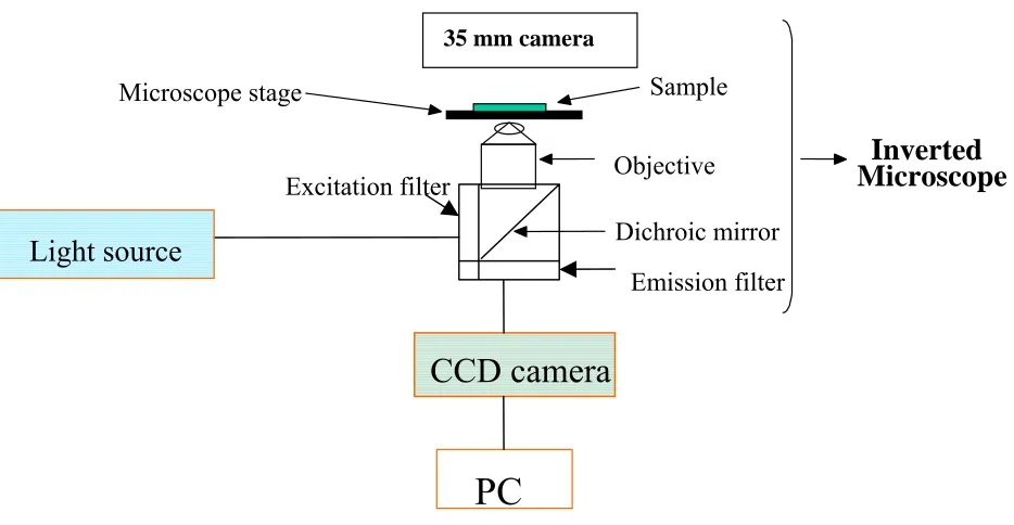
2.3.2 Digital fluorescence imaging microscopy
The digital fluorescence imaging microscopy system, which is used for fluorescence
measurement of particles and cell experiments are shown in Figure 2.1. The system consists an
inverted fluorescence microscopy (Olympus IX-70) equipped with three detection ports. A 100-W
mercury lamp is used as the light source. The fluorescence is collected by a 20x or 40x objective
with a numerical aperture of 0.50, 0.90 respectively. Filter cube containing a excitation filter, a
dichroic mirror and a emission filter was used to obtained the fluorescence images of particles and
cells. A slow scan, high performance charged-coupled device (CCD) camera (Andor technology,
Model DV 434) with a 1024 × 1024 pixel array is employed to collect digital fluorescence images of
the drug containing- particles. A PC-compatible microcomputer is employed for data acquisition
using Roper Scientific software winspec 3.2. The image ProPlus software was used for imaging
analysis.
Figure 2.1 Digital Fluorescence Imaging Microscopy System
2.3.3 Transmission Electron Microscopy
The size and morphology of particles were characterized using JEOL EM8291 electron microscopy. The operation voltage is 200 kV.
Excitation filter
Dichroic mirror
35 mm camera
Objective
Microscope
Inverted
Microscope stage Sample
Emission filter
PC
CHAPTER 3 TUMOR TARGETED DELIVERY OF
ANTICANCER DRUG-DOXORUBICIN USING POLYMERIC
NANOPARTICLES AS CARRIERS
3.1 Introduction
In the present study, we have entrapped the anti-cancer drug Doxorubicin (DXR) in poly
(lactide-co-glycolide) PLGA nanoparticles and evaluated the cytotoxicity on MCF-7 cells.
Doxorubicin (DXR) is one of the most widely prescribed antracycline agents. It has been proven
to be effective against variety of human malignancies, such as leukemia and breast cancer [70].
The mechanism of action of DXR has been extensively investigated. DXR is a
DNA-intercalating agent and a topo-isomerase inhibitor [71]. However, its use is restricted by
problems associated with its cardio and systemic toxicity. To improve the therapeutic index of
DXR, considerable interests have been drawn to develop submicron carriers-associated DXR
formulation such as liposomes [72], micelles [73, 74], and nanoparticles [75-78]. PLGA is the
most commonly used biodegradable polymer in drug delivery systems. It was approved by the
Food and Drug Administration (FDA) in the United States. Also it’s a slow release polymer and
often used to sustain a constant level of drug overtime. Yoo et al conjugated DXR with PLGA,
then formed nanoparticles and achieved high drug loading. They showed maintained drug
conditions to prepare DXR-loaded PLGA nanoparticles and evaluated the potential of PLGA
nanoaprticles as drug carriers for the delivery of doxorubicin into MCF-7 breast cancer cells.
3.2 Specific Experimental and Technical Details
3.2.1 Characterization of nanoparticles
The Shape, surface and size of particles were characterized using Transmission Electron
Microscopy (TEM) (JEOL EM8291). Samples were stained with 2% phosphotungstic acid,
immobilized on copper grids and dried overnight for viewing. Particle size and size distribution
were analyzed by image ProPlus from TEM images taken from different fields.
3.2.2 Fluorescence measurements
Fluorescence spectra of DXR were measured using a PTI international (model QM1)
fluorimeter. A 75-W continuous Xe arc Lamp was used as a light source. Emission spectra of
3.2.3. Loading studies of DXR
DXR-containing nanoparticles were prepared by the method described in chapter 2.
These nanoparticles were washed twice with DI water and collected by centrifugation at 4 oC.
The amount of drug loaded was determined by analyzing the amount of drug present in
supernatant. The supernatant concentration was calculated by measuring the emission at 590 nm
with excitation wavelength 480 nm. All measurements were performed in triplicates.
Encapsulation efficiency was calculated using formula shown below:
% Encapsulation efficiency = (Total DXR- free DXR) /Total DXR
3.2.4 In vitro drug release studies
The in vitro release studies of DXR from nanoparticles were carried out in phosphate
buffered saline of pH 7.4 and pH 5 buffer solutions at 37oC. 1 mL DXR-containing nanoparticle
suspensions were incubated in a water bath at 37 oC. At various time intervals, the supernatants
were isolated by centrifugation for 15 min and their fluorescence spectrum was measured using
the fluorimeter. The concentration of doxorubicin in the supernatants was calculated using
calibration curves constructed using standard doxorubicin solutions in pH 7.4 and pH 5 buffer
3.2.5 In vitro cytotoxic measurements
In vitro cytoxicity against the human breast cancer cell line MCF-7 was determined using
tetrazolium dye (MTT) cell proliferation assay [79], which involved the conversion of 3-(4,
5-dimethyl-2-yl )-2, 5-diphenyltetrazolium bromide into an insoluble formazan by metabolically
active cells. MCF-7 cells were seeded into 96-well plates at a density 1×10⁴
cells/well. One day
later, the medium was removed and DXR-loaded PLGA nanoparticle suspensions were added to
the wells. The cultures were incubated with different concentration DXR-containing PLGA
nanoparticles for 24 hours. After incubation, the cultures were washed three times with sterile
PBS buffer and returned to incubator for a further 48 hours. Then, the cell medium was changed
with 100ul fresh medium and 10µl MTT (5 mg/ ml in sterile PBS buffer) solution. These plates
were reincubated for 4hours. The formed formazan crystals were dissolved in DMSO. The
absorbance was measured at 540nm using a microplate reader. Cells grown in medium alone
were used as a reference as 100% viability. All samples were made in sextuplicates.
3.2.6 Cellular uptake studies by fluorescence microscopy
The cellular uptake of DXR-containing nanoparticles was studied using fluorescence
microscopy. Briefly, MCF-7 cells were grown on coverslips for one day to adhere to the surface.
The cell cultures were then incubated with DXR-containing nanoparticles at final concentration
of DXR at 5 µM for 2 hours. After washed with PBS buffer, the cells were viewed by using a
filter. A slow scan, high performance charged-coupled device (CCD) camera (Andor technology,
Model DV 434) with a 1024 × 1024 pixel array was employed to collect digital fluorescence
images of the cells.
3.3 Results and Discussion
3.3.1 Characterization of PLGA nanopatricles
The ability of nanometric drug carriers to change the biodistribution and pharmacokinetics
of drugs has been found in both vitro and vivo therapeutic applications. Among many factors,
particle size and surface properties have been shown to be of primary importance in determining
the pharmaceutical characteristics of these drug delivery systems. Administered particles of
several micrometers in diameter for example, become entrapped within the lung capillaries.
Smaller particles (< 200 nm) are unique in their ability to benefit from EPR effect and avoid
spleen-filtering effects [80]. In the present study, PLGA nanoparticles were prepared, using an
interfacial polymer deposition (nanoprecipitation) method based on the procedure previous
reported by Fessi et al [68]. The PLGA copolymers assembled in aqueous media following
precipatation from water-miscible organic solvent. Two different mean size particles (less than
200 nm) were obtained by formulating nanoparticles with different polymer concentration
(shown in Table 3.1). TEM analysis revealed that all these particles had a dense, spherical
morphology and size distribution is relatively broad (Fig.3.1). A trend of increasing size with
slightly with increasing polymer concentration. However, the encapsulation efficiency was less
than 10% in both cases. The low drug loading was due to the hydrophilic nature of Doxorubicin
hydrocloride. When drugs were added during the preparation process, they tend to diffuse into
aqueous phase.
3.3.2 Fluorescence studies
The inherent fluorescence of DXR was used to characterize the release properties, cellular
uptake and intracellular distribution of drug-encapsulated nanoparticles. When excited at 480nm,
DXR has fluorescence emission at 590 nm and 560 nm (Fig 3.2a). Fig 3.2b showed the
fluorescence image of DXR- loaded PLGA nanoparticles. Clearly, we can see that DXR was
entrapped in the nanoaprticles. The fluorescence spectrum of DXR-loaded PLGA nanoparticles
was obtained by adding particles into the PBS solution to form the uniform suspension. Fig. 3.3
showed the fluorescence spectra of DXR-loaded PLGA nanoparticles, free DXR and the released
DXR from the nanoparticles.The emission spectrum of the released Doxorubicin was similar to
that of free Doxorubicin. This indicated that the encapsulation of doxorubicin in PLGA
nanoparticles did not affect their structures and spectral properties. The emission of Doxorubicin
encapsulated in PLGA nanoparticles showed a change of the ratio between the two characteristic
emission peaks of doxorubicin. This could be attributed to the change in the chemical
Table 3.1 Nanoparticles formulation and characterization
Concentration of
polymer (mg/mL)
Particle size (nm) Encapsulation
efficiency (%)
a 17 182 8.8 ± 2.1
b 10 110 6.2 ± 1.8
Formulation a Formulation b
520 540 560 580 600 620 640 660 680 700 0
100000 200000 300000 400000 500000
Fl
uor
es
ce
nce I
n
te
ns
it
y
(
a.
u
.)
wavelenth(nm)
Fig. 3.2 a) Fluorescence emission spectra of DXR in PBS solution
Fig 3.2 b) Fluorescence image of DXR-loaded nanoaprticles . a
Fig 3.3 Emission spectra of a) Released Doxorubicin from PLGA nanoparticles, b) free Doxorubicin c) Doxorubicin encapsulated in PLGA nanoparticles. (The fluorescence intensities are not comparable since these samples do not have the same concentration.)
520 540 560 580 600 620 640 660 680 700 0
100000 200000 300000 400000 500000
Flu
o
rescen
c
e Intensi
ty
(a.u.)
Wavelength (nm)
a
b
3.3.3 In vitro release Studies
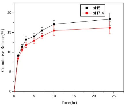
The in vitro release profile of DXR was obtained at 37 oC by representing the percentage
of released DXR with respect to the amount of DXR encapsulated in nanoparticles. Fig. 3.4
showed the release profile of DXR loaded nanoparticles that were prepared with different
polymer concentrations. The profiles exhibited initial burst release, followed by a slow release in
both cases. The release percentage of smaller particles was higher than that of larger particles.
But the difference was not significant. Generally, there are mainly two release mechanisms from
polymeric nanoparticles: diffusion through the microchannels that were formed during
nanoparticles preparation and polymer erosion. In this system, the initial burst release effect is
probably dominated by diffusion, rather than polymer erosion.
To examine the effect of pH on the drug release, we also performed the release studies
in pH 5 buffer solutions keeping all of other conditions the same. At pH 5, the release rate was
slightly enhanced (Fig 3.5). Around 18% and 15% drug content was released in 24 hours at pH 5
and pH 7, respectively. The slight difference in release rate could be attributed to the enhanced
solubility of drug at pH 5, since pH have an effect on the drug solubility due to its influence on
the ionization of the drug. However, it must be noted that the difference between 18% and 15%
in our experiments is too small to come to a clear conclusion. More studies are needed to better
0 5 10 15 20 25 0 5 10 15 20 25 % cu lm u lat iv e re le as e Time(hr)
polymer concentration 10mg/ml polymer concentration 17mg/ml
Fig 3.4 In vitro release profile of doxorubicin from PLGA nanoparticles (pH 7.4, 37oC)
Fig 3.5 Release of Doxorubicin at different pH (polymer concentration 17 mg/ml, 37oC)
0 5 10 15 20 25
3.3.4 Cellular uptake and In vitro cytotoxicity studies
Cellular uptake of DXR-loaded nanoparticles was evidenced by fluorescence microscopy
(Fig 3.6). Fig 3.6a is the transmission image of intact cells, which were incubated with free DXR
and Fig 3.6 b is the corresponding fluorescence image. Fig 3.6c and 3.6d are the images of cells,
which were incubated with DXR-loaded nanoaprticles. These images were taken after 2hours
incubation. It is interesting to note here, free DXR was localized within the cell nucleus and
DXR-loaded nanoaprticles were localized in the perinuclear region. It could be evidence that
DXR-loaded naoparticles were internalized through nonspecific endocytosis due to their small
size.
The cytotoxicity of DXR-loaded nanoparticles and free drug were tested against human
breast cancer cell line MCF-7 by MTT assay [79]. Blank particles and untreated cell culture were
used as control. Fig 3.7 showed cell viability after the cells were exposed to drug-containing
nanoparticles with different concentration of DXR for 24 hours at 37oC under 5% CO2
Fig 3.6 Transmission images of MCF-7 cells treated with (a) free DXR (c) DXR-loaded PLGA nanoaprticles. Fluorescence images of (b) free DXR (d) nanoparticles. In image (b), the drugs were localized within the cell nucleus and in image (d) the drugs were localized in the perinuclear region. The cells were incubated with 5µM equivalent DXR concentration. All images were taken with 400x magnification.
a b
c d
b
d
a
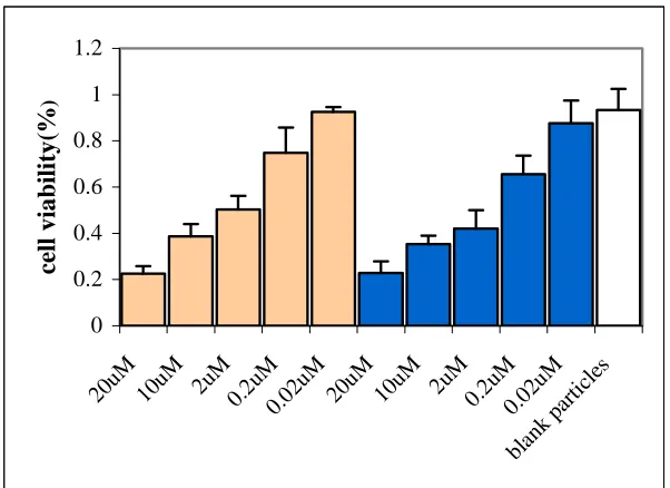
Fig 3.7 The cytocoxicity of DXR-loaded PLGA nanoparticles (mean size 110nm)( ) and free DXR ( ). The density of blank particles is the same as that of drug-load nanoaprticles. Untreated cell cultures were used as a reference as 100% viability.
0 0.2 0.4 0.6 0.8 1 1.2
20uM 10uM 2u M
0.2u M
0.02 uM
20uM 10uM 2u M 0.2u M 0.02 uM blan k pa
As we can see in Fig 3.7, no cytotoxicity was observed when the cells were incubated
with highest concentration of blank particles. But both drug-loaded nanoparticles and free drug
showed concentration dependence cytotoxicity. In both cases, 50% inhibition of cell growth was
achieved with around 2µM DXR (IC50). There is no significant difference about drug activity
between drug-loaded particles and free drug, which indicated that the cytotoxicity of the drug has
been maintained in the nanoparticles.
Numerous studies aimed to develop DXR-loaded colloidal carriers. Some showed
enhanced cytotoxicity of drug-loaded carrier compared to free drug [72, 73, 75]. Other studies
like ours showed comparable cytotoxicity between drug-loaded carrier and free drugs [74, 76].
R. Tomlinson et al. conjugated DXR with polyacetal and in fact reported decreased cytotoxicity
[81]. It seems that cytotoxicity of drug-loaded carrier is mainly affected by the rate of the cellular
uptake and the nature of drug carriers, which determine the release properties in cells.
Doxorubicin is a DNA-intercalating agent, which targets the cells nucleus. Generally, to travel
through the nuclear pore complex, there are two mechanisms depending on molecular size. Small
molecules (< 10 nm) can pass across the nuclear envelope by diffusion. Macromolecule and
particles (< 25 nm) can only pass through by an energy-dependent process [82]. Therefore, to
realize the cytotoxicity of drug-loaded carriers, which is usually larger than 25 nm, drugs must
be released from carrier and diffuse into nucleus. The fact that our 110nm nanoparticles
maintained drug activity suggested that the nanoparticles first attached and then encapsulated in
cellular endosomes. The drugs are then released and diffuse into the cell nucleus. However,
Further studies about determining subcellular drug distribution and delivery kinetics in real time
3.4 Summary and Conclusions
This study systematically evaluated the potential of PLGA nanoparticles as drug carriers
for anticancer drug Doxorubicin for the first time. Doxorubicin was encapsulated into
biodegradable PLGA nanoparticles by simple nanoprecipitation method. The size of particles can
be changed by changing the polymer concentration. Fluorescence studies indicated that the
encapsulation of doxorubicin in PLGA nanoparticles did not affect their structures and spectral
properties. Therefore, we were able to characterize the release properties of system by
fluorescence measurements. The release profiles showed that small particles had a higher release
rate compared to larger particles. Fluorescence images indicated that these particles can be
internalized by human breast cancer MCF-7 cells by non-specific endocytosis. In vitro
cytotoxicity study showed the drug activity of DXR-containing PLGA nanoparticles has been
CHAPTER 4 NOVEL SILICA NANOPARTICLES-BASED DRUG
DELIVERY SYSTEM TRIGGERED BY ANTIMICROBIAL
PEPTIDES
4.1 Introduction
In recent years researchers in the field of drug delivery have attempted to induce
drug release from drug carriers using physical or chemical stimuli [10-14]. For the most part the
studies involved the use of liposomes [83-86]. Drug release from the liposomes was induced by
a change in pH (83), temperature (84), light (85) and magnetic field (86). Polymer nanoparticles
were recently developed as alternative drug carriers to replace liposomes since liposomes often
show limited capability as drug carriers (87). Efficient drug loading into liposomes has proved
difficult. Additionally, drug molecules tend to leak out of liposomes prior to their localization in
a targeted tissue. This decreases the therapeutic efficiency and causes undesirable side effects.
Recently, several groups explored the use of porous silica nanoparticles as drug delivery
carriers [63-66]. Although silica particles are not biodegradable, the potential advantages of
porous silica nanoparticles as drug carriers cannot be ignored. First, it has been shown that silica
nanoparticles are biocompatible [61]. Second, porous silica materials have a large surface area
surface properties of mesoporous silica particles are easily controlled by changing conditions
during their synthesis [88, 89]. Recent studies demonstrated that drug and protein molecules
could be encapsulated with high loading efficiency in porous silica nanoparticles [63, 65, 90].
However, while the loading efficiency was indeed high the leakage rate of the encapsulated
molecules was high as well. A mechanism to minimize leakage of encapsulated molecules from
porous silica particles and trigger the release by a chemical or physical stimulation is needed to
facilitate their use as drug carriers.
In a recent study Lin and coworkers addressed the issue of minimizing leakage and
triggered release from porous silica nanoparticles [66]. Following the encapsulation of ATP
molecules in porous silica nanoparticles the pores of the particles were blocked by the covalent
attachment of CdS nanocrystals to the surface of the particles. This effectively blocked leakage
of ATP from the particles. ATP release was then triggered by incubating the CdS capped silica
particles in a solution that contained reducing agents like dithiothreitol and mercaptoethanol. Lin
and coworkers showed that the CdS nanocrystals-coated silica nanoparticles did not affect the
growth of astrocytes. However, it is reasonable to expect that CdS nanocrstals would not be
ideal plugs to contain drug molecules in porous silica particles. Furthermore, the use of high
levels of reducing agents to induce drug release in vivo may not be a viable option due to
expected cytoxicity of the reducing agents themselves. In this chapter we describe the use of a
phospholipid membrane to block the release of drug molecules from porous silica nanoparticles
(Fig. 4.1) and a new triggering mechanism, based on the use of low levels of antimicrobial
peptides to induce drug release from these particles. The advantages and limitations of this
4.2 Specific Experimental and Technical Details
4.2.1 Characterization of silica nanoparticles
The morphology and size of particles were characterized using Transmission electron
microscopy (TEM). Specimens were prepared by dispersing the as-obtained powder in alcohol
and then placing a drop of suspension on a copper grid coated with transparent graphite,
followed by drying. Particles size and size distribution were analyzed by image ProPlus from
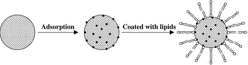
Fig. 4.1. Schematic representation of formation of phospholipid coated mesoporous silica nanoparticles as drug carriers.
4.2.2 Studies of TMR-Dex release profiles
The in vitro release studies of TMR-Dex from nanoparticles were carried out in 10 mM
HEPES buffer solution. 17.5mg TMR-Dex loaded silica lipobeads were dispersed in 10mL
buffer. The solution was sonicated for minute to obtain monodispersity. Then the suspension was
divided into 1 ml ×10 aliquots. At various time intervals, 1 ml suspension was taken and the free
TMR-Dex was washed by centrifugation for 5 min at 12000 rpm. The particles were then
resuspended in 1 mL buffer solution and the fluorescence spectrum of the suspension was
recorded using the fluorimeter. The retained percentage of TMR-Dex at various times was
calculated by comparing the fluorescence intensity of the suspension at t = t’ and t= 0, where t’
represents aspecific time interval. Samples were kept in the dark all the time. All samples were
made in triplicates to enable quantitative data analysis.
To study the effect of temperature, the release was carried out in HEPES buffer solution
at pH 7.4 at room temperature and 42oC.
4.2.3 The effects of antimicrobial peptide cecropin-melittin
The dependence of the %-released contents on peptide concentration was determined in 10
mM HEPES buffer solution at pH 7.4. Different amounts of cecropin-melittin were added to 500
µL TMR-DEX loaded silica lipobeads. Then, the total volume was brought to 1mL by adding
particles were resuspended in 1mL fresh HEPES buffer. The percentage of retained dye was
calculated as described above.
4.3 Results and Discussion
4.3.1 Synthesis of mesoporous silica nanoparticles
Silica nanoparticles are attractive candidates for many applications such as chemical
sensing, ion-exchange coating and chromatography [91]. Extensive studies were carried out
using silica materials which were prepared by the sol-gel process. In a typical sol-gel process,
tetraalkylsilane is mixed with water followed by the addition of catalyst. Tetraalkylsilane can be
hydrolyzed and condensed to form the sol and the sol is further crosslinked through
polycondensation to form a rigid, porous network-gel. However, the limitation of sol-gel
materials is their variability in pore size, which cannot be tailored for specific molecules.
Discovery of mesoporous silica materials made it possible to tailor both pore size and structure
of these materials for specific hosts. Mesoporous silica materials are synthesized by
self-assembly of silica-surfactant in which inorganic species simultaneously condense, giving rise to
mesoscopiclly ordered composites formation [69, 92]. Well-defined pore size depends mainly on
the surfactant, which is employed as a template in the synthesis. In our study, mesoprous silica
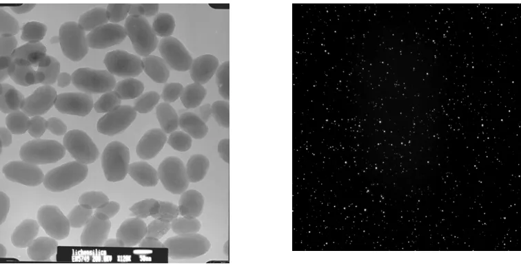
nanoparticles were synthesized using a method developed by Cai [69]. TEM images revealed
that the average size of nanoparticles was around 90 nm with narrow size distribution. The
Fig. 4.2 (A) TEM (B) Fluorescence images of TMR-DEX loaded silica nanoparticles. The average size of the nanoparticles was around 90 nm.
4.3.2 Loading studies of TMR-Dex into silica nanoparticles
To easily monitor the drug- loading and release properties of the particles, we chose the
fluorescent dye-tetramethylrhodamine dextran (3000Da)(TMR-Dex), which has high extinction
coefficient, stability and pH insensitivity, as a drug analog. To load the dye into the particles,
mesoporous silica nanoparticles were dispersed in TMR-Dex solution at pH 3 for 24 hours.
Electrostatic attraction between the protonated TMR-Dex due to the acidic conditions and the
negatively charged silica enhanced the entrapment efficiency of TMR-Dex in the silica particles.
The dye content in the particles was measured by monitoring the difference in the fluorescence
of the preparation solution prior and following dye entrapment into the particles. The loading
efficiency was about 28±2%. However, TMR-Dex molecules diffused out of the particles under
4.3.3 Lipids membrane coating of TMR-DEX-loaded silica nanoparticles
Liposomes were extensively studied for drug delivery and chemical sensing. They have a
flexible, cell-like lipid bilayer surface, which acts as a permeability barrier such that compounds
can be entrapped in their aqueous interior. However, liposomes can be mechanically unstable
and their loading capacity is relatively low compared to solid particles. Previously, lipid bilayers
supported on various solid surfaces, such as glass [93], plastic [94], and metal [95] as well as
modified polymers [96] have been shown to provide a stable and well-defined
cell-membrane-like environment. Based on this knowledge, several groups proposed hybrid vesicle systems,
which have polymer nanoparticles as core and lipid bilayer as shell [97-100], termed lipobeads.
In our group, lipobeads were used for intracellular sensing [99,100]. Our studies showed that the
phospholipid membrane protected sensing elements from cellular environment.
The objective of our study was to realize stable drug containing nanoparticles that only
release their content instantly when stimulated. To this end the particles were coated with a
phospholipids membrane using a procedure previously developed in our laboratory for the
fabrication of lipobead-based nanosensors. The silica lipobeads were washed several times to
remove dye molecules that leaked out of the particles prior to applying the phospholipid
membrane and re-adsorbed to the membrane following the formation of the membranal coating.
Temporal release profiles shown in figure 4.3 indicate that the phospholipids membrane
efficiently blocked leakage of dye molecules from the silica lipobeads. Almost 95% of the
90% of the TMR-Dex molecules leaked from uncoated silica matrix in 12 hours following their
preparation.
To examine the effect of temperature, the release experiment also carried out at 42 oC
with silica DMPC-lipobeads. We expected an increased release since DMPC had a phase
transition at 24 oC [101]. Above the phase transition temperature, lipid bilayers undergo a change
in structure, switching from a gel state where acyl chains of the lipids molecules are closely
organized, to a sol state where the acyl chins are disorganized. Therefore, the lipid bilayer is
more fluid. Surprisingly, the release was the same as that of room temperature within 4 hours
(Fig. 4.4). After this time, the release increased rapidly. The possible reason is that the TMR-Dex
molecule were too large to pass through bilayer membrane even at sol state. But overheating
(longer than 4 hours) could destabilize the lipid membrane. The same experiments were carried
0 100 200 300 400 500 600 700 800 0.0 0.1 0.2 0.3 0.4 0.5 0.6 0.7 0.8 0.9 1.0 1.1 Frac tio n o f re taine d Time (min) silica lipobeads silica partilces
Fig. 4.3 Release profile of TMR-Dex loaded silica particles in 10mM HEPES pH7.4 buffer solution.
0 100 200 300 400 500 600 700 800
0.0 0.1 0.2 0.3 0.4 0.5 0.6 0.7 0.8 0.9 1.0
Fraction of tetained
Time (min)
4.3.4 The induced release of TMR-DEX by antimicrobial peptide
As mentioned previously, the development of drug carriers that effectively encapsulate
the drug with minimal leakage and only release it when triggered chemically or physically would
be highly beneficial to many clinical situations. The selection of an appropriate triggering
strategy is crucial. For example, pH- sensitive drug carriers would release their content as a
result of a pH change in a tissue. In many cases the pH required for effective release is
unreachable. Similarly, drug carriers that rely on temperature changes to induce release are
limited because of the narrow temperature range under physiological conditions and the
relatively wide range of body temperature of normal patients. In this study we explored the use
of low concentrations of antimicrobial peptides to induce the release of our fluorescent drug
analog TMR-Dex from the silica lipobeads. Antimicrobial peptides are a group of small peptides
that show a broad range of activity against Gram-negative and Gram-positive bacteria, fungi,
mycobacteria and some enveloped viruses [102]. These peptides attracted increasing attention in
recent years since they represent a promising new alternative for conventional antibiotic drugs.
These peptides could provide a solution to the growing problem of antibiotic resistance [102].
The mechanism of action of these peptides involves increasing cell membrane permeability
either by forming aqueous channels which span the membrane bilayer or by disrupting
membrane organization [102,103]. We predicted that antimicrobial peptides could disrupt the
membranal coating of the silica lipobeads to facilitate the release of TMR-Dex from the
particles. To test this triggered release strategy, our TMR-Dex containing lipobeads were
incubated with solutions of the antimicrobial peptide- Cecropin A(1-8)-Melittin(1-18) hybrid.
1,2-Dimyristoyl-sn-Glycero-3-Phosphocholine (DMPC). Temporal release profiles of TMR-Dex silica lipobeads
upon addition of antimicrobial peptide are shown in figure 4.5. Curve a shows a control
experiment in the absence of the peptides in which no release is seen, indicating that the
membrane remained intact throughout the experiment. Curves b shows that the release was
rapidly triggered after the addition of 0.175 mg/mL Cecropin A-Melittin. About 70% of dye
contents were released. Also, the dependence of the %-released contents on peptide
concentration was studied. As Fig. 5 showed, a maximum release was obtained at 0.350mg/mL
peptides and minimum peptide concentration 0.044mg/mL was required to induce the release
effectively.
The effect of Cecropin A(1-8)-Melittin(1-18) hybrid on the negatively charged
phospholipids Bovine brain phosphatidylserine (PS) coated silica particles was also tested. As
Fig.4.6 and 4.7 showed, the cecropin A-melittin is more effective on the negatively charged PS
lipobeads than on the neutral DMPC lipobeads. This may result from the cationic nature of the
peptide at pH 7. Further studies about dependence of the %-released contents of PS lipobeads on
0 50 100 150 200 250 0.0 0.1 0.2 0.3 0.4 0.5 0.6 0.7 0.8 0.9 1.0 1.1 Fraction of retained Time (min)
silica particles coated with DMPC lipids
silica particles coated with DMPC +Cecropin A-melittin
Fig. 4.5 Release profiles of silica DMPC-lipobeads treated with 0.175 mg/mL Cecropin A-melittin peptide. 0 0.2 0.4 0.6 0.8 1 1.2 0 Triton X-100
0.35 0.175 0.875 0.044 0.022 0.008 0.004
Peptide concentration (mg/mL)
Fr a c ti on of r e ta in e d
Fig. 4.6 Peptide concentration-dependent release. (The measurements were taken 2 hours after addition of cecropin-melittin peptide).
a
540 560 580 600 620 640 660 680 700 0 2 4 6 8 10 12 14 16 silica DMPC-lipobeads
silica DMPC-lipobeads +1% triton x-100 silica DMPC-lipobeads +0.175 mg/mL peptides
Fluorescence Intensity
(a.u.)
wavelength (nm)
Fig. 4.7 Fluorescence spectra of silica DMPC-lipobeads (Measurements were taken 2 hours after peptides were added)
540 560 580 600 620 640 660 680 700 0 2 4 6 8 10 12 silica PS-lipobeads
silica PS-lipobeads + 0.175mg/mL peptides silica PS-lipobeads + 1% Triton X-100
Fl uor es cen ce I n te ns ity ( a.u.) wavelength (nm)
4.4 Summary and Conclusions
In summary, we have developed a novel silica nanoparticles-based drug delivery system,
which enables high drug loading and regulated drug release by adjusting the level of an adjuvant
drug. While this approach show clear advantages over physical stimulation like temperature
change or other non-selective and unregulated pH changes the toxicity of antimicrobial peptides
still remained a concern. Future work is in progress to seek a lipid membrane which would be
CHAPTER 5 SUMMARY AND CONCLUSIONS
Controlled drug delivery can influence the performance of a drug by manipulating its
concentration, location and duration. Therefore, it provides a promising way to minimize side
effects and increase therapeutic efficacy. In the past decades, controlled drug delivery technology
has represented one of the frontier areas of science, which involves multidisciplinary scientific
approach. This thesis describes the development and characterization of new nanoparticles-based
drug carriers with unique release properties.
Chapter 3 describes the development of a polymeric nanoparticle-based drug delivery
system for the anticancer drug doxorubicin. Biodegradable poly (lactide-co-glycolide) (PLGA)
particles of various sizes were prepared by changing the polymer concentration. Transmission
electron microscopy (TEM) images revealed that the particles had smooth spherical morphology
with a mean size ranging from 110nm to 180nm. The in vitro release profile showed that small
particles (110nm) had a higher release rate compared to larger particles (180nm). For all
nanoparticles prepared with different polymer concentration, an initial burst release, followed by
a slow release, were observed. These results indicated that the drugs were released through a
diffusion-controlled release mechanism. Fluorescence imaging studies showed that the
nanoparticles could be internalized by MCF-7 human breast cancer cells. Interestingly, different