Theses and Dissertations
2017
The Role Of Inflammation In Atherosclerosis
Fatma SaaoudUniversity of South Carolina
Follow this and additional works at:https://scholarcommons.sc.edu/etd
Part of theBiomedical and Dental Materials Commons
This Open Access Dissertation is brought to you by Scholar Commons. It has been accepted for inclusion in Theses and Dissertations by an authorized administrator of Scholar Commons. For more information, please [email protected].
Recommended Citation
Saaoud, F.(2017).The Role Of Inflammation In Atherosclerosis.(Doctoral dissertation). Retrieved from
By Fatma Saaoud
Bachelor of Medicine and Surgery Zawia University, 2005
Master of Science
University of South Carolina, 2012
Submitted in Partial Fulfillment of the Requirements For the Degree of Doctor of Philosophy in
Biomedical Science School of Medicine University of South Carolina
2017 Accepted by:
Daping Fan, Major Professor Susan Lessner, Committee Member
Holly LaVoie, Committee Member Angela Murphy, Committee Member Saurabh Chatterjee, Committee Member
To the best people I have ever had in my life, my parents, Elmokhtar Saaoud and Salma Saaoud, for their endless love, support, and encouragements throughout my life pushing me always toward success.
To my dear husband, Ismail Ben Issa, I give my deepest expression of love and appreciation for your support, encouragement, and the sacrifice you made during this graduate program. Thank you for your practical and emotional support.
First and for most, I would like to thank my advisor, Dr. Daping Fan, for his admirable guidance, excellent advice, and patience over the past several years. I am very grateful to him for providing me such opportunity to work in his laboratory and great training that is valuable to my future development. I would like to thank all current and past members of Dr. Fan’s lab, in particularly Xuemei Jia and Junfeng Wang for their invaluable training and assistance since the first day I joined the group. I would like to thank Yuzhen Wang for her help with the histological analysis. I would like to thank my committee members Dr. Susan Lessner, Dr. Angela Murphy, Dr. Holly LaVoie, and Dr. Saurabh Chatterjee for serving on my dissertation committee and for their comments, help and advice.
Dedication ... iii
Acknowledgements ... iv
Abstract ... v
List of Tables ... ix
List of Figures ... x
Chapter I: Introduction ... 1
1.1 Atherosclerotic cardiovascular diseases ... 1
1.2 Structure of blood vessel wall ... 2
1.3 Pathogenesis of atherosclerosis ... 3
1.4Animal models of atherosclerosis ... 13
1.5 Bone marrow transplantation and total body irradiation ... 15
1.6 microRNA-155 in atherosclerosis ... 16
Chapter II: Inflammation markers in human atherosclerotic lesions and the role of microRNA-155 in vascular calcification ... 18
2.1 Background ... 18
2.2 Material and Methods ... 20
2.3 Results ... 25
2.4 Discussion ... 31
Chapter III: The role of Tristetraprolin (TTP) in inflammation and atherosclerosis ... 43
3.3 Results ... 55
3.4 Discussion ... 64
Chapter IV: The effect of macrophage-specific GP-96 deficiency on inflammation and atherosclerosis development ... 89
4.1 Background ... 89
4.2 Material and Methods ... 92
4.3 Results ... 98
4.4 Discussion ... 100
Chapter V: The therapeutic value of an anti-inflammatory Chinese herb compound, SsnB, for atherosclerosis ... 111
5.1 Background ... 111
5.2 Material and Methods ... 113
5.3 Results ... 116
5.4 Discussion ... 120
Chapter VI: Summary and conclusion ... 133
Table 2. 1 Primers that were used for qRT-PCR in this study ... 34
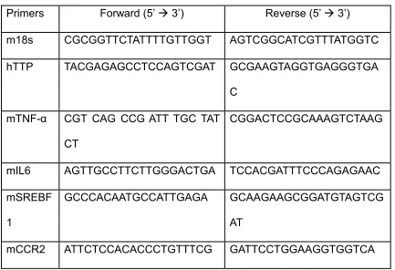
Table 3. 1 Primers that were used for qRT-PCR in this study……… 70
Table 4. 1 Primers that were used for qRT-PCR in this study……….. 103
Figure 2. 1 Increased expression of inflammation markers in human carotid atherosclerotic atheroma ... 36 Figure 2. 2 Increased expression of miR155 in human carotid
atherosclerotic plaques. ... 37 Figure 2. 3 Expression levels of miR155 correlate with the levels of
inflammatory cytokines and MMP-9 in human carotid artery atheroma ... 38 Figure 2. 4 The expression of miR155 and calcification gene in human
atherosclerotic carotid artery ... 39 Figure 2. 5 miR155 deficiency attenuates calcification in vascular
smooth muscle cells ... 40 Figure 2. 6 miR155 deficiency attenuates aortic calcification in vivo and ex vivo ... 42 Figure 3. 1 In vivo experimental design, mouse model and bone marrow
transplantation used for assessment of atherosclerosis ... 71 Figure 3. 2 TTP expression is increased in human carotid
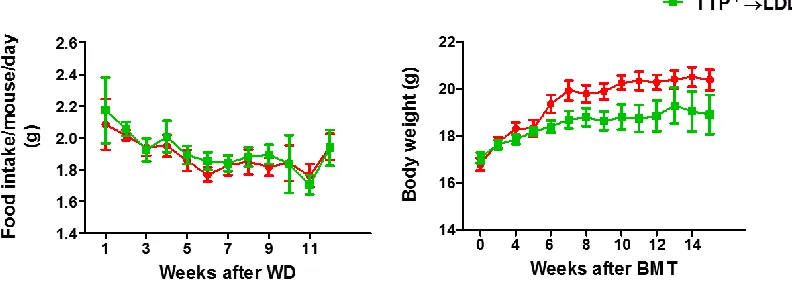
atherosclerotic lesions ... 72 Figure 3. 3 Bone marrow TTP deficiency causes growth retardation, but did not affect food intake ... 73 Figure 3. 4 Bone marrow TTP deficiency causes hematological disturbance ... 74 Figure 3. 5 Bone marrow TTP deficiency causes immunological disturbance .... 75 Figure 3. 6 TTP deficiency increased macrophage responses to LPS
stimulation, and bone marrow TTP deficiency resulted in extensive
Figure 3. 9 Bone marrow TTP deficiency alters expression of liver genes
involved in inflammation and lipid metabolism ... 82 Figure 3. 10 Efficiency of macrophage-specific TTPdeficiency in the bone
marrow transplantation model ... 84 Figure 3. 11 macrophage-specific TTP deficiency did not affect mouse food intake, but reduced body weight ... 85 Figure 3. 12 Effect of macrophage-specific TTP deficiency on inflammation. ... 86 Figure 3. 13 Effect of macrophage-specific TTP deficiency on plasma lipid levels and lipoprotein profile ... 87 Figure 3. 14 Effect of macrophage-specific TTP deficiency on
atherosclerosis ... 88 Figure 4. 1 In vivo experimental design, mouse model and bone marrow
Introduction
1.1 Atherosclerotic cardiovascular diseases
Cardiovascular diseases (CVDs), including myocardial infarction (heart attack) and angina pectoris (chest pain), represent the leading causes of mortality and long-term morbidity worldwide1. The incidence of CVDs continues to rise and
is projected to remain as the number one cause of death globally by 20202.
Atherosclerosis is by far the main underlying pathological process of coronary artery disease, carotid artery disease, and peripheral arterial disease — which are among the most prevalent CVDs3.
The term ‘atherosclerosis’ comes from Greek word "athero" meaning gruel,
and sclerosis means hard — which is hardening with loss of elasticity leading to
narrowing of the arteries. Atherosclerosis is both a lipid deposition disorder and a chronic inflammatory disease, characterized by formation of atherosclerotic plaque in the arterial wall4,5. It mainly affects medium-sized and large arteries such as: the
aorta, coronary arteries, carotid arteries, and renal arteries, in focal areas —
particularly regions in which laminar flow is disturbed (e.g. arterial branch points
and bifurcations)6,7. The disease begins early in life and goes undetected until
thrombotic complications, such as acute coronary syndromes and stroke occur8.
There are numerous genetic and environmental risk factors associated with
usually associated with hypercholesterolemia, smoking, diabetes mellitus, hypertension, obesity, physical inactivity, family history, and high-fat diet (HFD)8-9.
It has been shown that hypercholesterolemia alone is sufficient to drive the development of atherosclerosis. However, the presence of other risk factors appears to accelerate disease progression, driven by the atherogenic lipoproteins (i.e. low-density lipoprotein; LDL)10.
1.2 Structure of blood vessel wall
The normal wall of blood vessels consists of endothelial cells, vascular smooth muscle cells (VSMCs), and extracellular matrix (ECM) arranged in three different layers (from innermost to the outermost): the intima, media, and adventitia11. The tunica intima is composed of a single monolayer of simple
squamous cells known as endothelial cells. It lies on the internal elastic lamina, a layer of elastic fibers, which defines the outermost boundary of the intimal space and separates the intima from the media. The endothelial layer represents the interface between circulating blood flow and the vascular wall — providing a smooth lining for blood vessels12. The endothelial cells have a significant role in
regulation of vascular permeability, regulation of immune responses, modulation of blood flow and vascular resistance, platelet aggregation, and thrombosis prevention. These are achieved by releasing platelet, coagulant, anti-thrombogenic, and fibrinolytic factors in addition to the production of the potent vasodilator nitric oxide (NO)13. Besides the vasodilators, endothelial cells also
to plaque formation . Therefore, the ability of the endothelial cell layer to repair itself and to maintain functional and structural integrity, is key to the prevention of atherosclerosis16,17.
The middle layer of the arterial wall is the tunica media, which is composed primarily of VSMCs surrounded by a network of ECM proteins including elastic fibers, collagen, and proteoglycans. This layer provides strength and elasticity to the blood vessels so they can stretch or expand without tearing18. The outermost
layer is the tunica adventitia, which is primarily composed of irregularly arranged collagen bundles, scattered fibroblasts, a few elastic fibers, and small blood vessels called vasa vasorum. Vasa vasorum provides oxygen and nutrients to outer layers of the vascular wall. The elastic collagen fibers allow arteries to stretch to prevent over-expansion due to higher pressures in these blood vessels19-20.
Understanding the structure and function relationship of each layer in the arterial wall provides insight into the pathophysiology of arterial wall diseases, such as atherosclerosis, and allows for better predictions for prevention and treatment strategies.
1.3 Pathogenesis of atherosclerosis
Atherosclerosis is a complex and multifactorial disease characterized by systemic and vascular wall inflammation and activation of immune pathways, dyslipidemia, endothelial dysfunction, foam cell formation, VSMC activation, platelet activation, and thrombosis21,22. A number of hypotheses have been
hypothesis25, lipid hypothesis26, and inflammation hypothesis27. A better
understanding of the pathogenesis of atherosclerosis will aid in formulating preventive and therapeutic strategies to reduce the mortality rate of cardiovascular events.
1.3.1 Response to injury hypothesis
The response to injury hypothesis suggests that atherosclerosis is initiated with injury-induced damage and dysfunction of the endothelial cells lining the intima. This damage caused by local disturbances of blood flow at branch points, along with systemic risk factors, perpetuates a series of events that culminate in the development of atherosclerotic plaque. Endothelial dysfunction is the initial step that allows entry and retention of cholesterol-containing LDL particles and inflammatory cells (i.e. monocytes, T lymphocytes) into the sub-endothelial spaces, altering normal homeostasis28,29,30. Under normal homeostatic
conditions, the white blood cells do not adhere to the endothelial cells lining the intima. However, under atherogenic conditions, the damaged endothelial cells express surface adhesion molecules such as vascular adhesion molecule-1 (VCAM-1), leading to adhesion of circulating blood monocytes to the endothelial surfaces. Those monocytes ultimately migrate to the sub-endothelial space. When acting together with imbalance between vasoconstriction and vasodilation, these processes promote increased vascular permeability and platelet aggregation31.
The response to retention hypothesis of atherosclerosis was initially proposed in 199524. Since then there has been several line of experimental
evidence supporting this hypothesis. The response to retention hypothesis suggests that retention of cholesterol-rich, atherogenic lipoproteins within the sub-endothelial space is the key initiating event in early atherosclerosis. Once retained, these lipoproteins increased susceptibility for modification/oxidation, and subsequently provoke the inflammatory response with increasing smooth muscle cell migration and phenotype switching as well as infiltration of inflammatory cells including macrophages24,30.
1.3.3 LDL oxidation hypothesis
The LDL oxidation hypothesis suggests that oxidative modification of lipoproteins, in particular LDL, within the arterial wall by reactive oxygen species (ROS) plays an important role in the pathogenesis of atherosclerosis25.
Monocytes/macrophages express scavenger receptors that engulf oxidized oxLDL in an uncontrolled manner. In turn, uptake of oxLDL leads to accumulation of lipids and lipid-laden foam cells to form a fatty streaks — an early feature of atherosclerotic plaque formation25,33,34. Experimental data indicate that oxLDL
within the arterial wall promotes the development of atherogenesis25. Additionally,
involved in LDL oxidation and uptake of oxLDL (e.g. 12/15 lipoxygenase and scavenger receptors (SRA and SRB)) in various atherogenic murine models significantly attenuated the severity of atherosclerosis36–38.
1.3.4 Lipid hypothesis
The lipid hypothesis of atherosclerosis states that both lesion initiation and progression of atherosclerosis is primarily associated with hyperlipidemia26. In the
last decade, considerable advances in the understanding of cardiovascular risk factors have been established, and dyslipidemia has been shown as one of the most powerful risk factors. Controlling blood cholesterol is efficient at reducing cardiovascular risk and an important therapeutic decision, as the rate of CVDs were significantly reduced with decreased blood cholesterol39. The link between
hyperlipidemia and atherosclerosis predominate, until the 1970s, based on strong experimental and clinical relationships between hypercholesterolemia and severity of atherosclerotic lesions. This was further supported by the fact that statins, lipid lowering agents, were able to significantly reduce atherosclerotic diseases40.
The first description of an atherosclerotic lesion was recorded in 1908 when Alexander I. Ignatowski, an experimental pathologist, fed rabbits cholesterol for 17 weeks followed by chow diet for 14 weeks. These rabbits developed pronounced aortic atherosclerosis41. In 1913, Anitschkow and colleagues proposed the
atherosclerotic lesions similar to those of human atherosclerosis .
In order to examine the lipid hypothesis of atherosclerosis induced by cholesterol, Clarkson and Newburgh (1926) fed rabbits a normal diet with various cholesterol doses for 47- 87 days. They discovered atherosclerosis in rabbits fed high cholesterol doses43. These observations strongly supported the theory that
cholesterol was the main driving force in the development of atherosclerotic CVDs44.
In the early stages of atherosclerosis, plasma LDL crosses the endothelial barrier and enters the intima. Once accumulated in the sub-endothelial arterial intima, LDL undergoes oxidative modification via the reactive oxygen species produced by macrophages and damaged endothelial cells, forming oxLDL34,22,45.
oxLDL is a pivotal molecule representing the initial event in atherosclerotic lesion formation and progression by promoting inflammation, foam cell formation, and smooth muscle cell migration/proliferation46. oxLDL is also considered a potent
chemo-attractant and strong pro-inflammatory stimulus, which recruits the circulating blood monocytes and T-cells to the sub-endothelial space47. Once
retained in the intima, oxLDL activates endothelial cells and up-regulates the expression of adhesion molecules, such as VCAM-1 and intercellular adhesion molecule-1 (ICAM-1), and the secretion of chemokine/cytokines that contribute to the recruitment of circulating leukocytes48,49. oxLDL taken up by activated
more monocytes to the intima and potentiate the inflammatory response . The role of plasma lipoproteins in atherosclerosis development has gained significant attention and has been demonstrated by randomized controlled trials. These studies revealed the rate of cardiovascular events were significantly reduced via inhibition of cholesterol biosynthesis using lipid lowering agents, mainly statins. Statins act by inhibiting 3-hydroxy-3-methyl-glutaryl-coenzyme A reductase (HMG-CoA Reductase); the rate limiting step in the cholesterol biosynthesis pathway40. The lipid hypothesis of atherosclerosis is now considered
fact, supported by: 1) epidemiological studies that have shown a positive relationship between total cholesterol concentrations and the mortality rate of CVDs, and 2) the success of statin drug therapy in the past 40 years; statins significantly reduced atherosclerotic disease mortality through reducing plasma LDL levels39.
1.3.5 Inflammation hypothesis
In the last century, it was believed that atherosclerosis was merely a lipid storage disease, associated with hyperlipidemia and accumulation of cholesterol and fatty streaks in the arterial wall. Over the past two decades, studies demonstrated the complexity of atherosclerosis and the involvement of both the innate and adaptive immune systems by detecting different immune cells (e.g. monocytes, macrophages, dendritic cells (DCs) and T cells) associated with production of immune mediators within the arterial wall21. Presently, the role of
atherosclerosis from lesion initiation, through progression, and ultimately the thrombotic complications of atherosclerosis51.
Inflammation in the arterial wall proceeds as a cascade, which begins with endothelial cell activation and expression of surface adhesion molecules such as VCAM-1, ICAM-1, selectins, and integrins that increase the adhesion of circulating blood monocytes to the endothelial cell layer lining the blood vessel wall52,53. Furthermore, the activated endothelial cells will secrete
chemo-attractants, such as monocyte chemoattractant protein-1 (MCP-1) also known as CCL2, that interact with chemokine receptors on blood monocytes and promote their recruitment and entry into sub-endothelial intimal space in a phenomenon known as diapedesis54,55,56. Once the blood monocytes migrate into the
sub-endothelial space, they activate and differentiate into macrophages, stimulated by macrophage colony stimulating factors (M-CSF)57–59. These activated macrophages exhibit high expression levels of surface recognition receptors called scavenger receptors (SR-A and CD36), which have the ability to engulf oxLDL and form lipid-laden foam cells within the sub-endothelial space. Further, these activated macrophages secrete inflammatory cytokines/chemokines including MCP-1, interleukin-1 (IL-1), and tumor necrosis factor-α (TNF-α) that increase adhesion and recruitment of monocytes into the lesion, potentiating the inflammatory response of atherosclerosis60,60,61. Over time, the lipid-laden foam
of collagen fibers, and migration of smooth muscle cells from media into the intima. This ultimately leads to the irreversible formation of fibrous atherosclerotic plaques. Fibrous lesions usually have a fibrous cap composed of smooth muscle cells and ECM, which encloses a lipid-rich, necrotic core62. In addition to macrophages,
atherosclerotic lesions contain other immune cells including dendritic cells, B lymphocytes, and T lymphocytes, notably CD4+ T cells and regulatory T cells,
which regulate many innate immune pathways63–65.
Vascular smooth muscle cells (VSMCs) play a fundamental role in atherosclerosis development66. The inflammatory mediators such as
cytokines/chemokines and growth factors released by injured endothelial cells and inflammatory cells stimulate migration of VSMCs from the underlying media to the sub-endothelial space resulting in intimal area expansion. Additionally, VSMCs will switch from quiescent contractile phenotype to the proliferative, synthetic phenotype, characterized by excess production of ECM. This results in deposition of ECM proteins and formation of a fibrotic cap that covers the necrotic core and stabilizes/prevents plaque rupture66–69. Over time, these lesions become calcified and grow toward the adventitia. At a certain point, they begin to encroach to the lumen leading to narrowing of the blood vessels and the complications associated with atherosclerosis70.
alternatively activated macrophages in advanced atherosclerosis leads to impaired resolution71,72. One important function of M2 macrophages is the clearance of
apoptotic cells via a process called efferocytosis.Efferocytosis is a term refer to the engulfment or phagocytosis of antigen presenting cells such as macrophages and dendritic cells73. As macrophages engulf oxLDL in the arterial wall during early
stages of atherosclerosis, these macrophages undergo apoptosis74. Apoptosis is
a physiological, programmed, and energy-dependent cell death cascade. In early atherosclerosis, macrophage apoptosis is associated with reduced atherosclerosis progression. This is most likely due to effective efferocytosis by M2 macrophages75,76. Efficient efferocytosis has been shown to induce
anti-inflammatory mediators, such as interleukin-10 (IL-10) and transforming growth factor-β (TGF-β)77. As atherosclerosis progresses, efferocytosis is thought to
become impaired. A lack of efferocytosis leads to secondary necrosis, where macrophages die and release their cellular contents including oxidized lipids and pro-inflammatory mediators. Secondary necrosis amplifies the inflammatory response and leads to the development of a necrotic core in the plaque78.
atherosclerosis. Therefore, several anti-inflammatory compounds have been examined as potential therapeutic strategies for the prevention of atherosclerotic complications using various atherosclerotic animal models82. Despite strong
evidence arising from animal studies that lowering inflammation may be a promising strategy for decreasing atherosclerosis and its complications, results have yet to be recapitulated in humans. Various protein therapeutic strategies, such as anti-cytokine therapies, have received noticeable appreciation for clinical application because of their potential direct anti-inflammatory effects. However, these strategies will require extensive clinical evaluation and testing in randomized trials before implementation into practice.
There are two large randomized controlled clinical trials testing the inflammation hypothesis of human atherosclerosis. These studies aim to evaluate whether inhibiting inflammation will decrease event rates and improve prognosis among patients with heart disease using anti-inflammatory agents. The Canakinumab Anti-inflammatory Thrombosis Outcomes Study (CANTOS) is evaluating whether interleukin-1β (IL-1β) inhibition by anti-human IL-1β monoclonal antibody (Canakinumab) can reduce the rates of myocardial infarction, stroke, and cardiovascular death among patients with a history of previous myocardial infarction and elevated levels of high-sensitivity C-reactive protein (hs-CRP) — a clinical marker for chronic inflammatory conditions83. This study showed
levels and provide a novel cytokine-based therapy for the secondary prevention of CVDs84. The second clinical trial that is still on-going is Cardiovascular
Inflammation Reduction Trial (CIRT) which aims to elucidate whether treatment with low-dose methotrexate (a common drug used in the treatment of autoimmune disorders such as rheumatoid arthritis and psoriasis arthritis) will reduce major vascular events among patients with a history of myocardial infarction85.
1.4Animal models of atherosclerosis
Atherosclerosis animal models have been developed to understand the cellular and molecular mechanisms of atherosclerosis pathogenesis, and to drive research that evaluates the efficacy of newly developed preventive and therapeutic atherosclerotic drugs86. Animal models used to study atherosclerosis and
thrombosis include: mouse, rat, rabbit, and pig87. The majority of animal models
are based on the presence or induction of hyperlipidemia, primarily hypercholesterolemia, which is the most important risk factor influencing atherosclerosis development. In 1908, Ignatowski demonstrated that the experimental atherosclerosis could be induced in animals, when he fed rabbits a diet enriched in animal proteins including milk, eggs, and meat. At the experimental endpoint, these rabbits presented with atherosclerotic lesions in the aortic wall41,44.
After that, several animal models were employed to understand the mechanisms involved in both induction and regression of atherosclerotic lesions87,88.
manipulate . The development of atherosclerosis in mice is typically based on genetic modifications of lipoprotein metabolism, including deletion of LDL-receptor (LDLR) or apolipoprotein-E (apoE) gene, followed by administration of a high fat diet to induce hyperlipidemia. High-fat diet (HFD) in experimental research has been found to elevate serum LDL and atherosclerosis in different atherosclerotic animal models, including mice91. The apoE-deficient (apoE−/−) mice and
LDLR-deficient (LDLR−/−) mice are the most widely used murine models92,93.
Apolipoprotein-E is a plasma lipoprotein secreted from liver and promotes hepatic binding, uptake, catabolism, and clearance of triglyceride-rich lipoproteins such as very low density lipoprotein (VLDL) and low density lipoprotein (LDL)94. In apoE−/−
mice, the total plasma cholesterol levels are dramatically increased and extensive atherosclerotic lesions form (widely distributed throughout the aorta) even in the absence of cholesterol– enriched diet, where lesions progress and advance with age95. LDLR is a cell-surface receptor expressed in different cell types, where they
consequently, massive accumulation of plasma LDL93. Thus, the LDLR−/− mouse
model is characterized by elevated plasma cholesterol levels and develop atherosclerotic lesions slowly when fed a normal chow diet. When fed HFD, LDLR−/− mice develop hypercholesterolemia and extensive lesions throughout the entire aorta and aortic root97.
1.5 Bone marrow transplantation and total body irradiation
Lethal total body irradiation of atherosclerotic mice followed by bone marrow transplantation from donor mice with transgenic alterations in the innate and adaptive immune systems is a common method utilized to reconstitute the immune system and experimentally used to identify the role of bone marrow-derived cells in atherosclerosis. The experimental mice are compared to control mice that have undergone comparable irradiation and bone marrow transplantation with syngeneic wild-type bone marrow cells98. This technique is commonly used in
atherosclerosis research to determine the contribution of different hematopoietic cells of interested genotype to the pathogenesis of atherosclerosis99. The high
produced by the transplanted bone marrow cells are sufficient to reverse the atherogenic phenotype101. After bone marrow transplantation, mice were allowed
4 – 5 weeks to reconstitute the hematopoietic and immune system. After this period, majority of the myeloid cells in peripheral blood and bone marrow will be a donor derived cells98.
1.6 microRNA-155 in atherosclerosis
MicroRNAs (miRNAs) are small, noncoding RNA molecules that regulate gene expression at the post-transcriptional level through base pairing with mRNAs, resulting in either translational repression or mRNA degradation. The expression and targets of miRNAs are cell type dependent, which determines the biological function of miRNAs102,103. microRNA-155 (miR155), a typical multi-functional
miRNA, has recently emerged to play a significant role in atherosclerosis development104. It has been shown that a hematopoietic miR-155 deficiency
reduced atherosclerosis in partial carotid artery ligation Apoe–/– mouse model by increasing the expression of B-cell leukemia/lymphoma 6 (Bcl6) in macrophages thus reducing vascular inflammation105. Additionally, miR155 inhibition by
antagomirs in Apoe–/– mice significantly reduced lesion formation after high-fat diet feeding106. However, hematopoietic miR155 deficiency increased atherosclerosis
in LDLR−/− mice fed a high-fat diet by generating a more pro-atherogenic immune cell profile and a more pro-inflammatory monocyte/macrophage phenotype107.
Chapter II
Inflammation markers in human atherosclerotic lesions and the role of
microRNA-155 in vascular calcification
2.1 Background
Inflammation plays a critical role in all stages of atherosclerosis: endothelial activation resulting in a chemokine-mediated recruitment of different immune cells; modification and uptake of oxLDL by macrophages and defective cholesterol efflux leading to formation of foam cells and fatty streaks in the early stage of atherosclerosis. Over time, the disease progresses and complex fibrotic plaques are produced as a result of foam cell death, migration and proliferation of VSMCs and a continued inflammatory response. The production of ECM by VSMCs leads to stabilization of plaques, whereas the production of matrix metalloproteinases (MMPs) from macrophages resulted in destabilization and rupture of unstable plaques and subsequently thrombosis and clinical complications (e.g. myocardial infarction)31,51,109. The inflammatory response in atherosclerosis is regulated by
both the innate and adaptive immune system coordinated by different cytokines110.
Knowledge of the roles of cytokines in all stages of atherosclerosis has recently advanced considerably mainly by studies using mouse model systems51,110,111.
approach for prevention and treatment of atherosclerotic vascular diseases. The inflammatory cytokine IL1β is the central cytokine in the inflammatory response in atherogenesis which drives IL6 signaling pathway. The CANTOS study using Canakinumab, IL-1β neutralizing antibody validated the role of inflammation in CVDs and revealed a significant 15% reduction of major adverse cardiovascular events in patient with a prior heart attack and inflammatory atherosclerosis83,84.
The expression of inflammatory markers in human atherosclerotic plaques has been studied since 1985 using different techniques including RT-PCR, immunohistochemistry, and in situ hybridization112. Interleukin 1-β (IL-1β) and
interleukin 6 (IL-6) expression have been observed in atherosclerotic lesions in humans, hypercholesterolemic rabbits, and apoE−/− mice and their roles in nearly all phases of atherosclerosis have been widely documented, indicating the pro-inflammatory role of these cytokines113,114. We recently demonstrated a
pro-atherogenic role of microRNA-155 (miR155) in mouse models115. However, if the
media of the carotid plaque and that miR155 positively correlated with IL-6, IL-1β, and MMP-9. We showed that miR155 expression was significantly increased in the fibrous caps of lesions in symptomatic patients compared to those in a- symptomatic patients. We also showed that miR155 expression is increased in calcified media and its expression levels positively correlates with osteogenic genes, which are confirmed in cell culture and in vivo animal studies. We found that miR155 deficiency significantly attenuated VSMCs and aortic calcification induced by calcification media and reduced osteogenic gene expression. Compared to wildtype mice, miR155deficient mice showed a significant decrease in vascular calcification induced by vitamin D3 (vitD3).
2.2 Material and Methods
Human carotid atherosclerotic specimens
Human carotid artery specimens were collected from symptomatic or a symptomatic carotid atherosclerotic patients undergoing carotid endarterectomy (n = 81) with patients' informed consent at Greenville Memorial Hospital (Greenville health system, Greenville, SC, USA) and Palmetto Hospital (Palmetto Health System, Columbia, SC, USA). Specimens were sliced transversely into 7 mm segments. One segment from each specimen was dissected into separate samples representing normal media, atheroma, underlying diseased media, and fibrous cap.
Vascular smooth muscle cell isolation and culture
were cultured in DMEM supplemented with 10% fetal bovine serum, penicillin-streptomycin, and 2 mM L-glutamine. Cells passage five were used in all of the experiments. To induce calcification, cells were cultured in calcification media (2 mM CaCl2, and 2 mM inorganic phosphate) for 7 days. Calcification media was
changed every other day.
RNA extraction, cDNA synthesis, and quantitative real-time PCR
level the greater the higher expression of target gene in the sample). Primers used are listed in table 2.1.
Aortic ring culture
Aortas were dissected from wild-type or miR155−/− mice, cut into small rings, then cultured in calcification media which was changed every other day. After 7 days, aortic rings were harvested for calcium content quantification and embedded in optimal cutting temperature (OCT) compound (Sakura Finetek USA, Inc., Torrance, CA, USA) for frozen section and Alizarin Red staining for calcium deposition.
Induction of vascular calcification in mice
Twenty week-old male C57BL/6J mice and miR155−/− mice (n=5 for each group) were used to induce vascular calcification by administration of Vitamin D3 (VitD3) — Cholecalciferol (Sigma Aldrich, St. Louis, MO, USA). VitD3 was dissolved in absolute ethanol (5 mg per 40 μL) then diluted to a concentration of 3.75 mg/mL with highly refined olive oil (Sigma Aldrich, St. Louis, MO, USA). Stock solutions of VitD3 were prepared fresh for each 3-day injection cycle and then placed in foil wrapped containers and stored at 4°C. The mice were intraperitoneally injected with a dose of VitD3 (100 μL/30 g body weight, 500000 IU/kg, per day) or vehicle (100 μL olive oil, per day) for 10 days. On the 14th day after injection, blood and aorta tissues were harvested for further analysis.
Calcium content quantification
assay with calcium assay kit (QuantiChromTM Calcium Assay Kit, BioAssay Systems, Hayward, CA, USA) according to the manufacturer’s instructions. Briefly, 5 μL of the samples was added to a 96-well plate. 200 μL of working reagent was added and absorbance was measured at 570 nm using a microplate ELISA reader (BioTek Instruments, Winooski, VT, USA). After decalcification, cells were washed three times with PBS and solubilized with 0.1 N NaOH/0.1% SDS. The protein content was measured with a DCTM Protein Assay kit (Bio-Rad). The calcium content of VSMCs was then normalized to the protein content, whereas that of the tissues was normalized to tissue dry weight.
For serum calcium measurement, blood was collected from mice by retro-orbital venous plexus puncture, serum obtained via centrifugation at 4000 x g for 20 minutes. Serum calcium concentration was measured using the QuantiChrom calcium assay kit (BioAssay Systems, Hayward, CA, USA) according to the manufacturer’s instructions.
Alizarin Red S staining
Western blot analysis
To obtain the total cell lysates, cells were incubated and lysed for 30 minutes on ice in RIPA buffer (PierceTM, Rockford, IL, USA) supplemented with
protease inhibitor cocktail and phosphatase inhibitors (Sigma, Sigma Aldrich, St. Louis, MO, USA). Equal amount of total cell proteins (30 µg) were separated by 4-20% SDS–polyacrylamide gel electrophoresis (Bio-Rad) and transferred onto nitrocellulose membranes (Millipore Corp., Bedford, MA, USA). After blocking with 5% fat-free milk solution, primary antibodies and HRP-conjugated secondary antibodies were used to detect the protein of interest. The primary antibodies used were as follows: anti-RUNX2 (Santa Cruz Biotechnology, Santa Cruz, CA, USA), anti-OPN (Bioworld Technology, MN, USA), and anti-GAPDH (GeneTex, Irvine, CA, USA). Signal was detected using Pierce ECL Western Blotting Substrate (PierceTM, Rockford, IL, USA) and exposed to X-ray film to obtain optimal results in the dark room. The density of blots was quantified by Image-Pro Plus 6.0 software. For stripping, membranes were submerged for 30 minutes at 55 – 60°C in a buffer containing 100 mM 2-mercaptoethanol, 2% SDS and 62.5 mM Tris-HCl, pH 6.7, and washed three times with PBST.
Statistical analyses
were expressed as the mean ± standard error of the mean (SEM). Statistical differences were determined using a Student’s t-test for two group comparisons and one-way analysis of variance (ANOVA) for multiple group comparison. Data were considered statistically significant at *P < 0.05, **P<0.01, and ***P<0.001.
2.3 Results
Increased expression of inflammatory cytokines in human carotid
atherosclerotic lesions
First, we examined the expression levels of IL-6 and IL1-β in human atherosclerotic carotid artery using qRT-PCR. Atheroma, underlying diseased media, fibrous cap covering the atheroma, and the surrounding normal media from human atherosclerotic carotid artery were dissected and total RNA was extracted for measurement of human IL-6 and IL1-β expression. In agreement with the growing body of literature that demonstrates the higher expression of inflammatory cytokines in atherosclerotic plaques114,117,118, our results showed that 6 and
IL-1β mRNA expression levels were significantly increased in human carotid plaques. The expression of IL-6 (Figure 2.1A and 2.1B) and IL-1β (Figure 2.1C and 2.1D) in different locations of the lesion — atheroma, underlying diseased media and fibrous cap were significantly higher compared to surrounding normal carotid media in atherosclerotic carotid artery for the same patient (p<0.05).
Increased expression of matrix metalloproteinase-9 (MMP-9) in human
carotid atherosclerotic lesions
a critical role in atherosclerotic plaque progression22. Collagen, a key component
in the fibrous cap of atherosclerotic plaques, helps maintain the plaque stability. MMPs degrade collagen, leading to increased plaque instability119. Therefore,
inflammation and MMPs contribute to all phases of atherosclerosis, from initiation through progression and eventually to the thrombotic complications. MMP-9 represents a novel inflammation marker in coronary artery disease patients and its expression is found to be increased in advanced lesions in atherosclerotic mouse models and in progressive carotid atherosclerotic plaques obtained from humans undergoing carotid endarterectomy, especially in the fibrous cap regions where macrophages accumulate, implicating MMP-9 and macrophages in plaque rupture120,121. Our data showed that MMP-9 expression was significantly increased
in atheromatous tissue, diseased media and fibrous caps of human atherosclerotic carotid plaques compared to the adjacent normal media of the same patient (Figure 2.1E and 2.1F) (p<0.001).
Increased expression of Adiporedoxin in atheroma tissue of human carotid
atherosclerotic plaques
Adiporedoxin (Adrx) is a redox regulatory protein that is expressed in adipose tissue and plays a critical role in the adipocyte function and regulates metabolism by modulating secretion of adiponectin that protect from the development of diabetes and heart disease122. Adrx has been shown to have
anti-inflammatory effects by negatively regulating macrophage inflammation123.
TNF-α stimulation124. In this study, we examined expression of Adrx in atheroma
tissue from human carotid atherosclerotic plaques. We found that Adrx expression was significantly increased in the atheroma compared to normal tissue from the same carotid arteries (Figure 2.1G) (p<0.05). These results suggest induction of Adrx expression may serve as an intrinsic anti-atherogenic mechanism which can be exploited for atherosclerosis treatment.
miR155 expression is up-regulated in human carotid atherosclerotic lesions
miR155 has been shown to enhance macrophage inflammation and proved to be pro-atherogenic115. Recently, we and others showed that the macrophage
miR155 may be pro-atherogenic115,125. We therefore examined the miR155
Expression levels of miR155 positively correlate with the expression of
inflammatory cytokines and MMP-9 in human carotid plaques
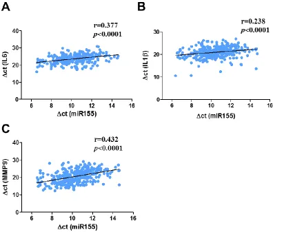
To test whether the expression levels of miR155 are correlated with pro-inflammatory cytokine expression, we analyzed the correlation between miR155 expression and the expression of inflammation markers; IL-1β, IL-6, and MMP-9 using linear regression. Gene expression levels of inflammation markers, 6, IL-1β as well as MMP-9 in human carotid plaques showed highly significant positive correlations with miR155 expression levels (r=0.377, r=0.238, r=0.432 respectively, p<0.0001) (Figure 2.3A, 2.3B, and 2.3C) suggesting that increased miR155 expression is associated with more inflammation and plaque instability in human carotid atherosclerotic diseases.
miR155 expression is up-regulated in calcified media and positively
correlated with osteogenic genes
Cardiovascular calcification is a major feature of chronic inflammatory disorders — such as atherosclerosis and calcific aortic valve disease (CAVD) — that associate with increased mortality and morbidity126. There has been evidence
adjacent normal non-calcified media, p=0.0356 (Figure 2.4A). Concurrently, we examined the expression levels of some calcification (osteogenic) genes, such as Osteopontin (OPN), Bone morphogenetic protein-2 (BMP-2), Runt-related transcription factor-2 (RUNX-2), and Collagen Type I Alpha 1 (COL-1). The expression levels of these genes were significantly up-regulated in calcified media compared to adjacent non-calcific media, p<0.05 and p<0.01 (Figures 2.4B, 2.4C 2.4D and 2.4E). Next, we analyzed the correlation between expression of miR155 and osteogenic genes. We plotted the correlation of miR155 levels and OPN, BMP-2, or RUNX-2 levels, showing a clear positive correlation between expression levels of miR155 with OPN and RUNX-2 expression levels (r=0.446, r=0.322 respectively) (Figure 2.4F). Thus, miR155 seems to influence plaque stability by influencing the expression levels of osteogenic genes, and thus the grade of calcification.
miR155 deficiency attenuates VSMCs calcification in vitro
To further study the role of miR155 in vascular calcification and to confirm the data obtained using human samples, mouse VSMCs were isolated from aorta of wild-type or miR155−/− mice, cultured in DMEM media supplemented with 5% FBS, penicillin/streptomycin, and treated with calcification media containing 2 mM CaCl2 and 2 mM of inorganic phosphate (Pi) to induce calcification. Calcification
osteogenic genes including BMP-2, RUNX-2, OPN and OCN were detected. The results showed that the expression of these genes was significantly increased after calcification media treatment and that the miR155−/− VSMCs had a lower expression than the wild-type VSMCs, p-value<0.05 (Figure 2.5B). This result indicated that miR155 deficiency significantly reduced expression of osteogenic genes in calcified VSMCs. Furthermore, we measured the protein levels of RUNX-2 and OPN. The results showed that miR155 deficiency significantly reduced protein expression levels of RUNX-2 and OPN (Figure 2.5C). These data indicated that miR155 deficiency attenuated VSMCs calcification by reducing expression of osteogenic genes.
miR155 deficiency attenuates aortic calcification ex vivo and in vivo
The effect of miR155 on vascular calcification was further assessed ex vivo
and in vivo. The aortas were dissected from miR155−/− or wild-type mice, cut into
miR155 deficiency reduced aortic calcification ex vivo and in the aorta of vitD3 injected mice.
2.4 Discussion
Inflammation plays a significant role in all stages of atherosclerosis. The cytokines secreted by innate and adaptive immune cells regulate the inflammatory response in atherosclerosis22,31. The expression of inflammatory cytokines in
mouse models of atherosclerosis has been extensively studied. A previous study found that the expression of the pro-inflammatory cytokines in apoE−/− mice after 4 weeks of high fat diet was significantly increased132. The expression levels of
several pro-inflammatory cytokines in the atherosclerotic plaques were studied and reported from the mid-1980s mainly in human carotid endarterectomy specimens112. Better understanding of cytokine gene expression is important for
successful development of therapeutic approaches that decrease the inflammatory response. Inhibiting cytokine-induced inflammation and promoting the anti-inflammatory cytokine response represent potential therapeutic approaches for the prevention of disease development and progression. Currently available therapies against atherosclerosis are mainly lipid modulators. While lipid lowering drugs are successful at reducing the risk of death result from cardiovascular diseases, there is a need for new therapies that directly target the inflammation to further attenuate atherosclerosis and improve cardiovascular outcome133.
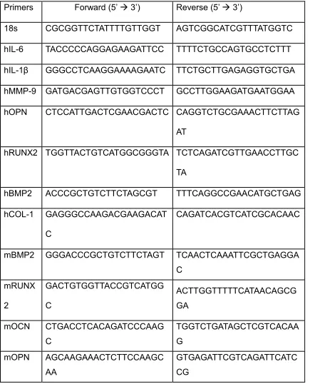
Table 2. 1 Primers that were used for qRT-PCR in this study
Primers Forward (5’ 3’) Reverse (5’ 3’)
18s CGCGGTTCTATTTTGTTGGT AGTCGGCATCGTTTATGGTC hIL-6 TACCCCCAGGAGAAGATTCC TTTTCTGCCAGTGCCTCTTT hIL-1β GGGCCTCAAGGAAAAGAATC TTCTGCTTGAGAGGTGCTGA hMMP-9 GATGACGAGTTGTGGTCCCT GCCTTGGAAGATGAATGGAA hOPN CTCCATTGACTCGAACGACTC CAGGTCTGCGAAACTTCTTAG
AT
hRUNX2 TGGTTACTGTCATGGCGGGTA TCTCAGATCGTTGAACCTTGC TA
hBMP2 ACCCGCTGTCTTCTAGCGT TTTCAGGCCGAACATGCTGAG hCOL-1 GAGGGCCAAGACGAAGACAT
C
CAGATCACGTCATCGCACAAC
mBMP2 GGGACCCGCTGTCTTCTAGT TCAACTCAAATTCGCTGAGGA C mRUNX 2 GACTGTGGTTACCGTCATGG C ACTTGGTTTTTCATAACAGCG GA
mOCN CTGACCTCACAGATCCCAAG C
TGGTCTGATAGCTCGTCACAA G
mOPN AGCAAGAAACTCTTCCAAGC AA
Figure 2. 1 Increased expression of inflammation markers in human carotid
atherosclerotic atheroma
Figure 2. 2Increased expression of miR155 in human carotid atherosclerotic
plaques
Figure 2. 3 Expression levels of miR155 correlate with the levels of inflammatory cytokines and MMP-9 in human carotid artery atheroma
Figure 2. 4 The expression of miR155 and calcification gene in human atherosclerotic carotid artery
Figure 2. 5 miR155 deficiency attenuates calcification in vascular smooth
muscle cells
(A) Calcium content of VSMCs was measured using colorimetric assay with a calcium assay kit. The expression levels of calcification genes; BMP-2, RUNX-2, OPN and OCN in VSMCs after treatment with calcification media were detected by qRT-PCR. (C) Protein levels of RUNX2 and OPN were assessed by western blotting. *p<0.05
Figure 2. 6 miR155 deficiency attenuates aortic calcification in vivo and ex
vivo
(A) Calcium deposition in cultured aortic rings from wild-type or miR155−/− mice was detected by alizarin Red staining. (B) Quantification of calcium content in the aortic rings using colorimetric assays with calcium assay kit. (C) Calcium deposition in the aortas of wild-type or miR155−/− mice injected with VitD3 was determined by Alizarin Red staining. (n=5 for each group). (D) Quantification of aortic calcium content. (E) Serum calcium concentration was measured by colorimetric assay using a calcium assay kit. Scale bar, 500 μm. *P<0.05,
Chapter III
The role of Tristetraprolin (TTP) in inflammation and atherosclerosis
3.1 Background
Tristetraprolin (TTP) — also known as ZFP36 (zinc finger protein of 36 kDa)134, Nup 475135, TIS11136, or G0S24137 — is member of a small family of
mRNA-binding proteins containing a tandem Cys-Cys-Cys-His (CCCH) zinc finger (TZF) domain. The initial description of TTP was in 1990 in mouse 3T3-L1 fibroblasts stimulated with insulin and serum138. This protein is encoded by the
immediate – early response gene called Zinc Finger Protein 36 (ZFP36)139. It is
highly expressed in immune organs in a variety of cell types including monocytes and macrophages138. Normally, TTP protein levels are low and the protein is
primarily localized in the nucleus of quiescent cells. However, mitogenic stimulation of cells induces TTP expression and promotes rapid translocation from the nucleus into the cytoplasm140. Through the TZF domains, TTP can binds
TTP acts at the post-transcriptional level to inhibit the expression of a number of pro-inflammatory cytokine/chemokines and growth factors. The most apparent physiological targets of TTP are the mRNAs encoding TNF-α143–145,
granulocyte-macrophage colony-stimulating factor (GM-CSF)146, Interleukin-2 (IL-2)147,
interleukin-3 (IL-3)148, Interleukin-10 (IL-10)149, Interleukin-12 (IL-12)150, and
Interferon-γ (IFN-γ)151. The role of TTP as an mRNA destabilizing protein initially
comes from the characterization of TTP knockout mice, which appeared normal and healthy at birth, but after few weeks displayed a complex inflammatory syndrome manifested by growth retardation, erosive poly-articular arthritis, conjunctivitis, dermatitis, myeloid hyperplasia, splenomegaly, kidney lesions and autoimmunity152. This inflammatory phenotype was due to excess production of
the potent pro-inflammatory cytokine, TNF-α145. Furthermore, macrophages
isolated from the TTP knockout mice and stimulated with lipopolysaccharide (LPS) showed a significant increase in TNF-α mRNA and protein levels due to stabilization of TNF-α mRNA145,153. The inflammatory phenotype of TTP knockout
mice was significantly attenuated by injecting the mice, soon after birth, with anti-TNF-α antibodies or by breeding with anti-TNF-α receptor deficient mice143,154.
control mice. These studies demonstrated that myeloid cell-specific TTP deficiency did not completely phenocopy whole-body TTP deficiency in mice under normal laboratory conditions suggesting that other cell types were involved in the pathogenesis of the TTP deficiency syndrome155,156. Bone marrow transplantation
has been used to determine if it could transfer the inflammatory phenotype of TTP knockout mice to recipient mice. Recombination activating gene-2knockout (RAG-2−/−) mice transplanted with TTP deficient bone marrow cells reproduce the TTP deficiency inflammatory syndrome, which may indicate that hematopoietic cells play a significant role in excess production of TNF-α that leads to the pathology reported in TTPknockout mice153. Recently, it has been shown that TTP is a potent
modulator of TNF-α secretion from dendritic cells after LPS stimulation157,158.
TTP has been shown to be highly expressed in the vascular endothelium of atherosclerotic mice but not in the vascular endothelium of healthy mice. The anti-inflammatory effects of endothelial cell TTP might post it as a potential therapeutic target for the prevention and treatment of atherosclerosis159.
induce atherosclerosis. We demonstrate that TTP deficiency in bone marrow cells transferred the phenotype of TTP deficiencyincluding systemic and multi-organ inflammation. Surprisingly, we found that TTP deficiency in bone marrow cells did not affect atherosclerosis development. Unexpectedly, TTP deficiency in bone marrow cells dramatically reduced plasma lipid and hepatic lipid accumulation (hepatic steatosis). Increased inflammation and attenuated hyperlipidemia offset each other leading to unchanged atherosclerosis in LDLR−/− mice.
To define the role of macrophage-specific TTP deficiency on atherosclerosis, LDLR−/− mice were lethally irradiated and reconstituted with either macrophage-specific TTPdeficientbone marrow cells or WT bone marrow cells. Four weeks after bone marrow transplantation, mice were fed a western diet for 9 weeks. This study reveals that macrophage-specific TTPdeficiency did not mimic the inflammatory phenotype of whole myeloid cell TTP deficiency in mice. In addition, these mice had comparable lesion size in the aorta and aortic root. Interestingly, macrophage infiltration and lipid deposition in the aortic root were reduced in macrophage-specific TTP deficient mice, which might be due to efficient removal by healthy macrophages in early atherosclerosis.
3.2 Material and Methods
Human carotid atherosclerotic specimens
Human carotid artery samples were collected as described in chapter 2.
Mice
conditions at the University of South Carolina according to National Institutes of Health (NIH) guidelines. Animal care and experiments were approved by the Institutional Animal Care and Use Committee (IACUC) at the University of South Carolina.
Bone Marrow Transplantation (BMT) and atherosclerosis induction in mice
Bone marrow transplantation was performed as previously described160.
Briefly, bone marrow cells from donor WT or TTPdeficientmice were harvested from cleaned femurs and tibias by flushing with sterile RPMI-1640 media (Invitrogen Life Technologies, Grand Island, NY, USA). The cell suspension was centrifuged at 300 x g for 5 minutes and re-suspended in cold phosphate buffer saline (PBS). One week before and two weeks after BMT, recipient mice (LDLR−/− mice) were given autoclaved antibiotic water supplemented with 100 mg/L neomycin and 500,000 U/L polymyxin B sulfate. Early morning, all recipient mice were lethally irradiated (900 rad) using a cesium gamma source. Six hours after irradiation, mice were injected in the retro-orbital venous plexus with nucleated bone marrow cell (5 X 10⁶ in 200 µL PBS). After BMT, mice were observed for any
marrow cells or control (WT) bone marrow cells. Four weeks later, mice were put on western diet for 9 weeks (Figure 3.1B). During the western diet period, body weight and food intake were measured weekly. At the experimental endpoint, mice were euthanized by isoflurane and venous blood from the eyes was collected. Tissues including: heart, aorta, liver, lung, and spleen were collected and analyzed.
Peritoneal macrophage isolation and culture
Standard techniques were used to isolate thioglycollate-elicited peritoneal macrophages. Briefly, mice were injected intraperitoneally (i.p.) with 3 mL of 3% (w/v) sterile thioglycollate (BD Biosciences Clontech; Palo Alto CA, USA). After 3 days, peritoneal macrophages were collected by washing the peritoneal cavity with 10 mL of cold PBS twice. The cells were centrifuged at 300 x g for 5 minutes and re-suspended in high-glucose Dulbecco’s modified Eagle’s medium (DMEM, Invitrogen, Grand Island, NY, USA) supplemented with 10% fetal bovine serum (FBS), a combination of penicillin/streptomycin (Sigma-Aldrich, St. Louis, MO, USA), and 50 μM of β-mercaptoethanol. Two million cells/well were cultured in a 12-well plate. Two hours later, cells were washed with PBS to remove the non-adherent cells, and the non-adherent macrophages cultured in serum free media (SFM) overnight at 37⁰C and 5% CO2. Macrophages were stimulated with LPS (50 ng/mL,
VetScan hematology analysis
Blood was drawn into heparinized tubes by retro-orbital puncture. Blood samples were run on the VetScan HMT hematology analyzer (Abaxis, Union City, CA, USA). The following parameters were measured: total red blood cells (RBC), hemoglobin, mean corpuscular hemoglobin (MCH), mean corpuscular hemoglobin concentration (MCHC), hematocrit (Hct), platelet number, mean platelet volume (MPV), platelet distribution width (PDW), total white blood cells (WBC), lymphocytes, mean cell volume (MCV), and red cell distribution width (RDW).
Blood Glucose Measurement
Blood glucose concentration was measured using OneTouch Ultra Blood Glucose Monitoring System (LifeScan, Inc., Milpitas, CA, USA) following the manufacturer’s instructions.
Serum Lipid and Lipoprotein Analysis
liquid chromatography (FPLC) system (AKTA purifier, GE Healthcare Biosciences, Pittsburgh, PA, USA) equipped with a Superose 6 10/300 GL column (GE Healthcare). Pooled mouse serum (100 µL) was loaded onto the column, and eluted at a constant flow rate of 0.5 mL/min with 1 mM sodium EDTA and 0.15 M NaCI. Fractions of 0.5 mL were collected and cholesterol concentration from each fraction was measured.
Total RNA extraction, cDNA synthesis, and quantitative Real-Time PCR
Total RNAs were extracted from liver tissues and cultured peritoneal macrophages incubated in SFM with or without LPS stimulation using Trizol reagent (Invitrogen, Grand Island, NY, USA) following the manufacturer’s instructions. One microgram of isolated RNA of each sample was reverse transcribed into complementary DNA (cDNA) in 20 μL reactions using iScriptTM
cDNA synthesis kit (Bio-Rad, Hercules, CA, USA). qRT-PCR was performed on a Bio-Rad CFX96 system using iQ™ SYBR® Green Supermix (Bio-Rad) according to the manufacturer’s instructions. The relative amount of target mRNA was calculated using the 2(-ΔΔCt) method by normalizing target mRNA Ct values to those of housekeeping gene, 18S rRNA expression. PCR thermal cycling conditions were 3 minutes at 95°C, and 40 cycles of 15 s at 95°C and 58 s at 60°C. Samples were run in triplicate. Primers used are listed in table 3.1
Flow cytometry analysis
single cell suspensions, tissues were passed through a 70 µM cell strainer (Life Sciences, Tewksbury, MA, USA). After red blood cell lysis, cells were stained for 30 minutes on ice in dark with anti-CD19 mAb, anti-CD3 PE mAb, anti-CD4 FITC mAb, anti-CD8a FITC mAb, anti-FOXP3 mAb, anti-ly6C FITC mAb, anti-CD11b PE mAb, anti-ly6G FITC mAb, and anti F4/80 FITC mAb (eBioscienceTM,
Invitrogen, Grand Island, NY, USA) in staining buffer. Stained cells were acquired on a Cytomics FC 500 flow cytometer and CXP software version 2.2 (Beckman Coulter, Brea, CA, USA). Data were collected for 10,000 live events per sample.
Enzyme-linked immunosorbent assay (ELISA)
ELISA assays were performed per the manufacturer’s protocol. Briefly, blood was collected and centrifuged at 4,000 x g for 20 minute. Serum layer was removed and diluted 1:5. 96-well ELISA plates were coated with 100 µL of TNF-α capture antibody (2 µg/mL) and incubated overnight at 4°C. The cells were incubated with 300 µL of blocking solution (1% BSA, 5% sucrose, and 0.05% NaN3) for 1 hour at room temperature. Next, 100 µL of each sample were loaded to each well and incubated for 2 hour at room temperature. After washing three times, the plates were incubated with biotinylated anti-mouse TNF-α (250 ng/mL), 1 µg/mL horseradish peroxidase streptavidin and substrate solution. The reaction was stopped by adding 50 µL of 1M H2SO4 solution. Recombinant mouse TNF-α
Bioplex analysis of serum cytokine/chemokines
Serum levels of 23 cytokine/chemokines from mice were measured using a Bio-Plex ProTM Mouse Cytokine 23-plex Assay (Bio-Rad, Hercules, CA, USA) on
a Bio-Plex system following the manufacturer’s instructions.
Quantification of atherosclerosis
At the experimental endpoint, all mice were euthanized and the vasculature was perfused with cold phosphate buffer solution (PBS). The aorta from arch to bifurcation was fixed in 10% neutral buffered formalin. Then, it was opened longitudinally, pinned onto black wax plates, and stained with Sudan‐IV (Sigma‐
Aldrich, St. Louis, MO, USA) for aortic enface analysis. Aortic lesion areas were quantified and analyzed by ImageJ software. The aortic root was isolated and embedded in Optimal Cutting Temperature (OCT; Tissue-tek) compound (Sakura Finetek USA, Inc., Torrance, CA, USA) in a plastic mold, stored at - 20 ⁰C. Ten‐
After washing with PBS three times, the biotinylated goat anti-rabbit IgG (Vector Laboratories, Burlingame, CA) was used as the secondary antibody. Immunoreactivity was amplified using the Vectastain ABC kit (Vector Laboratories), and signal was enhanced by peroxidase enhancer (GeneTex, Irvine, CA; USA) and reacted with the substrate from AEC Chromogen/FRP substrate kit (GeneTex Irvine, CA; USA). Nuclei and cytoplasm were counterstained with haematoxylin for 1 min. Finally, sections washed with deionized water and air-dry before getting covered with coverslips. Macrophage infiltration was analyzed via microscope. Inflammatory cell infiltration was expressed as Moma-2 positive area. Oil red O staining was performed to detect lipid content in atherosclerotic plaques. Slides placed in oil red O (ORO; Sigma‐
slides were dehydrated and cleared through 95% ethanol, 100% ethanol, and xylenes, 2 changes each, mounted with permount, and coversliped. All images were recorded with a Nikon E600 Wide field Epifluorescence microscope and Micropublisher digital camera with Q-imaging software. Plaque size, lipid content, collagen percentage, and macrophage cell content were quantified by computerized image Pro-Plus.
Histological analysis
Tissue samples (Liver, lung, kidney and spleen) were fixed in 10% neutral buffered formalin. H&E staining and microscopy were implemented to detect infiltrating inflammatory cells. All tissue sections were visualized with a Nikon E 600 microscope.
Microarray analysis
molecules were purified using Qiagen’s RNeasy Mini Kit (Valencia, CA). After spectrophotometric assessment of dye incorporation and cRNA yield, samples were hybridized to Agilent whole mouse genome microarrays 8 × 60,000 using a gene expression hybridization kit (Agilent) according to the manufacturer’s recommendation. Microarray analysis was performed using an Agilent DNA microarray scanner system. After washes, arrays were scanned using a High Resolution Agilent DNA Microarray Scanner and images saved in TIFF format. A heat map of genes from relevant pathways identified by Ingenuity pathway analysis was generated using R function heatmap.2.
Statistical analysis
Data were presented as mean ± standard error of the mean (SEM). Statistical analysis was performed with Graph-Pad Prism 6.0 software (Graph-Pad Software Inc, San Diego, CA, USA). Statistical significance was determined by one-way analysis of variance (ANOVA) for multi-group comparison and Student’s
t test for two-group comparison. Data were considered statistically significant at *P
< 0.05, **P<0.01, and ***P<0.001. 3.3 Results
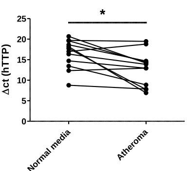
TTP is up-regulated in human carotid atherosclerotic lesions
study159, we demonstrated that TTP is highly expressed in atheromatous tissue
compared to the adjacent normal media in the carotid atherosclerotic plaques p -value <0.05 (Figure 3.2).
TTP deficiency in bone marrow-derived cells causes growth retardation, but
did not affect food intake
Five million bone marrow cells from either TTPdeficient mice or WT mice were transplanted into lethally irradiated LDLR−/− mice. After 4 weeks recovery, the recipient mice were challenged with a western diet for 12 weeks, after which mice were sacrificed. During western diet period, mouse food intake and body weight were measured weekly. None of these mice showed any signs or symptoms of bone marrow transplant rejection. We did not observe significant differences in food intake between bone marrow TTPdeficient mice and control mice (Figure 3.3A). There was, however, a reduction in the mouse body weight with bone marrow TTPdeficiency (Figure 3.3B). This reduction in body weight and cachexia was the most important characteristic phenotype of TTPknockoutmice161.
TTP deficiency in bone marrow-derived cells causes hematological and
Immunological disturbance
monocytes, but these changes did not reach statistical significance, perhaps because of the relatively small sample number (n=5-6). There was, however, a significant increase in the number and percentage of neutrophils (P=0.0037, P=0.0336 respectively) of bone marrow TTP deficient mice compared to the control mice (Figure 3.4A). Total platelet number, platelet hematocrit were significantly increased (P=0.0107, P=0.0053 respectively) in bone marrow TTP deficient mice compared to the control mice (Figure 3.4B). On the other hand, we did not observe any differences in the red blood cell count, hemoglobin concentration, hematocrit, mean corpuscular volume, mean corpuscular hemoglobin, MCHC, and the RDWC between the two groups (Figure 3.4C). Flow cytometric analysis of the leukocytes population in the spleen showed a significant decrease in the number and percentage of CD3+/CD4+ T lymphocytes, and a
significant increase in CD11b+/GR1+ myeloid cells, CD11b+/Ly6G+ (neutrophils),
CD11b+/Ly6Chigh (inflammatory monocytes). Whereas, the number and
percentage of CD19+ B lymphocytes, CD3+/CD8a+ T lymphocytes, CD4+/FoxP3+,
CD4+/IL17+, CD11C+, F4/80+, and CD11b+/Ly6Clow were comparable between the