Copyright 0 1984 by the Genetics Society of America
ANALYSIS OF GENETIC MOSAICS
OF T H E NEMATODE
CAENORHABDITIS ELEGANS
ROBERT K. HERMAN
Department of Genetics and Cell Biology, University of Minnesota, St. Paul, Minnesota 55108 (current address), and MRC Laboratory of Molecular Biology, Cambridge CB2 2QH, England
Manuscript received February 20, 1984 Revised copy accepted May 12, 1984
ABSTRACT
A new method for producing genetic mosaics, which involves the spontaneous somatic loss of free chromosome fragments, is demonstrated. Four genes that affect the behavior of C. elegans were studied in mosaic animals. The analysis was greatly aided by the fact that the complete cell lineage of wild-type animals is known. Two of the mutant genes affect certain sensory responses and prevent uptake of fluorescein isothiocyanate (FITC) by certain sensory neurons. Mosaic analysis indicated that one of these mutant genes is cell autonomous with respect to its effect on FITC uptake and the other is cell nonautonomous. In the latter case, the genotype of a non-neuronal supporting cell that surrounds the processes of the neurons that normally take up FITC probably is critical. The other two mutant genes affect animal movement. Mosaic analysis indicated that the expres- sion of one of these genes is specific to certain neurons (motor neurons of the ventral and dorsal nerve cords are prime candidates) and the expression of the other gene is specific to muscle cells.
HE analysis of genetic mosaics and chimeras has been a powerful tool in
T
developmental genetics, primarily in work with Drosophila and the mouse (for reviews, see GEHRING 1978). In Drosophila, in which the analysis of genetic mosaics has been most extensive, mosaics have been used to elucidate cell lineage and to ascertain the anatomical foci of mutations affecting behavior. In cases in which cellular abnormalities in mutants have been discernible, genetic mosaics have been used to assess the cell autonomy of mutant phenes; cell autonomy implies that the action of a mutation on cell differentiation is intrinsic to the cell, whereas nonautonomy indicates that cell-cell interactions are involved. Such interactions can also be studied by mosaic analysis (see, for example, MEYEROWITZ and KANKEL 1978). Genetic mosaics have also been used to set limits on the times of action of wild-type genes: if a wild-type gene is removed from a cell and as a consequence a descendant cell shows a recessive mutant phenotype, the implication is that the wild-type gene was needed after the time at which it was removed.T h e development of the nematode Caenorhabditis elegans is under intensive investigation: the complete cell lineage of the wild-type animal has been worked out (SULSTON 1976; SULSTON and HORVITZ 1977; DEPPE et al. 1978; KIMBLE and HIRSH 1979; SULSTON et al. 1983), and the neuroanatomy of the animal has
166 R. K . HERMAN
been reconstructed from serial section electron micrographs (WARD et al. 1975; WARE et al. 1975; WHITE et a l . 1976; ALBERTSON and THOMSON 1976; J. WHITE, personal communication). One reason for the choice of C. elegans as a develop- mental model is its suitability for many methods of genetic analysis (BRENNER
1974; HERMAN and HORVITZ 1980). It would clearly be desirable to add mosaic analysis to the methods available. Genetic mosaics are not needed to work out C. eleguns cell lineages because they can be followed by direct observation, but the other applications of mosaic analysis, dealing with the cell specificity of gene expression, would be very useful. SIDDIQUI and BABU (1980) have reported the production of C. elegans mosaics by X irradiation of embryos heterozygous for
flu-?, a gene that alters the autofluorescence of intestinal cells under ultraviolet light. T h e disadvantages of their method are that the frequency of mosaicism is low (less than 0.1% mosaic animals), the radiation (2000 rads) is likely to cause cell death and other abnormalities and the mechanism by which the mosaics arise is not clear. Here, I report a new method for producing mosaics: the spontaneous loss of a free chromosome fragment present as a duplication. Duplication loss thus generates a cell with a normal chromosome complement; because the cell lineages are rigidly specified and completely known, it should be possible with appropriate cell markers to pinpoint precisely the division at which a duplication loss took place and, hence, predict the exact cell composition of the duplication- free clone in the mosaic animal. Free chromosome duplications representing several different regions of the genome have been identified (HERMAN, ALBERT-
SON and BRENNER, 1976; HERMAN, MADL and KARI 1979; HODCKIN 1980; P.
ANDERSON, personal communication; A. ROSE and D. BAILLIE, personal com-
munication). Because of the holokinetic nature of the C. eleguns chromosomes (ALBERTSON and THOMSON 1982), suitable free duplications of most regions of the genome may eventually become available and help make the approach introduced here more generally applicable.
MATERIALS AND METHODS
Genes, alleles and general procedures: C. elegans var. Bristol strain N2 was the wild-type parent for all strains used in this work. The following genes and mutations were used: LG 11: dpy-lO(el28); LG 111: unc-93(e1500); LG V: dpy-1 l(e224); LG X: unc-9(elOl), let-4(mn105), unc-3(el51), daf6(e1377), let-l(mnl19), unc-7(e139), sup-lO(n183,mn219), osm-l(p808). The derivation of sup-lO(mn219) is described under Strain constructions. The sources of the other mutations are either cited in RESULTS (or under Strain constructions in MATERIALS AND METHODS) or they were described by BRENNER ( 1 974). Media, culture and mating techniques were as described by BRENNER ( 1 974) and HERMAN (1978). Genetic nomenclature follows the guidelines described by HORVITZ et al. (1979).
Duplications: The derivations and characterizations of mnDpl(X;V), mnDp2(X;f) and mnDp3(X;f)
have been described (HERMAN, ALBERTSON and BRENNER 1976; HERMAN, MADL and KARI 1979). In addition, CHALFIE and SULSTON (1981) have shown that mnDp2 does not suppress mec-5 or mec-4
mutations, which map to the right of the region covered by mnDp2, and I have shown that mnDp2
does not suppress osm-l(p808), which maps near mec-4. The free X chromosome duplications
mnD#l2(X,f) and m n D p l l ( X ; f ) were induced, identified and characterized by C. KARI (personal communication) by methods previously described (HERMAN, ALBERTSON and BRENNER 1976) except that y rays were used rather than X rays. y Radiation was supplied by '"Cs in a Shepherd irradiator (model 143-45). Doses of 7200 roentgens (r) were used at a dose rate of 600 r/min. Both m n D p l 2
GENETIC MOSAICS O F C. ELEGANS 167
duplication self-progeny of duplication-bearing hermaphrodites determined for each duplication (at least 1000 total progeny per measurement) were 59% for mnDp2,45% for mnDP3,76% for m n D p l 2 , 44% for m n D p l 3 and 52% for m n D p l 4 .
FlTC staining: The fluorescein isothiocyanate (F1TC)-staining protocol of E. HEDGECOCK (personal communication) was followed: animals were put on an agar growth plate seeded with bacteria to which FITC had been added to a final concentration of 0.1 mg/ml. Worms were removed from the plate 3-12 hr later, put on a seeded plate without dye for at least 10 min and then put on a 5% agar pad on a microscope slide (SULSTON, ALBERTSON and THOMSON 1980) and viewed by epifluorescence. Genetic mapping of daf-6 and osm-1 : Fifteen independent Unc-7 non-Unc-3 recombinant self- progeny of unc-3 unc-7/duf-6 hermaphrodites were picked. Segregants homozygous for the recom- binant chromosome (giving no Unc-3 Unc-7 self-progeny) were found in each case. FITC staining was used to ascertain the status of the daf-6 locus: five chromosomes were daf-6+ and ten were daf- 6 ; thus, daf-6 maps between unc-3 and unc-7. The osm-1 gene, previously mapped by CULOTTI and RUSSELL (1 978), was further localized by complementation testing against deficiencies mnDf41, mnDf42 and mnDf43 (for descriptions of these deficiencies and the complementation methods, see MENEELY and HERMAN 1981); osm-1 complemented mnDf43 and failed to complement mnDf41 and mnDf42. This places it to the right of sup-10 (MENEELY and HERMAN 1981).
Strain constructions: Most of the strains were constructed by standard methods that need not be described. The unc-3 daf-6 double mutant was constructed as follows: Males of genotype mnDP1-
(X;V)/+; unc-3 let-110 (MENEELY and HERMAN 1981) were mated with dpy-10; daf-6 hermaphrodites. Wild-type hermaphrodite progeny were picked. Their progeny were screened for viable Unc recombinants, which were picked. Their progeny were then screened by FITC staining for unc-3 daf-6 homozygotes.
The let-4 unc-3 osm-1; m n D p l 3 strain was made as follows. Males of genotype m n D p l / + ; let-4 unc- 310 (MENEELY and HERMAN 1981) were crossed with dpy-11; unc-?; mnDPl3 hermaphrodites. Wild- type hermaphrodite progeny were picked, and those that did not carry mnDpl were identified by the absence of pseudolinkage between dpy-11 and unc-3. Wild-type hermaphrodite progeny were picked, and hermaphrodites of genotype let-4 unc-?; mnDpl? were identified by the absence of viable Unc animals among their self-progeny. Males of genotype let-4 unc-3/0; m n D p l 3 were then mated with dpy-1 1; unc-3 osm-1 hermaphrodites, and wild-type hermaphrodite progeny were picked. More than 100 wild-type hermaphrodite self-progeny of these animals were then picked. Self-progeny broods produced by these animals in which both viable Unc and arrested larvae were represented were identified and stained with FITC. A brood in which all Unc animals did not stain with FITC (but wild-type animals did stain) was found, and from it a wild-type descendant of genotype let-4 unc-3 osm-1; m n D p l 3 was readily identified.
The unc-93; unc-3 sup-10 osm-1 mutant was generated as follows. Rather than try to produce the sup-10 osm-1 double mutant by recombination, a new spontaneous sup-10 mutation was sought in an unc-93; osm-1 background, which was produced by standard methods. The unc-93; osm-1 strain was grown on dozens of 100-mm agar plates to select for spontaneous reversion of the Unc-93 phenotype. Revertants were then tested for the possession of an extragenic suppressor mutation by crossing them with N2 males, picking wild-type hermaphrodites from plates in which mating had been efficient (as judged by the presence of many male progeny) and looking for the segregation of Unc-93 self- progeny. In those cases in which an external suppressor was present, linkage of the suppressor to osm-1 was tested by FITC staining of both Unc-93 and non-Unc-93 segregants. Among the first five revertants identified, two carried an external suppressor, and one of these (mn219) was closely linked to osm-1. Finally, a complementation test established the allelism of mn219 and sup-lO(n183). Next, unc-93/+; sup-10 osm-llunc-3 osm-1 hermaphrodites were produced by crossing mnDpl/+; unc-3 osm- 110 males with unc-93; sup-10 osm-1 hermaphrodites. Unc-93 segregants of these were picked, and then wild-type self-progeny (homozygous for both unc-93 and sup-10) of the Unc-93 animals were picked. Finally, an Unc-3 segregant (genotype unc-93; unc-3 sup-10 osm-1) was identified.
RESULTS
R . K. HERMAN
AB
-10
n n
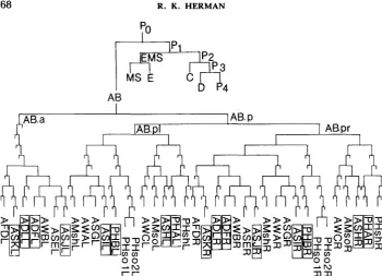
FIGURE 1 .-Lineages of cells comprising amphids and phasmids (SULSTON et al. 1983). Lineage tree conventions and cell nomenclature follow SUISTON and HORVITZ (1977), DEPPE et al. (1978) and SULSTON et al. (1983). The fertilized egg is called PO. Each of four successive asymmetric cleavages generates a larger somatic precursor cell (AB in the first cleavage) and a smaller P cell (DEPPE et al. 1978). P, is the precursor of the germ line. AB.a and AB.p are the anterior and posterior daughters of AB, respectively; and AB.pl and AB.pr are the left and right daughters of AB.p, respectively. Nearly all unmarked divisions shown give anterior and posterior daughters; an anterior duaghter is represented by a left branch in the lineage tree. Thus, for example, the right-most cell shown in the diagram (PHshR) has the lineage designation AB.prpppapaa. Lineages leading to cells not comprising the amphids and phasmids have been terminated in this figure; see SULSTON et al. (1 983) for complete lineages. Differentiated cell names beginning with A and PH refer to amphid and phasmid, respec- tively; sheath cell is abbreviated "sh" and socket cell is abbreviated "so"; all other cells are neurons. The cells that are stained in wild-type animals by FITC (E. HEDGECOCK, personal communication) are outlined.
GENETIC MOSAICS O F C. ELEGANS 169
FIGURE 9.--Fluoresccncc micrographs showing l a ~ c ~ ~ l v i e w of cell txxiics of (a) two neurons of
one phasmid of a wild-type animal stained with F I ' K and (b) one stained phasmid neuron in an o n -
I mosaic animal. The large area of fluorescence is part of the intestine. Animals were anesthetized with I -phenoxy-2-propanol ( S U L ~ N . ALBERTSON and WHITE 1980) for photography. Magnifications in both photographs are X560.
larvae (an alternative to the normal third stage larvae) in response to starvation or overcrowding have been shown to be abnormal in the structure of amphids or phasmids (WARD 1976; LEWIS and HODGKIN 1977; ALBERT, BROWN and RIDDLE 198 1 ; L. PERKINS, E. HEDGECOCK, N. THOMSON and J. CULOTTI, personal communication), presumably reflecting abnormal sensory function in these mu- tants. Furthermore, certain mutations, including alleles of osm-1 (CULOTTI and RUSSELL 1978) a n d daf-6 (ALBERT, BROWN a n d RIDDLE 1981). abolish the F I T C staining of amphid a n d phasmid neurons (E. HEDGECOCK, personal communica- tion).
170 R. HERMAN
TABLE 1
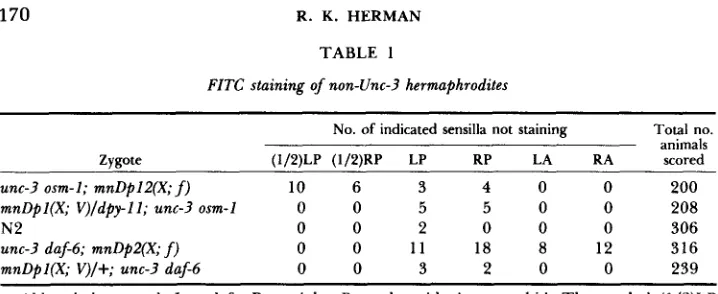
FITC staining of non-Unc-3 hermaphrodites
No. of indicated sensilla not staining Total no. animals Zvaote (1/2)LP (1/2)RP LP RP LA RA scored
unc-3 osm-1; mnDpl2(X; f ) 10 6 3 4 0 0 200 mnDpl(X; V)/dpy-ll; unc-3 osm-1 0 0 5 5 0 0 208
N2 0 0 2 0 0 0 306
unc-3 daf-6; mnDp2(X; f) 0 0 11 18 8 12 316
mnDpl(X; V)/+; unc-3 daf-6 0 0 3 2 0 0 239
Abbreviations used: L = left, R = right, P = phasmid, A = amphid. The symbol (1/2)LP signifies staining of one neuron of left phasmid. In only three animals did more than one sensillum show lack of staining: all were unc-3 daf-6; mnDp2 zygotes with two sensilla affected (RP and LA; RP and RA; LA and RA).
of a phasmid channel. T h e frequency of phasmid nonstaining may be higher in the duplication-bearing strains than in N 2 (Table 1); it is possible that this is related to the fact that duplication-bearing animals tend to be slightly smaller and less vigorous than N2. In any case, this is not a problem in the staining of amphid neurons: none of the free duplication-bearing animals or the control animals showed absence of staining of all six neurons of an amphid. No attempt was made to score absence of staining of a single amphid neuron, which is technically more difficult than for the phasmid neurons.
A similar set of experiments with daf-6 in place of osm-1 gave quite different results (Table 1). In this case, the free duplication-bearing strain (carrying mnDp2)
GENETIC MOSAICS OF C. ELEGANS
TABLE 2
FITC staining of Unc-? duplication mosaics
171
No. of animals with indi-
cated sensilla not staining Uric-3 duplica- tion mosaic per
LP RP LA duplication-bear- RA LP RP RA ina sib"
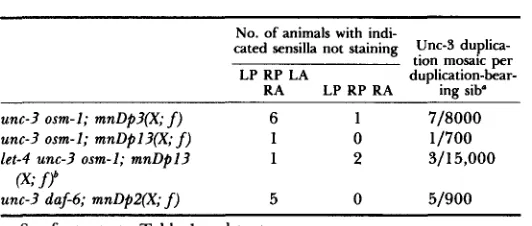
unc-3 osm-I; mnDp?(X; f) 6 1 7/8000 unc-3 osm-I; mnDpl?(X; f) 1 0 11700
let-4 unc-? osm-I; mnDpl? 1 2 3115,000
unc-3 daf-6; mnDPZ(X;
(x;
f) 5 0 51900f?
See footnote to Table 1 and text.
For the first two strains and the last strain listed, Unc-3 animals were picked, usually five or ten per plate. From a plate with young non-Unc progeny, the Unc-3 parents were picked individually to iden- tify the one animal responsible. The observed frequency of Unc-3 du- plication mosaics per Unc-3 animal for each strain was multiplied by the appropriate ratio of Unc-3 to wild-type animals characteristic of the duplication (see MATERIALS AND METHODS) to obtain the ratios given in this column.
' T h e recessive larval lethal mutant gene let-4 was used to select against Unc-3 animals devoid of mnDpl? (which carries let-4+ unc-3+ osm-I+). The fact that fertile animals were found whose cells descend- ing from AB or AB.p lacked let-4+ indicates that the let-4+ gene is not absolutely required by the cells; on the other hand, the relatively low incidence of these duplication mosaics may reflect a low level require- ment for let-4+ product among AB descendants, which is occasionally inherited in sufficient amount from PO.
with the results is the possibility that a socket cell lacking daf-6+ would abolish staining.
172 R. HERMAN
(Figure 1). Thus, in all 16 animals made Unc-3 by virtue of somatic duplication loss, the duplication loss occurred no later than at AB.p. We have seen that later duplication losses occur among non-Unc-3 animals (Table 1). This suggests that more than one descendent of AB.p must lack unc-?+ in order to
give
rise to an Unc-3 animal; that is, unc-? has a diffuse focus of action, which seems to be at least primarily localized among the descendents of AB.p.This interpretation suggests that a loss occurring after AB.p might give a semi- Unc-3 phenotype. Clear examples of semi-Unc-3 animals have been found. T h e distinction between Unc-3 and semi-Unc-3 animals was obvious. When touched on the head, Unc-3 animals do not back up but coil their tails. By contrast, semi- Unc-3 animals are able to back up, albeit in uncoordinated fashion; this pheno- type is very similar to that conferred by a weak allele of unc-3, e54. Many of the FITC-staining patterns found for semi-Unc-3 animals were consistent with du- plication loss occurring at AB.pl or AB.pr (Table 3). In the case of the osm-I- marked animals, loss at AB.pl should lead to absence of staining by both left phasmid neurons, and loss at AB.pr should lead to absence of staining by all right phasmid and right amphid neurons. For the duf-6-marked animals, on the other hand, loss at AB.pl should lead to absence of staining by all left phasmid and left amphid neurons, and loss at AB.pr should lead to absence of staining by all right amphid and right phasmid neurons. These predicted patterns were observed. In addition, many of the semi-Unc-3 animals showed later duplication losses or possibly double-event losses. The results thus indicate that the descend- ants of AB.pl and AB.pr are contributing about equally and additively to the Unc-3 phenotype.
Focus of sup-10 action: T h e mutation unc-9?(e1500) confers a phenotype very different from Unc-3: the animals have long bodies and assume abnormal postures when not moving; they move in a slow and uncoordinated fashion, and they recoil and then quickly relax when touched on the head (GREENWALD and HORVITZ 1980). Because a mild disorganization of the pattern of birefringence of body wall muscles is apparent, GREENWALD and HORVITZ (1980) have con- cluded that unc-93 animals are defective in muscle. T h e mutants are also egg- laying deficient, presumably because of defective vulval and uterine muscles (TRENT, TSUNG and HORVITZ 1983); as a result, progeny hatch inside the parent and devour it, giving small brood sizes (about 30 animals compared with about 300 for N2). All of the unc-9? phenes are suppressed by the recessive suppressor sup-10 (GREENWALD and HORVITZ 1980).
Because the free duplication mnDp? carries the dominant allele sup-lO+ (as well as unc-?+), hermaphrodites of genotype unc-93 ZZZ; unc-3 sup-ZO(nl8?)
X;
GENETIC
a 2
'$
.- E 4i
.-8 2
3
B
- e5
f
s
'5.Li
2
* 2
3
4 3
o4
g
z+ <
.."
F
C .e
e
t "
w c
E4 'I 0
s .- .I?
2
0
h
MOSAICS OF C . ELECANS 173
a
-1
:
2
a J %
c - x
2 8
P O
5
o o o ma ' Z
k
& 'E
-
w, m -
2
- 0 o mi t
p.
a
<
w, L e
b o 0
C O
cl
3
0 0 0 33 c
i$
0 - m m
$5
3
T
.:2
0 0 0 2._ a
cl
z z
.U 0I
& k
8 %
q 9
-
x E2 5
e o ,
gk
m=
* L E
3
o o o m d $ $3.5
U.5
e
e
2 n - - c o
Ma=a s 2-0 v1
z
s j
2.a
c:L o o 0
. ? $ Z
e
i
s"?
E - - - =
?q
:
v$ 2
Lz s
x
o s
a J a J
$ z o o o m
.E,
x Z +-1
CI-
m x c
u a
e
-
c.,:
v a n \ 6
m y E
-1
v) '$
0 O - O F
5 m o - m :RE
2 E m
z
.c
xz
aJz
aJL L
?
g
??2-
sea??
o
2 z = *
-
% % E %g 9 9 + ; s ,
N S E E S;;;;a.
y,-h
h ? Y $a a
E ' P3
3;;3
3 3 - 3z z 3 b . g M
4
g
+%.ca
2 . 5 m a J
E Y <
L O
2 aL
+ S E
$ 9 -
Q.-$ E $
0 5 c
- m y ,
r - 4 0
FIGURE 3.-Lineages of the 95 body wall muscle cells (0) and 16 vulval and uterine muscle cells
(0) present in the adult hermaphrodite. The newly hatched animal has 81 body muscle cells: 20 from
D, 32 from C, 28 from MS and one from AB (SULSTON et al. 1983). The mesoblast called M gives rise postembryonically to 14 additional body muscle cells, as well as to the vulval and uterine muscle cells (SULSTON and HORVITZ 1977). Pb is the germ line precursor cell.
All vulval and uterine muscles derive from PI, so that an animal produced in this way should also be wild type with respect to egg laying and, hence, brood size. All gametes also derive from PI; therefore, all self-progeny of such a wild-type duplication mosaic should be Unc-3 non-Unc-93. Four animals satisfying these predictions (i.e., non-Unc and giving at least 200 self-progeny per animal, all Unc-3) were found among about 8200 mnDp?-bearing sibs. In addition, three animals satisfying these predictions were found among about 5500 mnDp?- bearing progeny of unc-9? III; unc-? sup-lO(mn219) osm-1 X; mnDp? hermaphro- dites. T h e latter three animals were also tested for FITC staining and found to be wild type, as expected since the osm-l+ function would have been retained among the AB descendants. T h e overall incidence of the wild-type mnDp3 mosaics in these experiments (seven/l3,700) agrees with the estimated frequency of mnDp? loss by AB (six/8000; Table
2),
the sister of PI.Another class of wild-type segregant among the progeny of unc-93; unc-? sup- IO; mnDp3 (and unc-93; unc-? sup-IO osm-1; mnDp?) was also found, at a frequency of about 0.15% among mnDp3-bearing animals. These gave wild-type as well as Unc-3 (but not Unc-93) animals among their self-progeny. In every case, it was apparent from the self-progeny ratios that the wild-type animals carried a single unc-3+ allele and that it was carried by a free duplication. Thus, it appeared that the wild-type segregants were formed by virtue of loss of sup-lo+ (and not unc-
GENETIC MOSAICS OF C. ELEGANS 175
and they were seen not to segregate Unc-93 offspring. Six presumably altered free duplications generated in unc-93; unc-3 sup-10; mnDp3 animals were tested as follows for their ability to suppress osm-1, which is closely linked to sup-10.
Each duplication-bearing (non-Unc) stock was mated with
N2
males; wild-type male progeny were picked and crossed with dpy-11 V; unc-3 osm-1 X hermaphro- dites, and wild-type male progeny were picked and assayed for their FITC stainability. T w o of the six duplications failed to provide osm-l+ function. Because the events producing these altered duplications occurred in a strain that carried osm-l+ X chromosomes, it seems likely that, in at least two cases, the lossof sup-lO+ occurred through the formation of a deficiency that simultaneously
led to loss of the nearby osm-I+ gene.
There is another phenotype expected to be produced through somatic dupli- cation loss in unc-93; unc-3 sup-10; mnDp3 hermaphrodites. If duplication loss occurs after PI in a cell that is a precursor to the vulval and uterine muscle cells (Figure 3), the resulting mosaic would be expected to be largely Unc-93 with respect to movement but wild type with respect to egg laying. These animals would retain mnDp3 in their germ lines and, thus, should give both Unc-93 and Unc-3 self-progeny. Animals satisfying these predictions were also found; their relatively high incidence (1 5/5000) probably reflects the many divisions in which duplication loss can occur (presumably anywhere between EMS and one of the daughters of M; see Figure 3). I have also picked out three Unc-93 egg layers among the progeny of unc-93; unc-3 sup-10 osm-1; mnDp3 hermaphrodites and shown that they stained normally with FITC, as expected.
As a control for the experiments involving sup-10, I have looked for both wild- type hermaphrodites giving only Unc-3 self-progeny and Unc-93-moving egg- laying-proficient animals, as defined before, among the progeny of unc-93
III;mnDpl(X;V)/+ V; unc-3 sup-10 X hermaphrodites. Again, because mnDpl,
which carries sup-IO+ and unc-3+, is translocated to an autosome and is not free, no mosaic animals were expected. No wild-type hermaphrodite giving only Unc- 3 self-progeny was found among 13,000 mnDpl-bearing hermaphrodites, and no egg layer was found among 5000 Unc-93 animals scored for egg laying. These control experiments indicate that the exceptional animals identified in the
mnDp3 experiments were not simply the result of occasional incomplete expres-
sion of the single sup-IO+ allele but were in fact genetic mosaics. T h e experiments with sup-10 mosaics thus support the conclusion of GREENWALD and HORVITZ ( I 980) that the action of sup-10 is specific to muscle cells and provide evidence that the vulval and uterine muscles are able to function in egg laying even when the body wall muscles are largely abnormal; the results also indicate that unc-3+
and osm-l+ functions are not required in non-AB cells.
DISCUSSION
T h e view that osm-1 behaves cell autonomously with respect to FITC staining of sensory neurons was drawn first from the finding of unc-3 osm-1; Dp her- maphrodites with only one of two phasmid neurons stained by FITC, a pattern that was attributable to somatic duplication loss. T h e FITC-staining patterns of Unc-3 and semi-Unc-3 animals arising from unc-3 osm-1; D p zygotes also pointed to the cell autonomy of osm-1. This was most dramatically illustrated by the differences in staining between left and right amphids. Thus, among Unc-3 duplication mosaics, there were animals in which only left amphid neurons stained but no animals in which only right amphid neurons stained. Similarly, among semi-Unc-3 animals, there were several examples of nonstaining right amphids but no examples of nonstaining left amphids. These patterns were readily predicted from the known cell lineages of the amphid neurons (the lineages of these cells are not bilaterally symmetric).
T h e du.6 gene provides a good contrast to osm-1 because it is clearly not cell autonomous with respect to FITC staining. T h e results fit nicely the evidence of ALBERT, BROWN and RIDDLE (1981) that the duf-6 mutant has defective sheath cells, i.e., it appears that absence of duf-6+ from a sheath cell is sufficient to block staining of all sensory neurons of the corresponding sensillum. It is possible that a duf-6 socket cell would also abolish staining. T h e results are not consistent with a requirement for both a socket and the sheath cell to lack duf-6+ in order for the sensillum not to stain, however. T h e lineages of socket and sheath cells for the right phasmid, for example, diverge at AB.pr (Figure l), but the socket and sheath cells of the right amphid also diverge at AB.pr. Hence, it would be impossible by a single event to generate duplication-free socket and sheath cells for one right sensillum without simultaneously affecting the other right sensillum; many examples of single nonstaining sensilla were found (Table 1). T h e cell lineages of the sheath (and socket) cells are bilaterally symmetric, which is in accord with the bilaterally symmetric staining patterns of the Unc-3 and semi- Unc-3 animals arising from unc-3 du.6; mnDp2 zygotes.
T h e results clearly indicate that the unc-3 gene has a diffuse focus of action that is at least primarily localized among the descendants of AB.pl and AB.pr, which contribute about equally and additively to the Unc-3 phenotype. Thus, loss of unc-3+ at AB.p confers an Unc-3 phenotype and loss at AB.pl or AB.pr confers a semi-Unc-3 phenotype. Losses among the descendants of AB.pl or AB.pr can also lead to a semi-Unc-3 phenotype (Table 3), but presumably these losses generally either occur earlier in the lineage than do the more frequent losses that affect sensillum staining in non-Unc animals (Table 1) or they involve double event losses. In any case, the high incidence of early losses among semi- Unc-3 animals suggests that more than one descendant of both AB.pl and AB.pr is responsible for the unc-3 expression. Alternatively, unc-3+ product may be made early and perdure to rescue critical cells lacking the unc-3+ gene.
GENETIC MOSAICS OF C. ELEGANS
AB
177
AB.a 1AB.p
W - P l IAB.pr
I I I
\d:mh&$iEil
0 \ \ - \ \ \ U 0 0 0000000 \\A\\\ 00000000000Ul N -\PcDo3COCOCD>>OOW>> N~\POV3CODUICJJD>DWOO>
N 0- -mcD N P - O U I O ~ 0-r Oo3P-J-0cnNPCn-J
N N
FIGURE 4.-Lineages of ventral and dorsal cord motor neurons (SULSTON et al. 1983). Lineage tree conventions are the same as for Figure 1 . The names of all 22 motor neurons present in the newly hatched animal are given in the figure and begin with D. An additional 53 hermaphrodite cord motor neurons are generated through postembryonic lineages (not shown here; see SULSTON
1976; WHITE et al. 1976; SULSTON and HORVITZ 1977), which descend from 13 blast cells shown in the figure and called W and P1-P12. The cells P1-P12 are numbered (anterior to posterior) after their migration into the ventral cord, and, because there is some variability in the anterior-posterior order of a given left-right pair of P cells in the cord, each member of a pair is designated by the same symbol in the figure; thus, the two cells designated P1/P2 in the figure can be named P1 and P2
only after they have assumed their relative positions in the cord (SULSTON 1976; SULSTON and HORVITZ 1977).
lineages of all of the ventral and dorsal cord motor neurons, which drive the body muscles. All but one descend from AB.p; the lineages of these neurons make them, or a subset of them, prime candidates for the focus of unc-3 action. (Various interneurons derive from both AB.a and AB.p; a subset of interneurons deriving from AB.p are thus also possible candidates for the focus of unc-3
action.)
T h e predictions that were made in the sup-10 experiments depended on the assumption that sup-10 action is specific to muscle cells. This assumption follows from the conclusion of GREENWALD and HORVITZ (1980) that unc-93(e1500),
which sup-10 suppresses, confers a muscle defect. T h e fact that the predictions were borne out by the results is taken as strong support for the correctness of the assumption. T h e strongest prediction was that wild-type-moving her- maphrodites arising from unc-93; unc-3 sup-10; mnDp3 zygotes by virtue of somatic duplication loss would give only Unc-3 progeny. This result by itself only argues that the duplication was lost by a cell that is precursor to the germ line, i.e., Po-P4 (see Figure 3). But Po is excluded because it is precursor to the AB lineage, which was shown to have retained the duplication, and P4 can be
R. K.
non-AB cells had no discernible phenotypic effect. Additional results with sup- 10 indicated that loss of the duplication somewhere between EMS and the precursor to the vulval and uterine muscles (Figure 3) enables the animal to lay eggs even though most of the body muscle cells carry suP-l0+ and are Unc-93. Depending on where the loss occurs, these animals may be mosaic for sup-l0+
in either body muscle cells or vulval and uterine muscle cells. Unfortunately, the abnormality in birefringence conferred by unc-93 (GREENWALD and HORVITZ
1980) appears to be too subtle to use in identifying the phenotypes of individual cells.
An unexpected result in the sup-10 experiments was the finding of variant duplications that had lost sup-lO+ but retained unc-3+. One possible means by which such altered duplications might arise would be through picking up the chromosomal sup-10 mutation by recombination. But in at least two cases that mechanism seems unlikely, since the variant duplications in these cases were shown to be missing a nearby wild-type gene that was originally carried by the duplication as well as the chromosomes, i.e., in these cases the loss most likely occurred through the formation of a deficiency in the duplication. Indeed, it is possible that all of the losses of sup-l0+ occurred through deficiency formation rather than recombination. These events are not unique to the sup-10 region of
mnDp3. I have observed simultaneous losses of more than one wild-type gene from mnDp26 (HERMAN, MADL and KARI 1979), for example (unpublished observations), although the frequency of loss in that case was less than 1 O-4. T h e frequency of formation of variant duplications apparently can be much higher than the usual mutation frequencies; thus, one should be aware of the possibility of such events when working with free duplications.
Duplication loss occurred at many somatic cell divisions in the experiments reported here. Approximate estimates of the frequency of loss per cell division can be made at different cell divisions for mnDp2. Loss at AB or AB.p occurred at a frequency of about 0.3% per division (Table
2),
although loss at AB may be favored over loss at AB.p. If it is assumed that the action of duf-6 is specific to sheath cells but not sockets, loss at AB.pl and AB.pr occurs at a frequency of about 0.2% per cell division (Table 3). Finally, if it is assumed that loss of themnDp2 could occur at any of eight cell divisions after AB.p (and prior to formation of a given sheath cell) to give rise to nonstaining sensillum in a non- Unc-3 animal (this would include semi-Unc-3 animals but they were not neces- sarily excluded from the data of Table l), the overall average frequency of loss during these divisions would be about 0.4% per cell division. These are approx- imate estimates but suggest that the frequency of loss does not vary drastically over different parts of the lineage. mnDp3 was lost much less frequently than
GENETIC MOSAICS OF C. ELEGANS 179
involves using a duplication that carries a nonsense suppressor gene such as sup-
5 or sup-7 (WATERSTON and BRENNER 1978; WATERSTON 1980; WILLS et al. 1983), which could then be used to generate animals mosaic for expression of any mutant gene suppressible by a single dose of the suppressor (R. WATERSTON, personal communication).
I thank E. HEDGECOCK for teaching me his FITC staining procedure, J. HODGKIN, E. HEDGECOCK and others at the MRC Laboratory of Molecular Biology for very helpful discussions and encourage- ment, P. ANDERSON, R. HORVITZ and R. RUSSELL for useful suggestions, and C. KARI for able technical assistance. This work was supported in part by National Institutes of Health grants AGO2845 and GM22387.
LITERATURE CITED
ALBERT, P., S. BROWN and D. RIDDLE, 1981 Sensory control of dauer larva formation in Caenor- habditis elegans. J. Comp. Neurol. 1 9 8 435-451.
ALBERTSON, D. G. and J. N. THOMSON, 1976 The pharynx of Caenorhabditis elegans. Philos. Trans. R. Soc. Lond. (Biol. Sci.) 2 7 5 299-325.
ALBERTSON, D. G. and J. N. THOMSON, 1982 The kinetochores of Caenorhabditis elegans. Chro- mosoma 86: 409-428.
BRENNER, S., 1974
CHALFIE, M. and J. E. SULSTON, 198 1
Caenorhabditis elegans. Dev. Biol. 82: 258-270.
CULOTTI, J. G. and R. L. RUSSELL, 1978 Caenorhabditis elegans. Genetics 9 0 243-256.
DEPPE, U., E. SCHIERENBERG, T. COLE, C. KRIEG, D. SCHMITT, B. YODER and G. VON EHRENSTEIN, 1978 Cell lineages of the embryo of the nematode Caenorhabditis elegans. Proc. Natl. Acad. Sci. USA 7 5 376-380.
GEHRING, W. J. (Editor), 1978 Genetic Mosaics and Cell Dtfferentiation. Springer-Verlag, New York.
GREENWALD, I. S. and H. R. HORVITZ, 1980 unc-93(e1500): a behavioral mutant of Caenorhabditis
HERMAN, R. K., 1978 Crossover suppressors and balanced recessive lethals in Caenorhabditis elegans.
HERMAN, R. K., D. G. ALBERTSON and S. BRENNER, 1976 Chromosome rearrangements in Cuenor- habditis elegans. Genetics 83: 91-105.
HERMAN, R. K. and H. R. HORVITZ, 1980 Genetic analysis of Caenorhabditis elegans. pp. 227-261. In: Nematodes as Biological Models, Vol. 1, Edited by B. M. ZUCKERMAN. Academic Press, New York.
HERMAN, R. K., J. E. MADL and C. K. KARI, 1979 Duplications in Caenorhabditis elegans. Genetics
HODGKIN, J., 1980 More sex determination mutants of Caenorhabditis elegans. Genetics 9 6 649-
HORVITZ, H. R., S. BRENNER, J. HODGKIN and R. K. HERMAN, 1979 A uniform genetic nomencla-
KIMBLE, J. and D. HIRSH, 1979 The postembryonic cell lineages of the hermaphrodite and male
LEWIS, J. A. and J. A. HODGKIN, 1977 Specific neuroanatomical changes in chemosensory mutants The genetics of Caenorhabditis elegans. Genetics 77: 71-94.
Developmental genetics of the mechanosensory neurons of
Osmotic avoidance defective mutants of the nematode
elegans that defines a gene with a wild-type null phenotype. Genetics 96: 147-164.
Genetics 88: 49-65.
92: 4 19-435.
664.
ture for the nematode Caenorhabditis elegans. Mol. Gen. Genet. 1 7 5 129-133.
gonads in Caenorhabditis elegans. Dev. Biol. 7 0 396-4 17.
180 K . HERMAN
MENEELY, P. M. and R. K. HERMAN, 1981 Suppression and function of X-linked lethal and sterile
MEYEROWITZ, E. M. and D. R. KANKEL, 1978 A genetic analysis of visual system development in
SIDDIQUI, S. S. and P. BABU, 1980 Genetic mosaics of Caenorhabditis elegans: a tissue-specific
SULSTON, J. E., 1976 Postembryonic development in the ventral cord of Caenorhabditis elegans.
Philos. Trans. R. Soc. Lond. (Biol. Sci.) 275: 287-297.
SULSTON, J. E., D. G. ALBERTSON and J. N. THOMSON, 1980 T h e C . elegans male: postembryonic development of nongonadal structures. Dev. Biol. 78: 542-576.
SULSTON, J. E. and H. R. HORVITZ, 1977 Post-embryonic cell lineages of the nematode Caenorhab- ditis elegans. Dev. Biol. 56: 110-156.
SULSTON, J. E., E. SCHIERENBERG, J. G. WHITE and J. N. THOMSON, 1983 T h e embryonic cell lineage of the nematode Caenorhabditis elegans. Dev. Biol., 100: 64-1 19.
TRENT, C., N. TSUNG and H. R. HOROVITZ, 1983 Egg-laying defective mutants of the nematode
Caenorhabditis elegans. Genetics 104: 6 19-647.
WARD, S., 1976 T h e use of mutants to analyze the sensory nervous system of Caenorhabditis elegans.
pp. 365-382. In: The Organization of Nematodes, Edited by N. A. CROLL. Academic Press, New York.
Electron microscopical reconstruc- tion of the anterior sensory anatomy of the nematode Caenorhabditis elegans. J. Comp. Neurol.
T h e nerve ring of the nematode
A second informational suppressor, sup-7 X , in Caenorhabditis elegans.
A suppressor mutation in the nematode acting on
T h e structure of the ventral mutations in Caenorhabditis elegans. Genetics 97: 65-84.
Drosophila melanogaster. Dev. Biol. 62: 112-142.
fluorescent mutant. Science 2 1 0 330-332.
WARD, S., N. T . THOMSON, J. G. WHITE and S. BRENNER, 1975
160: 313-338.
WARE, R. W., D. CLARK, K. CROSSLAND and R. L. RUSSELL, 1975
Caenorhabditis elegans: sensory input and motor output. J. Comp. Neurol. 162: 71-1 10.
WATERSTON, R. H., 1980 Genetics 97: 307-325.
WATERSTON, R. H . and S. BRENNER, 1978
WHITE, J. G., E. SOUTHGATE, J. N. THOMSON and S. BRENNER, 1976
WILLS, N., R. F. GESTELAND, J. KARN, S. BOLTEN AND R. H. WATERSTON, 1983 specific alleles of many genes. Nature 275: 7 15-7 19.
nerve cord of Caenorhabditis elegans. Philos. Trans. R. Soc. Lond. (Biol. Sci.) 275: 327-348.
mediated suppression of nonsense mutations in C. elegans. Cell 33: 575-583.
Transfer RNA-