Volume 8, No. 5, May-June 2017
International Journal of Advanced Research in Computer Science
RESEARCH PAPER
Available Online at www.ijarcs.info
ISSN No. 0976-5697
Segmentation of Overlapping Elliptical Objects using K-Mean Clustering Method
Sukhjinder Kaur
Research Scholar
Computer Science and Engg. Department Punjab Technical University
Jalandhar,India
Puneet Mittal
Assistant Professor
Computer Science and Engg. Department Baba Banda Singh Bahadur Engineering College
Fatehgarh Sahib, India
Abstract: The aim of the Segmentation of overlapping objects is to direct the issue of representation of multiple objects with partial views. In different applications, overlapping or impeded articles happen, for example, morphology analysis of atomic or cell objects in biomedical and industrial imagery in which by the size and shape of quantitative investigation of individual items is desired. We propose an algorithm specialized in ellipse detection technique using k-mean clustering to detect elliptical objects. The techniques used in this new method are very simple and intuitive. The new technique mainly consists of four steps: (1) Selecting input image; (2) detect the centre of the ellipse by using bounded erosion and fast radial symmetry called as detection of seed points (3) find the edge points on the ellipse by contour evidence detection using specialized ellipse detection technique with K-Means clustering; and (4) performing contour estimation method. The proposed work is performed using two different images which give promising results. The number of detected objects in image 1 using previous method is 26 objects and with proposed method are 16 objects. Whereas the number of detected objects in image 2 using previous method is 14 objects and with proposed method is 20 objects.
Keywords: K-Mean clustering, elliptical object segmentation, Contour Detection.
I. INTRODUCTION
Image Segmentation is one of the important issues occurred in times of before computer visualization. Its capacity to maintain recognition, depiction of information structure and location of related items [8] has permitted various inquires about of the past to be concentrated towards it [9]. The fundamental objective of image segmentation is to segment a picture into its constituent areas [10] and accordingly, the action of handling the image can be essentially diminished [11]. These separations are the image objects that claim related texture. The image segmentation prepare brings about an arrangement of areas that mutually cover the image totally or deliver an arrangement of contours separated from the image. The pixels of image areas are connected through its trait or figured properties like color, intensity and texture. The same characteristics make neighboring regions to be different [12]. The most recurrent issue related with image segmentation is the requirement of an integrity measure that can independently assess its functioning. The reason for this trouble is the absence of total ground truth because of various manual segmentations of a similar image [13]. It is the issue of apportioning an image into its constituent parts. In carefully picking a segment that highlights the part and main properties of every segment, we get a solid description of an image regarding its valuable parts. Contingent upon the end application, the issue of segmentation can be subjective or objective. For instance, the issue of preparing a MRI image to separate pixels lying on the ventricle from everything else has a one of a kind arrangement and is very much characterized. It concentrates on the more broad issue of partitioning an image into main areas or "recognized things", an assignment which is much more subjective. Since there are as many suitable results as understandings of an image, it is a not well characterized
Figure 1: Basic steps used in Base paper for Segmentation of Elliptical Objects
The following categories are used:
• Threshold based segmentation: Histogram thresh holding and chopping procedures are used to phase the image. They could also be utilized straight to a photograph, but can even be mixed with pre- and publish-processing approaches.
• Facet headquartered segmentation: With this
procedure, detected edges in a photograph are assumed to symbolize object boundaries, and used to identify these objects.
• Neighborhood headquartered segmentation: Where an aspect based method could try and in finding the object boundaries after which locate the article itself by means of filling them in, a neighborhood based process takes the reverse technique, by means of (e.g.) starting within the center of an object after which “developing” outward until it meets the item boundaries.
• Clustering systems: Although clustering is oftentimes used as a synonym for (agglomerative) segmentation systems, we use it here to denote techniques that are mainly used in exploratory data analysis of excessive-dimensional size patterns. In this context, clustering ways try to staff together patterns which are an identical in some experience. This purpose is similar to what we are trying to do after we section a photograph, and certainly some clustering procedures can with ease be applied for snapshot segmentation.
• Matching: After we understand what an object we wish to identify in an image (approximately) appears like, we will use this knowledge to find the thing in a photo. This technique to segmentation is called matching .
Segmentation of partially overlapping objects
Segmentation of partially overlapping objects with a known shape is needed in an increasing amount of various machine vision applications. It presents a method for segmentation of clustered partially overlapping objects with a shape that can be approximated using an ellipse. The method utilizes silhouette images, which means that it requires only that the foreground (objects) and background can be distinguished from each other. The method starts with seed point extraction using bounded erosion and fast radial symmetry transform. Extracted seed points are then utilized to associate edge points to objects in order to create contour evidence. Finally, contours of the objects are estimated by fitting ellipses to the contour evidence. The experiments on one synthetic and two different real data sets showed that the proposed method outperforms two current state-of-art
approaches in overlapping objects segmentation. The goal is to analyze overlapping objects in an image, usually at microscopic level and for industrial and medical purposes. The task consists of the following research topics: to detect objects which are possibly attached and/or overlapping and to segment the objects, i.e. estimate their contours. The aim of the Segmentation of overlapping objects is to direct the issue of representation of multiple objects with partial views. In various applications, overlapping or occluded objects occur such as morphology analysis of molecular or cellular objects in biomedical and industrial imagery in which by the size and shape the quantitative analysis of individual objects is desired. In many such applications, the objects can often be assumed to contain approximately elliptical shape. For example, the most commonly measured properties of nano particles are their length and width, which can correspond to the major and minor axis of an ellipse fitted over the particle contour. Even with rather strong shape priors, segmentation of overlapping objects remains a challenging task.
Deficient information from the objects with occluded or overlapping parts introduces considerable complexity into the segmentation process. For example, in the context of contour estimation, the contours of objects intersecting with each other do not usually contain enough visible geometrical evidence, which can make contour estimation problematic and challenging. Frequently, the segmentation method has to rely purely on edges between the background and foreground, which makes the processed image essentially a silhouette image. Furthermore, the task involves simultaneous segmentation of multiple objects. A large number of objects in the image causes a large number of variations in pose, size and shape of the objects, and leads to a more complex segmentation problem.
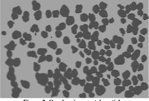
Figure 2: Overlapping crystal particles
[image:2.595.312.555.447.611.2]between seed and edge points combined with the cosine distance between the gradient and seed-to-edge vectors is sought. Once the contour evidence for each detected seed point is obtained, contour estimation is performed using numerically stable direct ellipse fitting.
II.LITERATUREREVIEW
Xue Yang et al . [1] presented a spatial-domain segmentation method in which both the value information of the pixels and the spatial relationship between them are utilized in the segmentation process and meanwhile the spatial information, such as the area, boundary or location, of the target is extracted, thus the target region to be segmented can be distinguished from the other parts in same value range by use of the spatial information. Furthermore, a parallel algorithm is designed and implemented on GPU for improving the computation efficiency of the spatial-domain segmentation method. Experiment results obtained in the work confirm the effectiveness of the new segmentation algorithm.
Hugo Leonardo Marcolino dos Santos et al. [2]
proposed a neural network based on by Auto Associative Pyramidal Neural Network and their architecture, which uses concepts of receptive fields and auto associative memory.. Furthermore, the proposed neural network also uses the concept of sharing weights aiming the applications on problems invariant translations. The neural network is able of perform implicit feature extraction and learns how to reconstruct a pattern of such features. The evaluation of the neural network is performed by two experiments. The first experiment is conducted with image processing problems. The Neural Network Auto associative learns about the transformation applied to the images, mapping a domain of images to another. In the second experiment the Auto Associative Neural Network gets satisfactory results in image segmentation. The second task uses the dataset of skin lesion images for segmentation. This work indicates that proposed model of neural network is valid due to the obtained results achieved in the performed experiments.
Sen Qian et al. [3] presented the noise is eliminated from background by median filtering and morphological filtering. Then the initial contour of the target is obtained through FCM algorithm. Finally the targets are segmented through multiple iterations of Level Set. The method has been tested on many images. Experimental results show that the proposed approach using FCM and Level Set algorithm for image segmentation is feasible and has a great effect.
Yu Linsen et al. [4] proposed a spatial correlation into mixture models by two ways: First, visual description and spatial position of pixels are considered as co-occurrence data and a joint Gaussian mixture models are built to make use of spatial constraint. Second, for the visual part of the mixture models, the pixels are sampled by the weights according to the visual similarity with their neighborhood. So the noise pixels rely more on the position and less on the visual description in the clustering procedure. Because the spatial distributions of parametric model cannot fit arbitrary shape of the segmented objects, the paper adopts an over-segmentation scheme and develops a fast EM algorithm. Experiments using
synthetic and natural images are presented to show the effectiveness of the proposed algorithm.
Riyanto Sigit et al. [5] proposed the segmentation of cardiac image for a heart disease. The method used Median High Boost Filter, Triangle Equation and Partial Monte Carlo. The first step is applying Median High Boost filter to eliminate noise. The second step is Triangle Equation to detect cardiac cavity and reconstruct the imprecise border. The third step is Partial Monte Carlo to measure the area of the heart cavity. This research used ultrasound to measure cardiac function. The experiments represented that the extended method is able to detect and improve the segmentation of cardiac cavity images with precise and faster. The performance segmentation for assessment errors cardiac cavity obtained an average triangle 8.18%, snake 19.94% and watershed 15.97%.
Vasily N. Vasyukov et al. [6] proposed a new approach to textured image segmentation based on applying finite-valued Hierarchical Markov Random Fields model. The difference of the approach from the formerly known ones is in use of three-valued Gibbs field for describing textured images. They demonstrate the performance of the developed algorithm with examples of modeled and real textured images processing.
Maedeh Sadat Fasihi et al. [7] proposed medical image processing is a very active and fast-growing field that has evolved into an established discipline. Accurate segmentation of medical images is a fundamental step in clinical studies for diagnosis, monitoring, and treatment planning. Manual segmentation of medical images is a time consuming and a tedious task. Therefore the automated segmentation algorithms with high accuracy are of interest. There are several critical factors that determine the performance of a segmentation algorithm. Examples are: the area of application of segmentation technique, reproducibility of the method, accuracy of the results, etc. The purpose of this review is to provide an overview of current image segmentation methods. Their relative efficiency, advantages, and the problems they encounter are discussed. In order to evaluate the segmentation results, some popular benchmark measurements are presented.
III.PROBLEMFORMULATION
we propose improvement in previous technique using K-Mean Clustering.
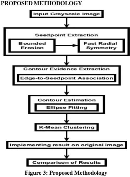
[image:4.595.49.270.89.390.2]IV.PROPOSEDMETHODOLOGY
Figure 3: Proposed Methodology
First of all gray scale image is selected and method for segmentation of clustered partially overlapping objects with a shape that can be approximated using an ellipse is performed. Segmentation method has various steps which are to be performed one by one starts with seed-point extraction using bounded erosion and fast radial symmetry transform. Extracted seed-points are then utilized to associate edge points to objects in order to create contour evidence. Then we perform contour evidence extraction using edge to seed point association in which overlapped objects edges are extracted. Then contours of the objects are estimated by fitting ellipses to the contour evidence. Finally K-Mean clustering is performed to detect any missed object or overlapped object showing as one object. Then final results are calculated on the basis of accuracy of both previous and proposed method and comparison of results is performed.
V.IMPLEMENTATION AND EXPERIMENTAL RESULTS
Elliptical objects are frequently seen in real world; hence, the detection of elliptical shapes from a digital image is important in computer vision and industrial applications. Most of these techniques seem to be complex and time consuming. Here, we propose a simple algorithm to detect elliptical objects. The techniques used in this new method are very simple and intuitive. The new method mainly consists of four steps: (1) Selecting input image; (2) detect the center of the ellipse by using bounded erosion and fast radial symmetry
called as detection of seedpoints (3) find the edge points on the ellipse by contour evidence detection using specialized elipse detection technique with K-Means clustering; and (4) performing contour estimation method. The simulation tool used for experimentation and implementation is MATLAB R2013B. The proposed and previous work is performed using two different images which gives promising results as shown in Table II and Table III below. The two images used for performing experiment are named as Original Input Image 1 and Original Input Image 2 are shown in Table I as Figure 4 and Figure 5.
[image:4.595.306.561.206.527.2]Table I: Original input images
Figure 4: Original Input Image 1
Figure 5: Original Input Image 2
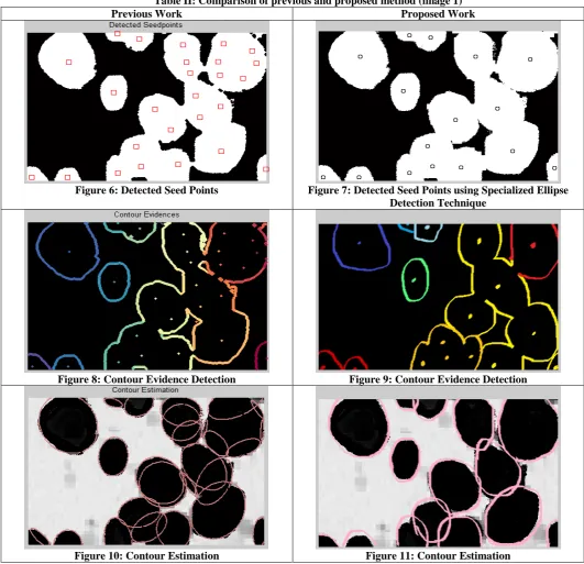
Table II: Comparison of previous and proposed method (image 1)
Previous Work Proposed Work
Figure 6: Detected Seed Points Figure 7: Detected Seed Points using Specialized Ellipse Detection Technique
Figure 8: Contour Evidence Detection Figure 9: Contour Evidence Detection
Figure 10: Contour Estimation Figure 11: Contour Estimation
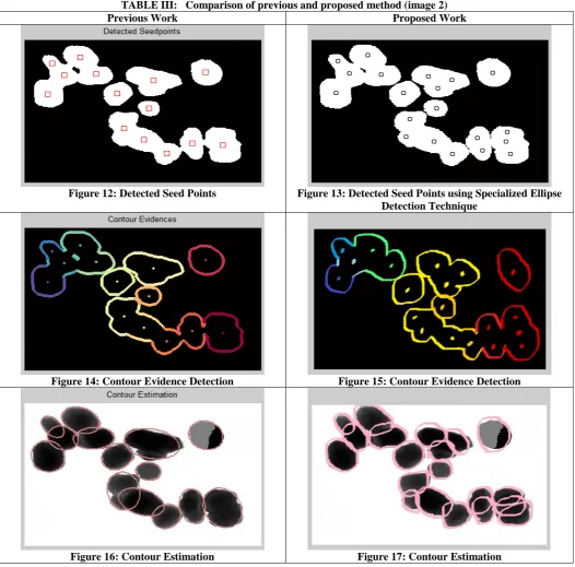
The comparison of Image 2 is shown in Table III. The detected seed points for proposed and previous work are shown in Figure 12 and Figure 13. The no.of seed points detected using previous method is 14 seedpoints and using proposed method is 20 seedpoints. The next step is to perform contour evidence detection which is shown in Figure
TABLE III: Comparison of previous and proposed method (image 2)
Previous Work Proposed Work
Figure 12: Detected Seed Points Figure 13: Detected Seed Points using Specialized Ellipse Detection Technique
Figure 14: Contour Evidence Detection Figure 15: Contour Evidence Detection
Figure 16: Contour Estimation Figure 17: Contour Estimation
[image:6.595.35.281.643.728.2]The comparison of previous and proposed ethods is shown in Table IV. The table shows comparison using image 1 and image 2 on the basis of number of detected elliptical objects.
Table IV: Comparison of previous and proposed method
Sr. No.
Image description
Actual No. of Objects
No. of Detected Objects Previous
Method
Proposed Method
1. Image 1 16 26 16 2. Image 2 20 14 20
VI.CONCLUSIONANDFUTURESCOPE
The detection of elliptical objects is frequently experienced in the real world situation. In this paper,
The future scope of proposed work would be to implement the concept of object detection to other shapes with ease of process the image to the detection different shapes.
REFERENCES
[1] X. Yang and C. Guo, "Parallel spatial-domain liver
segmentation of CT abdominal images, " In Intelligent Control and Information Processing (ICICIP), Seventh International Conference, IEEE, pp. 173-178, 2016.
[2] Y. Li, Y. Guo, Y. Kao and R. He, "Image Piece Learning for Weakly Supervised Semantic Segmentation, " IEEE Transactions on Systems, Man, and Cybernetics: Systems, 2016.
[3] S. Qian and G. Weng, " Medical image segmentation based on FCM and Level Set algorithm, " In Software Engineering and Service Science (ICSESS), 7th IEEE International Conference, pp. 225-228, 2016.
[4] Y. Linsen, L. Yanjun, C. Deyun and L. Peng, "A spatially compact mixture model for image segmentation," In Strategic Technology (IFOST), 11th International Forum IEEE, pp. 470-473, 2016.
[5] R. Sigit, A.R. Barakbah and I.A. Sulistijono, "Improved segmentation of cardiac image using triangle and partial Monte Carlo, " In Knowledge Creation and Intelligent Computing (KCIC), International Conference on IEEE, pp. 47-52, 2016. [6] V.N. Vasyukov and A.Y. Zaitseva,"Segmentation of textured
images described by hierarchical Gibbs model," In Strategic Technology (IFOST),11th International Forum, IEEE, pp. 452-455, 2016.
[7] M.S. Fasihi and W.B. Mikhael, "Overview of Current
Biomedical Image Segmentation Methods," In Computational Science and Computational Intelligence (CSCI), 2016. [8] G. Loy and A. Zelinsky, “Fast radial symmetry for detecting
points of interest,” IEEE Trans. Pattern Anal. Mach. Intell., vol. 25, no. 8, pp. 959–973, Aug. 2003.
[9] R. Fisker, J.M. Carstensen, M.F. Hansen, F. Bodker and
S.Morup, "Estimation of nanoparticle size distributions by image analysis," Journal of Nanoparticle Research, pp.267-277, 2000.
[10]J. Shu, H. Fu, G. Qiu, P. Kaye and M. Ilyas, "Segmenting overlapping cell nuclei in digital histopathology images," 35th Annual International Conference of the IEEE Engineering in Medicine and Biology Society, pp. 5445-5448, 2013.
[11]R.M. Haralick, X. Zhuang, C. Lin and J.S. Lee, "The digital morphological sampling theorem," IEEE Transactions on Acoustics, Speech, and Signal Processing, 37(12), pp.2067-2090, 1989.
[12] http://www.cs.uu.nl/. 2006. Segmentation. [ONLINE]
Available at: http://www.cs.uu.nl/docs/vakken/ibv/reader/chapter10.pdf[Acc
essed 1 January 2017].
[13]L. Barghout and L. Lee, W. Lawrence, "Perceptual information processing system, " U.S. Patent Application 10/618, 2003. [14]S. Chenand D. Zhang, "Robust image segmentation using FCM
with spatial constraints based on new kernel-induced distance measure," IEEE Transactions on Systems, Man, and Cybernetics, Part B (Cybernetics), 34(4), pp.1907-1916, 2004. [15] S. Zafari, T. Eerola, J. Sampo, H. Kalviainen and H. Haario,