University of Windsor University of Windsor
Scholarship at UWindsor
Scholarship at UWindsor
Electronic Theses and Dissertations Theses, Dissertations, and Major Papers
7-19-2018
Effect of Microplastics on the Accumulation of POPs in Fish
Effect of Microplastics on the Accumulation of POPs in Fish
Stefan Grigorakis University of Windsor
Follow this and additional works at: https://scholar.uwindsor.ca/etd
Recommended Citation Recommended Citation
Grigorakis, Stefan, "Effect of Microplastics on the Accumulation of POPs in Fish" (2018). Electronic Theses and Dissertations. 7523.
https://scholar.uwindsor.ca/etd/7523
This online database contains the full-text of PhD dissertations and Masters’ theses of University of Windsor students from 1954 forward. These documents are made available for personal study and research purposes only, in accordance with the Canadian Copyright Act and the Creative Commons license—CC BY-NC-ND (Attribution, Non-Commercial, No Derivative Works). Under this license, works must always be attributed to the copyright holder (original author), cannot be used for any commercial purposes, and may not be altered. Any other use would require the permission of the copyright holder. Students may inquire about withdrawing their dissertation and/or thesis from this database. For additional inquiries, please contact the repository administrator via email
Effect of Microplastics on the Accumulation of POPs in Fish
by
Stefan Grigorakis
A Thesis
Submitted to the Faculty of Graduate Studies
Through the Great Lakes Institute for Environmental Research
In Partial Fulfillment of the Requirements for
The Degree of Master of Science at the
University of Windsor
Windsor, Ontario, Canada
2018
EFFECT OF MICROPLASTICS ON THE ACCUMULATION OF POPS IN FISH
by
Stefan Grigorakis
APPROVED BY:
D Higgs
Department of Biological Sciences
C Weisener
Great Lakes Institute for Environmental Research
K Drouillard, Advisor
Great Lakes Institute for Environmental Research
iii
DECLARATION OF CO-AUTHORSHIP/ PREVIOUS PUBLICATION
I. Co-Authorship Declaration
I hereby declare this thesis incorporates material that is result of joint research, as follows:
I was the primary author and person responsible for the experimental implementation and
majority of writing for each chapter included in this thesis. Chapter 2 was co-authored by Dr.
Ken G. Drouillard and Dr. Sherri A. Mason. Ken G. Drouillard and Sherri A. Mason provided
intellectual guidance, and editorial support and it is published in the journal, Chemosphere.
Chapter 3 was co-authored by Dr. Ken G. Drouillard. Ken G. Drouillard provided intellectual
guidance, statistical guidance, and editorial support and is being prepared for submission to the
journal, Environmental Science and Technology.
I am aware of the University of Windsor Senate Policy on Authorship and I certify that I
have properly acknowledged the contribution of other researchers to my thesis, and have
obtained written permission from each of the co-author(s) to include the above material(s) in my
thesis.
I certify that, with the above qualification, this thesis, and the research to which it refers,
is the product of my own work.
II. Declaration of Previous Publication
This thesis includes three original papers that have been previously published/will be submitted
for publication in peer reviewed journals, as follows:
Thesis Chapter Publication title/full citation Publication status
Chapter 2 Grigorakis, S.; Mason, S. A.;
Drouillard, K. G.
iv
Determination of the gut
retention of plastic
microbeads and microfibers
in goldfish (Carasius auratus).
Chemosphere. 2017, 169,
233-238.
Chapter 3 Effect of microplastic
amendment to food on dietary
PCB assimilation efficiency
by fish
To be submitted
I certify that I have obtained a written permission from the copyright owner(s) to include
the above published material(s) in my thesis. I certify that the above material describes work
completed during my registration as a graduate student at the University of Windsor.
III. General
I declare that, to the best of my knowledge, my thesis does not infringe upon anyone’s
copyright nor violate any proprietary rights and that any ideas, techniques, quotations, or any
other material from the work of other people included in my thesis, published or otherwise, are
fully acknowledged in accordance with the standard referencing practices. Furthermore, to the
extent that I have included copyrighted material that surpasses the bounds of fair dealing within
the meaning of the Canada Copyright Act, I certify that I have obtained a written permission
v
I declare that this is a true copy of my thesis, including any final revisions, as approved
by my thesis committee and the Graduate Studies office, and that this thesis has not been
vi
ABSTRACT
Microplastic are ubiquitous in aquatic habitats and commonly found in the gut contents
of fish yet relatively little is known about the retention of microplastic particles by fish.
Microplastics also contribute to an anthropogenic organic phase in the environment capable of
absorbing hydrophobic organic compounds including persistent organic pollutants (POPs).
Relatively little is known about the potential interactions between microplastics and persistent
organic pollutant (POP) exposures to fish. In order to determine how microplastic particles affect
the accumulation of POPs in fish, I first determined the gut retention of two types of microplastic
particles (microbeads and microfibers) in goldfish. Although a small number of microplastic
particles were retained in fish GI-tracts after 6 days (0-3 particles/50), the retention of
microplastics was generally similar to the retention of bulk digesta contents. According to a
breakpoint regression model fitted to digesta contents and microplastic particles, the 50% and
90% evacuation times were 10 h and 33.4 h, respectively. The results of this study indicate that
neither microbeads nor microfibers are likely to accumulate within the gut contents of fish over
successive meals. In Chapter 3 of this thesis, I applied a duel-tracer design to quantify
polychlorinated biphenyl (PCB) dietary assimilation efficiencies (AE) in goldfish to compare
microplastic-associated PCB AEs with diet matrix-associated PCB AEs. Microplastic-associated
PCBs showed a 13.36% (12.27-14.49%) assimilation efficiency in goldfish while food
matrix-associated PCBs showed 51.64% (48.97-54.32%) assimilation efficiency; which is 3.9 fold
higher than measured for microplastic-associated PCBs. The joint findings from this thesis
indicate that microplastic particles, and POPs associated with them, are unlikely to significantly
vii
DEDICATION
To my parents who always encourage me to dream big and to my girlfriend Heather for
viii
ACKNOWLEDGEMENTS
Firstly, I would like to thank my supervisor, Dr. Ken Drouillard, for providing me the
opportunity to work as a MSc student under his tutelage. Dr. Drouillard not only provided me
with excellent supervision and guidance, but also compassion and patience when I needed it
most. I am endlessly grateful for the opportunity he presented me.
I would also like to thank Dr. Dennis Higgs and Dr. Chris Weisener for being a part of
my advisory committee. I appreciate their valuable input and discussion to my research projects.
I want to thank all of the members of the Drouillard Lab that I have had the pleasure to
work with: Bronson Goodfellow, Joe Lafontaine, Xin Sun, Jenna O’Brien, Joe Robinet, and
Shifeng Yao. A special thank you to Bronson and Joe L for our endless discussions, the vast
amount of time they spent helping me, and for their motivation they gave me to work hard and
never give up.
Whether it was soccer or school, my parents did everything in their power to ensure I was
always provided the best opportunities possible. Their endless supply of support and advice
carried me through this MSc project. I love them and do not know how to possibly thank them
enough for all they have done for me throughout my life.
Lastly, I would like to thank my girlfriend Heather. As my mental and physical health
took a toll on me, she was a rare source of happiness and motivation in my life. When I was
ready to quit, Heather picked me up by the bootstraps and carried me until I could carry myself
ix
TABLE OF CONTENTS
DECLARATION OF CO-AUTHORSHIP/PREVIOUS PUBLICATION………iii
ABSTRACT………...vi
DEDICATION………...vii
ACKNOWLEDGEMENTS………...…vii
LIST OF TABLES………..xi
LIST OF FIGURES………...xii
CHAPTER 1 General Introduction………..………1
1.1General Introduction……….1
1.2Polychlorinated Biphenyls as Model Persistent Organic Pollutants………..………..5
1.3Microplastic/POPs Interactions on POPS Bioaccumulation by Fish…………...……6
1.4Thesis Objectives………...9
1.5References………..……..10
Chapter 2 Determination of the gut retention of plastic microbeads and microfibers in goldfish (Carasius auratus)………...…20
2.1 Introduction……….20
2.2Methods………..………..22
2.3Results and Discussion……….………...27
2.4Conclusions………..31
x
Chapter 3 Effect of microplastic amendment to food on dietary PCB assimilation efficiency by
fish………..40
3.1Introduction………..40
3.2Methods………..………..42
3.3Results………..48
3.4Discussion……….………...52
3.5References………58
Chapter 4 General Discussion………71
4.1Thesis Objectives and Hypotheses Tested………71
4.2Implications of thesis findings……….………..………..74
4.3Data gaps and future studies ...………..…. 77
4.4References………78
xi
LIST OF TABLES
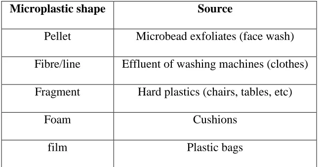
Table 1.1. Different shapes of microplastics and their sources (Eriksen et al., 2013)…………...18
Table 1.2. Examples of direct and indirect effects of microplastics on organisms………...19
Table 3.1. Geomean (95% CI) of fish body weights, lipid contents and concentrations of sum
PRC-PCB and sum Aroclor-PCBs in control and microplastic amended food……….65
Table 3.2. Principle Components Analysis (PCA) on PCB AEs in goldfish across
xii
LIST OF FIGURES
Figure 1.1. Documents explicitly mentioning microplastics by year (2017 data is up to date as of
April 5, 2017)……….17
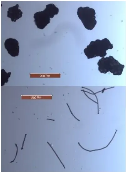
Figure 2.1. Image of microbeads (left) and microfibers (right) used for feeding trials (5 x
magnification)………38
Figure 2.2.Gut retention of digesta and microplastics in gold fish post feeding. Left graphic
presents mean microfiber (■) retention compared to digesta (O). Right graphic presents mean
microbeads (■) retention compared to digesta (O). Dashed line is the exponential fit to the
combined digesta retention data (Eq. 3). Error bars are standard error……….39
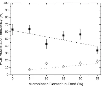
Figure 3.1. Squares – Diet-Matrix associated PCBs; circles PRC-PCBs sorbed to microplastics.
Model lines fitted to Eq. 5 and 6 for a normalized log KOW value of 6.25………68
Figure 3.2. Squares – Squares- Diet-Matrix associated PCBs; open circles are PRC-PCBs sorbed
to microplastics. Lines represent model fits to eq 5 and 6 under an assumed 5% microplastic
content……….…….….69
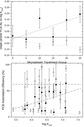
Figure 3.3. Top figure – AE vs KOW slopes for Aroclor PCBs square and PRC-PCBs; circle.
Bottom figure. PCB assimilation efficiency vs KOW for the 0% Aroclor (open square), 25%
1
CHAPTER 1
General Introduction
1.1General Introduction
In his review paper, Pruter (1987) made note of studies finding “tiny pellets” of plastic in the
environment. The first study to explicitly refer to these small plastic particles as microplastics
was published in 2004 (Thompson et al., 2004). The field of microplastic research dramatically
expanded after 2011 as demonstrated by a literature search. A Scopus database search performed
using the term “microplastic” yielded 444 publications (excluding non-environmentally relevant
database hits) over the years 1967—2017 (see Figure 1.1).
Microplastics are characterized as plastic particles under 5mm in size. As a result of this
broad definition, microplastics can be further categorized into plastic type, shape and degree of
weathering. Plastic types include Polyethylene (PE), polypropylene (PP), polystyrene (PS), and
polyvinyl chloride (PVC) which reflect some of the most commonly produced plastics on a
global basis. Microplastics can be further classified into 5 shapes (Table 1.1) (Eriksen et al.,
2013). Weathered categories of microplastics are sometimes designated as primary microplastics
which refer to plastics that have been intentionally manufactured as < 5mm in size and released
to the environment or secondary microplastics which were manufactured as macroplastics and
have broken down into microplastic size classes through UV radiation and mechanical stress
such as wave action or abrasion occurring in the environment (Cole et al., 2011).
Eriksen and colleagues estimate that there are at least 5.25 trillion microplastic particles
in marine waters (Eriksen et al., 2014). The shorelines of urban areas tend to have more
2
as 4843m, from the Porcupine Abyssal Plain, have been found to be contaminated with
microplastics (Van Cauwenberghe et al., 2013). Lake Hovsgal, northern Mongolia, is a
government protected park that has less than 10,000 people inhabiting a vast area surrounding
the lake. Despite this, Lake Hovsgal contains as much plastic as some heavily polluted areas
(Free et al., 2014). Any one particular area/body of water exhibits a great deal of heterogeneity in
terms of microplastic dispersion (Goldstein et al., 2013). A 2015 review of microplastics shows
that microplastics have contaminated both marine and freshwater systems; all while interacting
with various organisms (Eerkes-Medrano et al., 2015). This latter review of studies showcases
the extent of knowledge available in the field of microplastics: presence/distribution, transport
pathways, and proper methodology associated with environmental sampling for microplastics
detection and characterization.
Interactions between microplastics and aquatic organisms are an area of special concern,
considering the relative abundance and apparent global distribution of microplastic in water
systems. Various species of zooplankton are capable of ingesting microplastics and can further
transfer those plastic particles to the predators that consume them (Frias et al., 2014; Browne et
al., 2013; Setala et al., 2014). Mussels have also been shown to not only passively uptake and
accumulate microplastics, but also to transfer those plastics to higher trophic levels (Farrell and
Nelson 2013; von Moos et al., 2012; Collignon et al., 2012; Santana et al., 2017). While the
results of these studies are interesting, it is worth noting that the studies were designed to test the
possibility of the trophic transfer of microplastics. In essence, the prey items
(zooplankton/mussels) were exposed to high concentrations of microplastic and were fed to their
predators as soon as they were seen egesting microplastics. Such designs may not be indicative
3
been verified. Zooplankton, mussels, lugworms, and whales have been found to have
microplastics in their gut tracts upon capture (Van Cauwerberghe et al., 2015; Lusher et al.,
2012; Lusher et al., 2015). Thus, ingestion of microplastics by organisms has been documented
to occur but there remains a lack of knowledge in regard to how plastic exposures may affect the
health of organisms that consume them. Table 1.2 illustrates examples of potential direct and
indirect effects of microplastics on organism’s health.
Direct toxicity of microplastic on organisms in various trophic levels has been reported.
Cole and his colleagues put various zooplankton species in 20 mL of sea water and added
increasing amounts of microplastics that were in the size range of natural food items (Cole et al.,
2013). Feeding rate of algae was not lowered until the concentration of microplastics reached
4000 particles/mL and did not show a significant difference until a concentration of 7000
particles/mL. It was noted that microplastic particles were sticking to exterior appendages of
zooplankton; which impedes with the organism’s ability to swim and find food items.
Concentrations of microplastics higher than 4000 particles/mL are not environmentally relevant.
Zooplankton mitigate their risk of consuming microplastics by coupling two factors:
Zooplankton will avoid non-food items in search of preferred items and the encounter rate of
microplastics compared to prey items in the environment are very low (Lima et al., 2014).
Microplastics can be transferred up the planktonic food web in a lab setting (Setala et al., 2014).
Mysid shrimp were shown to uptake microplastics from zooplankton that were previously
exposed to high concentrations of microplastics. The Setala study highlights the possibility of
indirect microplastic exposure on organisms in higher trophic levels. Only 65% of Acartia
species exposed to 10000 particles/mL ingested microplastic particles; further showcasing
4
microplastic spheres of different sizes (20 nm and 1000 nm), but excreted the 1000 nm spheres
significantly more than the 20 nm spheres (Rosenkranz et al., 2009). This indicates that the 20
nm spheres may have translocated into tissues but adequate resolution was not attained to verify
such claims. Direct effects of microplastic toxicity has been better documented in lugworms
(Arenicola marina). The presence of microplastics, even at concentration as low as 0.074% dry weight, in sediments caused a significant decrease in feeding activity of lugworms compared to
the control group (Besseling et al., 2012). With a diet of 7.4% dry weight microplastic in
sediments, lugworms had 50% less tissue energy content than lugworms not exposed to
microplastics. Lugworm fitness in natural environments could therefore be lowered in the
presence of microplastics due to reduction in growth. Lugworms chronically exposed to
unplasticized PVC (UPVC) had increased phagocytic cell activity; which is an expensive
metabolic task and also caused a 50% reduction in available energy (Wright et al., 2013).
A potential secondary toxicity effect associated with microplastics may be related to how
these particles interact with toxic chemicals present in the environment. Phenanthrene and other
environmentally persistent, hydrophobic contaminants have a high affinity for plastic and the
fate of these chemicals in a natural environment can be influenced by the fate and distribution of
plastic (Teuten et al., 2007). Besseling and colleagues spiked microplastics with polychlorinated
biphenyls (PCBs) and observed significant uptake of PCBs by lugworms when in the presence of
contaminated microplastics (0.074, 0.74, and 7.4%) amended to sediments. Lugworms exposed
to microplastic spiked with triclosan had reduced survival and feeding activity (Browne et al.,
2013). Browne and his colleagues also showed that polybrominated diphenyl ethers (PBDEs)
dosed microplastics lowered feeding, while nonyphneyl- lowered immune response, and direct
5
determined to be due to a combination of reduced feeding, longer gut retention times of ingested
material (egestion events took 1.5 times longer during chronic exposure to UPVC), and
inflammation. Medaka fish (Oryzias latipes) exposed to microplastics naturally contaminated
with polycyclic aromatic hydrocarbons (PAHs), PCBs, and PBDEs had concentrations higher
than control and negative control groups; though it was only significant for PBDEs (Rochman et
al., 2013). In another study using medaka, Rochman and colleagues found that chronic exposure
to microplastics at environmentally relevant concentrations had endocrine disruption effects
(Rochman et al., 2014).
1.2Polychlorinated Biphenyls as Model of Persistent Organics Pollutants
PCBs are among the most common and widely distributed contaminants in the category of
persistent organic pollutants (POPs) and tend to be found in higher concentrations around
industrialized areas (Beyer and Biziuk, 2009). Their resistance to degradation from metabolism
and high degree of hydrophobicity make PCBs capable of bioaccumulation and food web
biomagnification in aquatic ecosystems (Hornebuckle et al., 2006). PCBs were banned over 30
years ago, but their presence in biota and sediment still persists today (Hornebuckle et al., 2006).
There are 209 possible congeners of PCB; all with varying physical-chemical properties, such as
hydrophobicity (Hornebuckle et al., 2006). The degree of hydrophobicity that a chemical
exhibits is measured by how the chemical partitions between octanol and water (Kow). As Kow
increases, so does the degree of hydrophobicity. Humans exposed to PCBs can suffer from both
acute and chronic symptoms. Human populations in Kyushu (Japan) and Yu-Cheng (Taiwan)
were both accidentally contaminated by large amounts of PCBs released into the food supply via
contaminated rice oil during industrial accidents. This caused Yusho disease with symptoms that
6
to child birth. Pregnant woman with Yusho disease had babies with a lower birth weight
(~160-250g) and at age 4, those children continued to exhibited lower weights in a dose-dependent
fashion (Jacobson and Jacobson, 1993). Human exposure to PCB is 90% from the diet with fish
contributing the bulk of human PCB exposures (Liem et al., 2000). Sub-populations of humans
can be exposed to higher levels of PCB as a result of high fish consumption in contaminated
areas (Liem et al., 2000). Considering the potential for microplastics to alter the bioavailability
of hydrophobic contaminants such as PCBs and ubiquitous presence of both microplastics and
PCBs in the environment, it is imperative that microplastic-PCB interactions be further studied.
1.3Microplastic/POPs Interactions on POPs Bioaccumulation by Fish
Microplastics have the potential to influence both the uptake and elimination of hydrophobic
contaminants, yet they are not accounted for in current bioaccumulation models. The potential of
microplastics altering the bioavailability of contaminants, such as PCBs, can be explained by
using the following equation for bioaccumulation:
𝑑𝐶𝑏
𝑑𝑡 = (𝐺𝑣 · 𝐸𝑤 · 𝐶𝑤𝑑+ 𝐺𝑓𝑒𝑒𝑑 · 𝐴𝐸𝑑𝐶𝑑)
− (𝐺𝑣 · 𝐸𝑤
𝐵𝐶𝐹 +
𝐺𝑓𝑒𝑒𝑑(1 − 𝐴𝐸𝑓𝑜𝑜𝑑) · 𝐴𝐸
𝐾𝑏, 𝑒𝑥 + 𝑘𝑚+ 𝑘𝑔) 𝐶𝑏
The variables Cb, Cd, and Cwd represent the concentration of chemical in the fish (ug/g), in
the diet (ug/g), and dissolved in water (ug/mL), respectively. Gv and Gfeed represent gill
ventilation rate (mL/gbw/d) and feeding rate (gfeed/gbw·d), respectively. BCF represents the
animal/water partition coefficient (L/d) and Kb.ex represents the animal/feces partition.
Metabollic biotransformation (km) and growth dilution (kg) have units of 1/d. Chemical exchange
7
unitless. A modified bioaccumulation model that considers microplastic/POPs interactions could
be constructed whereby microplastics are treated as a non-assimilated 3rd phase of organic
material that flows through the GI-tract via feeding and fecal egestion. Microplastics presence
within the GI-tract can be hypothesized to also alter multiple toxicokinetic parameters within
Equation 1. For example, microplastics could affect the magnitude of AE of hydrophobic
chemicals by acting as a non-assimilated sorptive pool or organic material in the digestive tract.
This would occur if PCB-microplastic sorptive interactions are capable of resisting PCB
solubilisation and transport across unstirred water layers as facilitated by mixed micelles (bile
salt/fatty acid vesicles generated in the small intestine). If POP/microplastic sorptive capacity
was high relative to miscelle vesical partition capacities, we would expect that high microplastic
contents in the diet would decrease the AE of POPs chemicals. The second toxicokinetic
parameter likely impacted by microplastic presence in the GI-tract is Kb,ex. This parameter refers
to the organism/feces partition coefficient and is estimated as the magnitude of the lipid
equivalent content of the organism compared to its feces and/or lower intestinal digesta contents.
Under the GI-magnification model, KB,ex progressively decreases from food to feces as lipids and
organic carbon from ingested food is assimilated by the organism. However, because
microplastics are considered a non-absorbable 3rd phase of high PCB sorptive capacity, K
B,ex will
be expected to be higher when animals are fed a microplastic containing diet compared to when
they are given non-microplastic diets. This would be theoretically similar to the effect mineral
oil has on the elimination of mirex/DDT (Rozman, 1983). If microplastics in food cause
decreases in both AEd and KB,Ex, then the POPs bioaccumulation and biomagnification potential
in the animal will be expected to decrease. Microplastics could also lower CWD because freely
8
across the gill surface. Finally, microplastic-adsorbed PCBs incorporated into food could
increase the Cfood of ingested food. This would potentially increase animal exposures to PCBs
and potentially counteract alterations in AEd or KB,Ex.
In order to assess the impact of microplastics on bioaccumulation of POPs by fish, the
effect of microplastics on POPs AE, KBEX and Cdiet need to be determined. Before we can study
all of the above, some baseline studies need also be completed. First, microplastic exposure needs
to be shown to be food web based. Considering the various organisms shown to naturally ingest
microplastic particles in their native environments, it can be safely concluded that microplastic
particles are a part of the aquatic food web (Eerkes-Medrano et al., 2015). Second, the partition
capacity of microplastics for POPs compounds and demonstration of POP-microplastic
contamination in natural environments need to be demonstrated. Several studies have, to date,
quantified partition capacity of different plastic types including polyethylene and polystyrene for
POPs compounds such as PCBs (Lee et al., 2013, 2017; Smedes et al., 2017). Other studies have
documented the presence of PCBs, PBDEs and PAHs present in environmental samples of
microplastics (Kalogerakis et al., 2014; Rios and Jones 2015). Third, the gut retention times of
different microplastic types (by shape and plastic type) need to be determined to establish if
microplastic retention in the gut tract is essentially the same as digesta, or if microplastics have
the capacity to accumulate within the gut tract over successive meals. The latter is necessary to
understand the potential size of the microplastic pool that can be accumulated within a fish’s gut
tract and whether this can be determined based directly on the microplastic content of ingested
food or if a separate microplastic GI-tract bioaccumulation model would be necessitated. Fourth,
AE values of microplastic-associated POPs need to be measured and compared with AEs of POPs
9
POPs AEs after loading chemicals on to microplastics (Granby et al., 2018; Wardrop et al., 2016).
Last, KBEX should be directly quantified in fish fed food containing microplastic particles and
compared with diets not containing microplastics (Drouillard et al. 2012). The latter would be
necessary to appropriately model enhanced elimination of POPs compounds by fish when fed
microplastic containing diets.
1.4Thesis Objectives
The objectives of this thesis were to characterise microplastic gut retention times and to
determine microplastic-diet matrix interactions on POPs dietary assimilation efficiencies by fish.
Chapter 2 of this thesis quantified the gastro-intestinal retention of two types of microplastics
(microbeads and microfibers) in gold fish after feeding fish food amended with each microplastic
type. The following specific hypotheses were tested in Chapter 2:
Hypothesis 1: microbeads, derived from personal care products, are retained and lost from the
gastrointestinal tract of fish at the same rate as food/digesta
Hypothesis 2: microfibers, derived from laundered textiles, are retained and lost from the
gastrointestinal tract of fish at the same rate of food/digesta
Hypothesis 3: microbeads have similar retention in the gastrointestinal tract of fish as microfibers
Chapter 3 of this thesis determined the dietary assimilation efficiencies (AE) of
polychlorinated biphenyls (PCBs) by fish in the presence and absence of microplastics (personal
care product derived microbeads) added to their food at five different microplastic treatment
concentrations. This study applied a unique duel tracer design whereby non-environmental PCBs
were adsorbed onto microbead particles and added to fish food pellets which had been previously
10
microplastic-associated PCB AEs and diet matrix associated PCB AEs and their potential
interactions across diet treatments. The specific hypotheses tested in Chapter 3 included the
following:
Hypothesis 1: microplastic-associated PCBs have similar AE values as diet matrix-associated
PCBs
Hypothesis 2: diet matrix-associated PCB AEs are unaffected by the presence of microplastic
particles in the diet across different treatments that vary microplastic contents (0 to 25%
microplastic content in food by weight)
Hypothesis 3: PCB AEs from microplastics or the diet matrix demonstrate similar relationships
with chemical hydrophobicity
1.5References
Besseling, E.; Wegner, A.; Foekema, E. M.; van den Heuval-Greve, M. J.; Koelmans, A. A.
Effects of microplastic on fitness and PCB bioaccumulation by the lugworm Arenicola
marina (L.). Environmental Science & Technology. 2012, 47, 593-600.
Beyer, A.; Wania, F.; Gouin, T.; Mackay, D.; Matthies, M. Selecting internally consistent
physiochemical properties of organics compounds. Environmental Toxicology and Chemistry.
2002, 21 (5), 941-953.
Browne, M. A.; Niven, S. J.; Galloway, T. S.; Rowland, S. J.; Thompson, R. C. Microplastic
moves pollutants and additives to worms, reducing functions linked to health and biodiversity.
11
Cole, M.; Lindeque, P.; Halsband, C.; Galloway, T. S. Microplastics as contaminants in the
marine environment: a review. Marine Pollution Bulletin. 2011, 62, 2588-2597.
Cole, M.; Lindeque, P.; Fileman, E.; Halsband, C.; Goodhead, R.; Moger, J.; Galloway, T. S.
Microplastic ingestion by zooplankton. Environmental Science & Technology. 2013, 47,
6646-6655.
Collignon, A.; Hecq, J. H.; Galgani, F.; Collard, F.; Goffart, A. Annual variation in
neustonic micro-and meso-plastic particles and zooplankton in the Bay of Calvi
(Mediterranean–Corsica). Marine Pollution Bulletin. 2014, 79, 293-298.
Drouillard, K. G.; Paterson, G.; Liu, J. Haffner, G. D. Calibration of the gastrointestinal
magnification model to predict maximum biomagnification potentials of polychlorinated
biphenyls in a bird and fish. Environmental Science & Technology. 2012, 46 (18),
10279-10286.
Eerkes-Medrano, D., Thompson, R. C., & Aldridge, D. C. Microplastics in freshwater
systems: A review of the emerging threats, identification of knowledge gaps and prioritization
of research needs. Water research. 2015, 75, 63-82.
Eriksen, M.; Mason, S. A.; Wilson, S.; Box, C.; Zellers, A.; Edwards, W.; Farley, H.; Amato, S.
Microplastic pollution in the surface waters of the Laurentian Great Lakes. Marine Pollution
Bulletin. 2013, 77, 177-182
12
Ryan, P. G.; Reisser. Plastic pollution in the world’s oceans: more than 5 trillion plastic
pieces weighing over 250,000 tons afloat at sea. Plos ONE. 2014, 9 (12), e111913.
Doi:10.1371/journal.pone.0111913
Farrell, P.; Nelson, K. Trophic level transfer of microplastic: mytillus edulis (L.) to Carcinus
maenas (L.). Environmental Pollution. 2013, 177, 1-3.
Free, C. M.; Jensen, O. P.; Mason, S. A.; Eriksen, M.; Williamson, N. J.; Boldgiv, B. High-levels
of microplastics pollution in a large, remote, mountain lake. Maine Pollution Bulletin. 2014,
85, 156-163.
Frias, J. P.; Otero, V.; Sobral, P. Evidence of microplastics in samples of zooplankton from
Portuguese coastal waters. Marine Environmental Research. 2014, 95, 89-95
Goldstein, M. C.; Rosenberg, M.; Cheng, L. Increased oceanic microplastic debris enhances
oviposition in an endemic pelagic insect. Biology Letters. 2012, 8 (23), 817-820.
Granby, K.; Rainieri, S.; Rasmussen, R.; Kotterman, M. J.; Sloth, J. J.; Cederberg, T. L.;
Barranco, A.; Marques, A.; Larsen, B. K. The influence of microplastics and halogenated
contaminants in feed on toxicokinetics and gene expression in European seabass
(Dicentrarchus labrax). Environmental Research 2018, 164, 430-443.
Hornebuckle, K. C.; Carlson, D. L.; Swackhamer, D. L.; Baker, J. E.; Eisenreich, S. J.
Polychlorinated biphenyls in the Great Lakes. Handbook of Environmental Chemistry. 2006,
5, 13-70.
13
Michigan fish. Journal of Great Lakes Research. 1993, 19 (4), 776-783.
Kalogerakis, N.; Kiparissis, S.; Yiantzi, E.; Perraki, T.; Kalogerakis, G. C.; Fodelianakis, S.;
Fava, F.; Psillakis, E. PCBs and PAHs on plastic pellets and microplastics collected on the
coastline of the island of Crete in Eastern Mediterranean Sea – monitoring and fate.
Proceedings of the 37th AMOP Technical Seminar on Environmental Contamination and
Response. 2014, 175-183.
Lee, H., S. Chang, S-K Kim, J-H. Kwon. Fugacity analysis of polycyclic aromatic
hydrocarbons between microplastics and seawater. Ocean Science Journal. 2017, 52, 43-55.
Leite, A. S.; Santos, L. L.; Costa, Y.; Hatje, V. Influence of proximity to an urban center in the
pattern of contamination by marine debris. Marine Pollution Bulletin. 2014, 81, 242-247.
Liem, A. K.; Furst, P.; Rappe, C. Exposure of populations to dioxins and related compounds.
Food Additives & Contaminants. 2000, 17 (4), 241-259.
Lima, A. R.; Costa, M. F.; Barletta, M. Distribution patterns of microplastics within the plankton
of a tropical estuary. Environmental Research. 2014, 132, 146-155.
Lusher, A. L.; McHugh, M.; Thompson, R. C. Occurrence of microplastics in the gastrointestinal
tract of pelagic and demersal fish from the English Channel. Marine Pollution Bulletin. 2013,
67, 64-99.
14
Microplastic and macroplastic ingestion by a deep diving, oceanic cetacean: the True’s
beaked whale Mesoplodon mirus. Environmental Pollution. 2015, 199, 185-191.
Onozuka, D.; Yoshimura, T.,; Kaneko, S.; Furue, M. Mortality after exposure to polychlorinated
biphenyls and polychlorinated dibenzofurans: a 40-year follow-up study of Yusho patients.
American Journal of epidemiology. 2009, 169 (1), 86-95.
Pruter, A. T. Sources, quantities, and distribution of persistent plastics in the marine environment.
Marine Pollution Bulletin. 1987, 18 (68), 305-310.
Rios, L. M.; Jones, P. R. Characterisation of microplastics and toxic chemicals extracted from
microplastic samples from the North Pacific Gyre. Environmental Chemistry. 2015, 12,
611-617.
Rochman, C. M.; Hoh, E.; Kurobe, T.; Teh, S. J. Ingested plastic transfers hazardous chemicals
to fish and induces hepatic stress. Scientific Reports. 2013, 3, 3263.
Rochman, C. M.; Kurobe, T.; Flores, I.; Teh, S. J. Early warning signs of endocrine disruption in
adult fish from the ingestion of polyethylene with and without sorbed chemical pollutants
from the marine environment. Science of the Total Environment. 2014, 493, 656-661.
Rosenkrantz, P.; Chaudhry, Q.; Stone, V.; Fernandes, T. F. A comparison of nanoparticle and
fine particle uptake by Daphnia magna. Environmental Toxicology and Chemistry. 2009, 28
(10), 2142-2149.
15
exposure scenario: insights on the likelihood of particles cascading along marine food webs.
Marine Pollution Bulletin 2017, 121, 154-159.
Setala, O.; Fleming-Lehtinen, V.; Lehtiniemi, M.; Ingestion and transfer of microplastics in the
planktonic food web. Environmental Pollution. 2014, 185, 77-83.
Smedes, F.; Rusina, T.P.; Beeltje, H.; Mayer, P. Partitioning of hydrophobic organic
contaminants between polymer and lipids for two silicones and low density polyethylene.
Chemosphere. 2017, 186, 948-957.
Teuten, E. L.; Rowland, S. J.; Galloway, T. S.; Thompson, R. C. Potential for plastics to transport
hydrophobic contaminants. Environmental Science & Technology. 2007, 41 (22), 7759-7764.
Thompson, R. C.; Olsen, Y.; Mitchell, R. P.; Davis, A.; Rowland, S. J. John, A. W.; McGonigle,
D.; Russell, A. E. Lost at sea: where is all the plastic?. Science. 2004, 304 (5672), 838.
Van Cauwenberghe, L.; Claessens, M.; Vandegehuchte, M. B.; Janssen, C. R. Microplastics are
taken up by mussels (Mytilus edulis) and lugworms (Arenicola marina) living in natural
habitats. Environmental Pollution. 2015, 199 10-17.
Von Moos N, Burkhardt-Holm P, Köhler A. Uptake and effects of microplastics on cells
and tissue of the blue mussel Mytilus edulis L. after an experimental exposure. Environmental
science & technology. 2012, 46, 11327-11335.
16
Chemical pollutants sorbed to ingested microbeads from personal care products accumulate in
fish. Environmental Science and Technology. 2016, 50, 4037-4044.
Wright, S. L.; Rowe, D.; Thompson, R. C.; Galloway, T. S. Microplastic ingestion decreases
17
Figure 1.1. Documents explicitly mentioning microplastics by year (2017 data is up to date as of
18
Microplastic shape Source
Pellet Microbead exfoliates (face wash)
Fibre/line Effluent of washing machines (clothes)
Fragment Hard plastics (chairs, tables, etc)
Foam Cushions
film Plastic bags
19
Direct Toxicity Indirect Toxicity
Clogging feeding apparatus
Gastric blockage
Gastric distention (leading to lower feeding)
Exposures to toxic substances associated with
microplastics
Leachates from plastic interior
Absorbed/partitioned chemicals
picked up from the environmnet by
microplastic particles
Reduced growth and fitness Oxidative stress, endocrine disruption,
reproductive toxicity
20
CHAPTER 2
Determination of the gut retention of plastic microbeads and microfibers in goldfish (Carassius auratus)
Reprinted with permission from (Grigorakis, S.; Mason, S. A.; Drouillard, K. G. Determination
of the gut retention of plastic microbeads and microfibers in goldfish (Carasius auratus). Chemosphere. 2017, 169, 233-238.
2.1 Introduction
Microplastics are a diverse array of synthetic polymer particles that vary in chemical
composition, size (from low micrometre scale to an upper size range variously defined between 1
nm and 5 mm), density and shape (Andrady, 2011). They have been observed in most freshwater
and marine environments (Eriksen et al., 2014; Corcoran, 2015; Eerkes-Medrano et al., 2015) to
such an extent that they were included as sedimentary geochemical markers of the Anthropocene
(Waters et al., 2016). Microplastics are often distinguished between those that are synthesized at
the defined sizes for an intended application (primary microplastics) relative to particles derived
from the breakdown of macroplastics (secondary microplastics). Microbeads are defined as
primary microplastics that range in size between 0.1 µm to <5 mm (Environment Canada, 2015)
and are used in a wide variety of industrial and consumer applications including personal care
products (PCPs). Legislation banning the production of microbeads in PCPs comes into effect in
2017 as passed by the U.S. federal government and similar legislation is under review in Canada.
While much of the legislative focus has been on microbeads used in PCPs, other common
21
textiles such as nylon and acrylics, henceforth referred to as microfibers, used in clothing
(Browne, 2011).
Concerns have been raised about the ecotoxicology of microplastics in the environment,
including their potential to bioaccumulate in organisms and subsequent transfer through food
webs (Sánchez et al., 2014). Zooplankton are capable of ingesting microplastics, potentially
mistaking them for food, and can further transfer these to tertiary consumers (Frias et al., 2014;
Browne et al., 2013; Setala et al., 2014; Rehse et al., 2016). Mussels have been shown to
accumulate microplastics and transfer them to higher trophic levels (Browne et al., 2013; von
Moos et al., 2012; Collignon et al., 2012). In a study examining 504 fish from the English
Channel that included benthic and pelagic species, 36.5% of specimens had microplastics in their
gastrointestinal (GI-) tracts (Lusher et al., 2013). Microplastics in the gut contents of field
collected fish have subsequently been widely reported in coastal and freshwaters (Sanchez et al.,
2014; Neves et al., 2015; Avio et al, 2015; Phillips and Bonner, 2015; Biginagwa et al., 2016;
Bellas et al., 2016). Considering microplastics are being found in fish, there are relatively few
studies focussing on the potential of microplastics to bioaccumulate.
Exposure to microplastics in water and food can interfere with normal digestive processes
due to intestinal blockage, causing reductions in animal feeding rates and energy assimilation
(Besseling et al., 2012), lead to histopathological alteration to intestinal and hepatic tissues of
fish (Pedà et al. 2016; Lu et al., 2016) and lower hatching success of fish eggs (Lönnstedt and
Eklöv, 2016). Translocation of microplastics from gut to the circulatory system has been
demonstrated in mussels (Browne et al., 2008; von Moos et al., 2012; Avio et al. 2015a)
22
some animals. Avio et al., (2015b) and Lu et al., (2016) confirmed microplastics accumulation in
hepatic tissues of fish exposed to microplastics at elevated concentrations in water.
Although microplastics are commonly detected in the intestinal tracts of fish, there is
limited information characterizing the retention of microplastics by fish. Particle size and shape
are likely to influence factors such as GI-retention but limited information is available comparing
microplastic types. Neves et al. (2015) observed a higher frequency of fibers in commercial fish
gut contents compared to plastic fragments. The above study further reported differences in
plastic types in benthic fish, which tended to accumulate a greater proportion of fibers, compared
to pelagic fish which contained more fragments. It is not known whether these differences are
related to emission patterns and fate of different particle types or whether particle shape might
influence the gut retention characteristics of these microplastic types. In this study, the GI-tract
retention of two microplastic types, microbeads and microfibers, was determined in goldfish with
the objective to determine if i) retention of microplastics by fish exceeds that of food digesta, i.e.
exhibits net accumulation in the GI-tract of fish, and ii) to determine if microfibers are retained
to a greater or lesser degree than PCP derived microbeads.
2.2 Methods
Microplastic source
Microfibers were extracted from clothing (35cm x 12cm cut out of a commercial
polyester fleece scarf) by mechanical agitation in hot water. Following agitation, the water was
sieved through stacked 500 µm, 250 µm and 63 µm sieves. Fibers retained on the 63µm sieve
were removed by tweezer under magnification and size graded to between 50-500 µm fiber
lengths under a dissecting microscope. Microplastic beads were extracted from a commercial
23
poured onto a 63 sieve and the soluble matrix associated with the product washed with water
until only microplastics remained. Microbeads were removed from the sieve under
magnification. Figure 2.1 provides images of isolated microbeads and microfibers under 5x
magnification.
Experimental
Goldfish were selected as a model fish species because they have been routinely used in
many bioaccumulation/toxicokinetic studies owing to the ease of husbandry, tolerance to
handling and willingness to accept artificial diets. In their wild state, goldfish are benthic feeders
and thus might be expected to accumulate microplastics similar to those reported for other
benthic feeders. Fish were exposed to microplastics via food. Commercial fish pellets
(0.18-0.21g, ~3 mm size) were placed in warm water to soften them. Treatment pellets were amended
with 50 microbeads or 50 microfibers per pellet by manual insertion of macroplastic particles
into each pellet under microscope. Pellets were air dried after manipulation. Control pellets were
wetted and dried in an identical manner but not amended with microplastics. The food was
prepared in this manner to ensure that every experimental fish consumed exactly 50 microplastic
particles to increase precision of gut retention characterization.
Twenty-Eight sexually mature goldfish were fasted for 48 h prior to exposing them to
prepared food in order to ensure complete evacuation of gut contents from previous meals and to
increase the likelihood that they would accept the microplastic amended pellet provided to them.
After fasting, fish were removed from their communal tank and placed in individual fish bowls.
Twenty four fish were allocated to the microbead and microfiber treatments, respectively. Five
fish were allocated as controls and fed non-amended pellets. Each fish was presented with a
24
the fish consumed the treatment pellet, non-amended fish pellets were added to the bowl and the
fish was allowed to consume to satiation for up to 60 minutes. Any remaining fish food in the
bowl was subsequently removed. Fish were fasted for the remainder of the experimental period.
Control fish were sacrificed after 1.5 h from feeding the control pellets. Triplicate animals from
each treatment were sacrificed after 1.5, 4, 8, 16, 32, 48, 96, and 144 h. The mean ± SE of water
temperatures was 14.2 ± 0.21 oC and exhibited no changes over the fasting duration. The mean ±
SE body weights of fish from the microbead and microfiber treatments were 24.80 ± 2.77 g and
27.07 ± 3.40 g and were not significantly different from one another (p>0.4; ANOVA). On
sacrifice, fish were euthanized by immersion in a solution of MS-222 (100 mg/L) and stored
frozen until subsequent analysis. This research was performed under ethics approval from the
University of Windsor's Animal Care Committee.
Microplastic analysis
On analysis, the gut tract of each fish was dissected and removed. The gut contents were
pushed thought the intestine using tweezers and a probe onto a pre-weighed aluminum weight
boat and the gut tract tissues were retained for further analysis. The weigh boat was dried at
110oC for 1 h and reweighed to determine dry food digesta weight. Subsequently, the dried
digesta and gut tissues were re-combined and placed into a 10% KOH solution on a hot plate set
at its lowest setting for 1 hour. The solution was taken off of the hot plate and after 2 additional
hours, 5mL of 30% H2O2 was added to the solution. The solution was poured through a vacuum
filtered Buchner funnel using WhatmanTM (55mm) filter papers (1 µm glass fiber filters). Fish
carcass samples were also digested in a similar manner. Filter papers from each digestion were
GI-25
tract/contents, fish carcass or digested food pellets. Quality control of the method was
established by measuring and verifying microbeads and microfibers in 5 amended pellets. The
mean ± standard deviation of recoveries of microplastic particles for the digested pellets was
98.8±1.8%.
Data analysis
Digesta contents weights were standardized to the mean body weight according to:
𝑋𝐷𝐺(𝑠𝑠) = 𝑋𝐷𝐺(𝑠)·𝐵𝑊(𝑚𝑒𝑎𝑛)
𝐵𝑊(𝑠) (1)
where XDG(ss) is the size standardized digesta weight (g), XDG(s) is the digesta weight measured in
an individual fish, BW(mean) is the mean body weight of fish from the treatment and BW(s) is the
body weight of the individual fish. The % remaining of digesta contents was calculating by
dividing XDG(ss) by the mean XDG(ss) generated for fish sampled at the first time point (1.5h) and
multiplying by 100. For microbeads and microfibers, %remaining was calculated by dividing the
number of microplastics measured in a fish's digestive tract by 50 and multiplying by 100.
Statistical analysis was performed using a general linear model (GLM) according to:
Model = Time + Group + Time * Group + Constant (1)
Where time is the time since feeding (h), group represents a categorical variable specified as
digesta retention treatment 1, digesta retention treatment 2, microfibers and microbeads. Under
cases where the interaction term (Time * Group) was non-significant, analysis of covariance
(ANCOVA) was performed to adjust for time as a covariate and increase the statistical power of
the group comparison test. Where the interaction term was found to be significant, GLMs were
26
GLM(1) tested for differences in digesta retention time between treatment 1 and treatment 2.
GLM(2) tested for differences in digesta retention time and microfiber retention from
measurements taken in treatment 1. GLM(3) tested for differences in digesta retention time and
microbead retention from measurements taken in treatment 2. Finally, GLM(4) tested for
differences in microfiber and microbead retention. Data transformation was necessary owing to
failure of normality of the % retention data on the combined data (digesta, microbeads and
microfibers). However, when the fist time point (1.5 h) was removed, transformation of %
retention data by natural log transformation yielded a normal data set (p>0.05; Lillefor's test).
Thus, statistical comparisons by GLM were performed with the 1.5 h time point removed and
applying a ln transformation. Non-transformed digesta retention data (inclusive of the 1.5 h time
point) for individual fish were subsequently fit to an exponential model using non-linear least
squares regression according to:
%𝑅𝑒𝑡𝑎𝑖𝑛𝑒𝑑 = 100 · 𝑒−𝐵·𝑡𝑖𝑚𝑒 (2)
Where 100 is constant forcing 100% of gut contents retention at time 0, b is the fitted coefficient
and time is time since feeding (h). The ability of Eq. 2 calibrated independently to gut contents
to predict microplastic retention was evaluated using goodness of fit tests by performing a linear
regression on observed (microplastic) vs model (Eq. 2) predicted digesta retention. The
goodness of fit result was evaluated by determining if the slope was significantly different from
1, the constant was significantly different from 0 and by evaluating the magnitude of the
coefficient of determination. All statistics were performed using Systat 13 statistical software.
Except where otherwise noted, measures of central tendency and variation are expressed as mean
27 2.3 Results and Discussion
Digesta retention
During experimental trials, all fish were observed to consume the microplastic amended
treatment pellet. No fish mortalities occurred nor were there apparent signs of distress following
exposure to the amended food pellet. Fish sacrificed at the 1.5 h time point had a mean XDG(ss)
weight of 0.60±0.04 g. This corresponds to a food consumption of 2.32% body weight across
the treatments and is consistent with expected food consumption rates in fasted fish.
A general linear model (GLM(1) as described in methods) was performed to compare %
retention of digesta between the two treatments. The GLM and ANCOVA revealed a
non-significant (F1,39 = 0.92; p>0.3; ANCOVA) difference in digesta retention between the
treatments after adjusting for time as a covariate. Given that digesta retention did not
significantly differ between the two treatments, the data were combined and fit to the exponential
model yielding the following solution:
%𝑅𝑒𝑡𝑎𝑖𝑛𝑒𝑑 = 100 · 𝑒−0.069·𝑡𝑖𝑚𝑒; R2 = 0.69 (3)
Based on Eq. 3, the time to evacuate 50% and 90% of digesta was 10.0 and 33.4 h, respectively.
Overall, the exponential model fit described the temporal trends of digesta contents well during
the first 24 h but tended to underestimate observed digesta contents at longer time points (Figure
2.2). This may be related to the method of separating gut contents from the intestinal tissues
which could have included residual gut secretions and/or sloughed cells/tissues generated from
the GI-tract processing method itself. However, the fitted model produced retention estimates
that were generally consistent with other studies on digesta retention in fish of similar size and
temperature. Stehlik et al., (2014) reported full clearance of gut contents from clearnose skate
28
evacuation coefficient of 0.035·time (h-1) based on a log linear model which implies a 50%
digest retention of 19.8 h and 90% retention of 65 h (Gringas and Boisclair, 2000).
Microplastic retention in GI-tracts
Control fish sacrificed after 1.5 h were examined for evidence of microplastics in gut
contents and carcass samples. No microplastics were found in control fish or within their gut
contents. In addition, 10 control fish pellets were examined for presence of microplastics.
Similar to control fish, microplastics were not observed in non-ammended food pellets.
During the first sampling point (1.5 h), there was good recovery of microplastics within
the gut contents of treatment fish. For microfibers, 2 fish had 50 microfibers recovered (100%
recovery) and the third fish had 48 fibers recovered in the GI-tract. For the microbeads, 40 to 44
particles (80-84% recovery) were recovered from fish during the first time point. Small numbers
of microplastics were recovered at the 144 h time point (1 to 3 microfibers in replicate 144h
sampled fish and 0 to 3 microbeads in triplicate fish).
A general linear model (GLM) was applied to test percent retention of all treatments
(digesta from each treatment, microbeads and microfibers) within the study. Both Time (F1,76 =
88.1; p<0.001) and the Group x Time (F3,76=3.09; p<0.05) interaction terms were significant but
group was not significant (F3,76 =0.212; p>0.8) in the overall GLM. Due to the significant
interaction terms, additional GLMs were applied to subsets of the data to evaluate for differences
in retention on selected measurements. GLM(2) and the ANCOVA revealed no significant
differences (F1,39=0.959; p>0.5; ANCOVA) in microfiber and digesta retention. Similarly,
GLM(3) and ANCOVA revealed non-significant (F1,39=4.00; p>0.05; ANCOVA) differences in
29
microbead retention yielded non-significant differences (F1,39=0.678; p>0.4; ANCOVA) from
one another. Microplastic and microfiber retention with time along with digesta contents trends
are presented in Figure 2.2.
For microfibers, the linear regression between %microfiber retention and gut digesta
model (Eq. 3) prediction yielded a slope of 0.96±0.09, constant of (7.33±4.07) and coefficient of
determination (R2) of 0.85. The above slope was not significantly different from unity
(t1,22=0.042; p>0.5; t-test) and the constant was not different from zero (t1,22=1.80; p>0.05;
t-test). For microbeads, the goodness of fit test produced a similar slope (0.94±0.04) that was not
significantly different from unity (t1,22 = 1.44; p>0.1; t-test) and constant (3.67±2.06) not
significantly different from zero (t1,22=1.79; p>0.05; t-test) with an R2 of 0.95. It is perhaps
notable that the digesta retention model (Eq. 3) which was calibrated only to digesta retention
data explained even more variation in microplastic retention than digesta contents itself. This
was mainly related to the better fit of model predictions to microplastic retention at the later time
points (Figure 2.2). The reason for the differences in model fit across measurements is attributed
to the fact that microplastic exposure was controlled with a high degree of precision compared to
gut contents. Although each fish was given exactly 50 microplastic particles, they were provided
with food ad libitum after verifying their consumption of the microplastic amended pellet. Thus,
digesta contents would have varied to a greater extent between fish compared to microplastic
exposures. Overall the goodness of fit tests indicates that the gut digesta retention model
adequately described the retention of both microplastic types.
Similar observations were generated for the marine isopod Idotea emerginata fed a diet spiked with microplastic particles and fibers (Hämer et al,. 2014). In the study by Hämer et al.,
30
readily egested with the feces. Mazurais et al. (2015) examined microplastic retention in
European sea bass (Dicentrarchus labrax) larvae when exposed to microplastics added to food. The above authors observed a correlation between microbeads in the gut of larvae with
concentration of microbeads added to the diet. However, the authors noted that microbeads were
fully cleared from the gut of larvae after 2 days post exposure and could be identified in feces
suggesting passive retention in the gut contents of fish. While fish are regularly feeding in their
native environments, microplastic particles retrieved from the gut contents of fish are still likely
to be a result of the most present feeding rather than an accumulation across successive meals.
Microplastics were also examined in carcass samples of treatment fish but were not
observed apart from the gut tissue and gut contents analyzed separately and discussed above.
This differs from the results of Avio et al., (2015b) who observed translocation of polyethylene
and polystyrene microplastics to liver of laboratory held mullet (Mugil cephalus) exposed to microplastics in water (nominal microplastic dose was 2.5x10³ particles/L of polyethylene or
polystyrene particles sized from 100 to 1000 µm) for 7 days. Between 1-2 microplastic particles
per individual were detected in liver of exposed fish, although the presence of microplastics in
liver was two orders of magnitude lower than what was observed in gut contents of fish.
Similarly, Lu et al. (2016) exposed zebrafish (Danio rerio) to solutions containing 5 or 20 µm diameter polystyrene microplastics at concentrations of between 4.5 x 10⁶ to 2.9 x10⁸ particles/L
for 7 days. The above authors reported that 5 µm microplastics accumulated in fish gills, liver
and gut, whereas larger microplastics (20 µm in diameter) accumulated only in fish gills and gut
but not in liver. Time to steady state of microplastics in zebra fish was reported to be 48 h,
implying rapid clearance from animals consistent with the gut retention data presented here (Lu
31
apart from their detection in the GI tract, this could be a function of exposures to different
microplastic types, different dosing strategies, levels of exposures and differences in the method
of detection of microplastics in exposed animals. The lack of translocation of larger (20 µm
plus) sized microbeads to liver in zebra fish as reported by Lu et al., (2016) is consistent with the
present work given that particles greater than 63 µm were utilized but is not consistent with Avio
et al. (2015b) who exposed fish to microplastics of comparable size to this research. Avio et al.,
(2015b) and Lu et al., (2016) provided continuous exposures of fish to microplastic contaminated
water for up to 7 d days compared to a single dose from a microplastic amended meal applied in
the present study. The above authors also used nominal microplastic concentrations in water that
were considerably higher than what is present in natural waters. Avio et al., (2015b) used a more
sensitive microplastic extraction/detection technique that employed a combination of density
gradient separation and oxidant treatment which was shown to yield higher recoveries of
microplastics from animal tissues then the oxidation treatment alone. Lu et al., (2016) utilized
microplastic particles with encapsulated fluorescent dies to facilitate their detection in tissues
which potentially yielded much lower detection limits then the visual method employed here.
Thus, even though microplastics had very good recovery in pellets and gut contents of early time
point sacrificed fish from the present work, translocation of smaller microplastic particles when
exposed at higher concentrations or under long term exposures cannot be ruled out based on the
results of this study.
2.4 Conclusions
Microplastics of two distinct particle shapes (microbeads and microfibers) exhibited
similar retention in the GI-tract of goldfish compared to bulk food and digesta. Although a small
32
bioaccumulation of microplastics in the GI-tract or internal translocation to tissues of fish post
exposure. This implies that the potential for long term entrainment and retention of textile
derived microfibers or PCP-derived microbeads in fish is relatively low and the detection of
microplastics in fish gut contents in the environment most likely represents recent exposures to
microplastics in the diet as opposed to cumulative retention across multiple meals. However, this
study was limited to evaluation of only two microplastic types and one species of fish. As such,
further research to characterize microplastic retention by fish species over different plastic types,
shapes and dietary concentrations may be warranted.
2.5 References
Andrady A.L. Microplastics in the marine environment. Marine Pollution Bulletin. 2011, 62,
1596-1605. DOI: 10.1016/j.marpolbul.2011.05.030
Avio, C.G., Gobri, S., Milan, M., Benedetti, M., Fattorini, D., D'Errico, G., Pauletto, M.,
Bargelloni, L., Regoli, F. Pollutant bioavailability and toxicologic risk from microplastics
to marine mussels. Environmental Pollution. 2015, 198, 211-222. DOI:
10.1016/j.envpol.2014.12.021
Avio, C.G., Gorbi, S., Regoli, F. Experimental development of a new protocol for extraction and
characterization of microplastics in fish tissues: first observations in commercial species
from Adriatic Sea. Marine Environmental Research. 2015, 111, 18-26. DOI:
10.1016/j.marenvres.2015.06.014
Bellas, J., Martinez-Armental, J., Martinez-Cámara, A., Basada, V., Martinez-Gómez, C.
Ingestion of microplastics by demersal fish from the Spanish Atlantic and Mediterranean
33
Besseling, E., Wegner, A., Foekema, E. M., van den Heuvel-Greve, M. J., & Koelmans, A. A.
Effects of microplastic on fitness and PCB bioaccumulation by the lugworm Arenicola
marina (L.). Environmetnal Science & Technology. 2012, 47, 593-600. DOI:
10.1021/es302763x
Biginagwa, F.J., Maymoa,B.S., Shashoua, Y., Syberg, K., Khan, F.R. First evidence of
microplastics in the African Great Lakes: Recovery from Lake Victoria Nile perch and
Nile tilapia. Journal of Great Lakes Research. 2016, 42, 146-149. DOI
10.1016/j.jglr.2015.10.012
Browne, M.A., Dissanyake, A., Galloway, T.S., Lowe, D.M., Thompson, R.C. Ingested
microscopic plastic translocates to the circulatory system of the mussel, Mytilus edulis
(L.). Environmental Science & Technology. 2008,42, 5026-5031. DOI:
10.1021/es800249a
Browne, M.A. Accumulation of microplastic on shorelines worldwide: sources and sinks.
Environmental Science & Technology. 2011, 45, 9175-9179. DOI: 10.1021/es201811s
Browne, M.A., Niven, S.J., Galloway, T.S., Rowland, S.J., Thompson, R.C. Microplastic moves
pollutants and additives to worms, reducing functions linked to health and
biodiversity. Current Biology. 2013, 23, 2388-2392. DOI: 10.1016/j.cub.2013.10.012
Collignon, A., Hecq, J.H., Galgani, F., Collard, F., Goffart, A. Annual variation in neustonic
micro-and meso-plastic particles and zooplankton in the Bay of Calvi (Mediterranean–
Corsica). Marine Pollution Bulletin. 2014, 79, 293-298. DOI:
34
Corcoran, P.L. Benthic plastic debris in marine and freshwater environments. Environmental
Science (Processes and Impacts). 2015, 17, 1363-1369. DOI: 10.1039/c5em00188a
Eerkes-Medrano, D., Thompson, R.C., Aldridge, D.C. Microplastics in freshwater systems: A
review of the emerging threats, identification of knowledge gaps and prioritisation of
research needs. Water Research. 2015, 75, 63-82. DOI: 10.1016/j.watres.2015.02.012
Environment Canada (2015). Microbeads – A science summary. Unpublished report.
http://www.ec.gc.ca/ese-ees/ADDA4C5F-F397-48D5-AD17-3F989EBD0E5/Microbeads_Science%20Summary_EN.pdf (Accessed March 23, 2016).
Eriksen, M., Lebreton, L.C.M., Carson, H.S., Theil, M., Mooree, C.J., Borerro, J.C., Galgani, F,
Ryan, P.G., Reisser, J. Plastic Pollution in the World’s Oceans: More than 5 Trillion
Plastic Pieces Weighing over 250,000 Tons Afloat at Sea. PLoS One. 2014, 9, 12. DOI:
10.1371/journal.pone.0111913
Frias, J.P.G.L., Otero, V., Sobral, P. Evidence of microplastics in samples of zooplankton from
Portuguese coastal waters. Marine Environmental Research. 2014, 95, 89-95. DOI:
10.1016/j.marenvres.2014.01.001
Gingras, J., Boisclair, D. Comparison between consumption rates of yellow perch (Perca
flavescens) estimated with a digestive tract model and with a radioisotope approach. Can Journal of Fish & Aquatic Science. 2000, 57, 2547-2557. DOI: 10.1139/cjfas-57-12-2547
Hämer, J., Gutow, L., Köhler, A., Sabrowoski, R. Fate of microplastics in the marine isopod
Idotea emerginata. Environmental Science & Technology. 2014, 48, 13541-13458. DOI:
35
Lönnstedt, O.M., Eklöv, P. Environmentally relevant concentrations of microplastic particles
influences larval fish ecology. Science. 2016, 352, 1213-1216. DOI:
10.1126/science.aad8828
Lusher A.L., McHugh, M., Thompson, R.C. Occurrence of microplastics in the gastrointestinal
tract of pelagic and demersal fish from the English Channel. Marine Pollution Bulletin.
2013, 67, 94-99. DOI: 10.1016/j.marpolbul.2012.11.028
Lu, Y., Zhang, Y., Deng, Y., Jiang, W., Zhao, Y., Geng, J., Ding, L., Ren, H. Uptake and
accumulation of polystyrene microplastics in zebrafish (Danio rerio) and toxic effects in
liver. Environmental Science & Technology. 2016, 50, 4054-4060. DOI
10.1021/acs.est.6b00183
Mazurais, D., Ernande, B., Quazuguel, P., Severe, A., Huelvan, C., Madec, L. Mouchel, O.,
Soudant, P., Robbens, J., Huvet, A., Zamonino-Infante, J. Evaluation of the impact of
polyethylene microbeads ingestion in European sea bass (Dicentrarchus labrax) larvae.
Marine Environmental Research. 2015, 112, 78-85. DOI:
10.1016/j.marevres.2015.09.009
Neves, D., Sobral, P. Ferreira, J.L., Pereira, T. Ingestion of microplastics by commercial fish off
the Portuguese coast. Marine Pollution Bulletin. 2015, 101, 119-126. DOI:
10.1016/j.marpolbul.2015.11.008
Pedà, C., Caccamo, L., Fossi, M.C., Gai, F., Andaloro, F., Genovese, L., Perdichizzi, A., Romeo,
T., Maricchiolo, G. Intestinal alterations in European sea bass Dicentrarchus labrax
(Linnaeus, 1758) exposed to microplastics: Preliminary results. Environmental Pollution.