Review
Bone loss
Factors that regulate osteoclast differentiation: an update
Sophie Roux* and Philippe Orcel
†*
†Lariboisière Hospital, Paris, and *Bicêtre Hospital, Bicêtre, France
Abstract
Osteoclast activation is a critical cellular process for pathological bone resorption, such as erosions in rheumatoid arthritis (RA) or generalized bone loss. Among many factors triggering excessive osteoclast activity, cytokines such as IL-1 or tumour necrosis factor (TNF)-αplay a central role. New members of the TNF receptor ligand family (namely receptor activator of nuclear factor-κB [RANK] and RANK ligand [RANKL]) have been discovered whose cross-interaction is mandatory for the differentiation of osteoclasts from hemopoietic precursors, in both physiological and pathological situations. Osteoprotegerin, a decoy receptor which blocks this interaction, decreases osteoclast activity and could have a fascinating therapeutic potential in conditions associated with upregulated bone resorption.
Keywords:bone cytokines, differentiation, osteoclast, osteoprotegerin, RANK, RANKL Received: 8 March 2000
Revisions requested: 29 June 2000 Revisions received: 7 August 2000 Accepted: 14 August 2000 Published: 6 September 2000
Arthritis Res2000, 2:451–456
The electronic version of this article can be found online at http://arthritis-research.com/content/2/6/451
© Current Science Ltd (Print ISSN 1465-9905; Online ISSN 1465-9913)
M-CSF = macrophage colony-stimulating factor; NF-κB = nuclear factor-κB; ODF = osteoclast differentiation factor; 1,25(OH)2D3= 1,25-dihydroxy-vitamin D3; PTH = parathyroid hormone; RA = rheumatoid arthritis; RANK = receptor activator of nuclear factor-κB; RANKL = receptor activator of nuclear factor-κB ligand; TNF = tumour necrosis factor.
Introduction
Bone remodelling is a continuous physiological process that occurs in adult skeleton in which bone resorption is followed by new bone formation, maintaining mechanical strength and structure. Bone cells that are responsible for this coupled process include bone-resorbing cells (osteo-clasts, which are derived from haematopoietic cells of the monocyte/macrophage lineage) and bone-forming cells (osteoblasts, which are of mesenchymal origin). The bone resorption process is involved in many clinical situations that are relevant to the work of rheumatologists, such as
focal bone destruction or erosion in RA and other inflam-matory arthritides, and the diffuse bone loss that is encountered in osteoporosis.
Osteoclast differentiation: basic mechanisms
and new insights
cells with bone-derived stromal cells, which give rise to large numbers of bone-resorbing osteoclasts [2]. Studies based on these models have found that mesenchymally derived stromal cells play a critical role in supporting and stimulating osteoclast differentiation, a process that prob-ably necessitates cell–cell contact between osteoclast precursors and stromal cells [3,4]. In some human models, however, a cellular interaction between osteoclast precur-sors and stromal cells is not always required [5–7].
Local and hormonal factors that are involved in osteoclast differentiation
Bone resorption is closely controlled in vivo by cellular and hormonal factors, which affect not only osteoclast activity, but also osteoclast formation. Parathyroid hormone (PTH) and 1,25-dihydroxyvitamin D3[1,25(OH)2D3] increase bone resorption, primarily via an indirect mecha-nism that is mediated by osteoblasts [2]. Oestrogens have a negative impact on osteoclast differentiation, and oestro-gen deficiency leads to increased osteoclast differentia-tion and activadifferentia-tion [8]. The cytokines IL-1, IL-6 and TNF-α are known to increase bone resorption by stimulating both osteoclast activity and differentiation. This effect involves, at least in part, prostaglandin production [9,10]. The major role of macrophage colony-stimulating factor (M-CSF) has been pointed out in M-CSF-deficient (op/op) mice, which develop an osteopetrosis that is characterized by the absence of osteoclasts [11]. Studies using murine cocul-tures [12] have shown that M-CSF acts both on prolifera-tion and on differentiaprolifera-tion of precursor cells. Local injections of M-CSF in rat metaphyseal bone also increase
in situosteoclast differentiation and bone resorption [13]. Other cytokines stimulate bone resorption at least partly by increasing osteoclast differentiation, such as leukaemia inhibiting factor and IL-11 [14,15]. Conversely, some cytokines such as IL-4 or IFN-γhave been shown to inhibit osteoclast differentiation in vitro [16]. The role of trans-forming growth factor-β is more complex; it decreases osteoclast precursor proliferation and bone resorption activity [17,18], but it also increases the expression of two osteoclastic markers – vitronectin receptor and calci-tonin receptor [19,20]. Most of the cytokines that regu-late osteoclast differentiation are produced in the bone
osteoprotegerin ligand (OPGL)
Because osteoblast–stromal cell interactions with osteo-clast precursors are required for subsequent osteoosteo-clast differentiation, an ODF expressed by these cells and rec-ognized by osteoclast precursors was suspected. Such a factor was identified as RANKL [23••,24••]. RANKL is a
membrane-bound TNF-related factor that is expressed by osteoblast/stromal cells. That the presence of RANKL is vital in osteoclast differentiation is now well established, and its soluble recombinant form has been tested in a number of in vitroand in vivostudies. In in vitromurine or human osteoclast differentiation models, soluble RANKL enables osteoclast precursors to differentiate in the pres-ence of M-CSF, even in the abspres-ence of osteoblast/stromal cells [25••,26]. Bone resorption activity is increased, as
well as the osteoclast survival [23••,26]. Mice that are defective for RANKL develop a form of osteopetrosis. They are characterized by the absence of osteoclasts, although osteoclast progenitors are present and are able to differentiate into bone-resorbing osteoclasts in the presence of normal osteoblast/stromal cells [27••].
In addition, the soluble form of RANKL has been shown to be produced by human fibroblasts transfected with an expression vector for RANKL and by in vitro activated murine T cells [23••,28]. However, it is not clear whether
this soluble form plays a role in vivoin normal bone homeo-stasis or in pathological processes that are characterized by increased bone resorption.
RANK
Osteoclast precursors express RANK, a membrane-bound TNF receptor that recognizes RANKL through a direct cell–cell interaction with osteoblast/stromal cells [29••].
Recent studies [29••,30] demonstrated that this receptor is
Osteoprotegerin
Osteoprotegerin is a member of the TNF receptor family that lacks a transmembrane domain and represents a secreted receptor. Osteoprotegerin recognizes RANKL, and this decoy receptor blocks the interaction between RANK and RANKL, leading to an inhibition of osteoclast differentiation and activation [32••,33••]. Overexpression of osteoprotegerin in transgenic mice results in a form of osteopetrosis that is characterized by a defect in osteo-clast differentiation [32••]. By contrast,
osteoprotegerin-deficient mice develop severe osteoporosis because of increased osteoclast differentiation and function [34••].In
vitrostudies have demonstrated the strong inhibitory action of osteoprotegerin on osteoclast differentiation, as well as on the bone-resorbing activity of osteoclasts [32••,33••].
Role of RANK/RANKL and osteoprotegerin in
osteoclast differentiation
RANKL/osteoprotegerin balance, signal transduction and osteoclast differentiation
Recent data suggest that M-CSF and RANKL are two major factors involved in osteoclast differentiation. M-CSF is required for both proliferation and differentiation, and RANKL (which is not a growth factor) is required for differ-entiation into mature osteoclasts and for osteoclast activ-ity [26]. In bone tissue, osteoprotegerin and RANKL are
expressed by osteoblast/stromal cells, and the ratio of these products may modulate the ability of these cells to stimulate osteoclast differentiation/activity, as well as the rate of bone resorption [21•].
In addition, it has been shown [26] that the interaction between RANKL and RANK results in a transduction signal in preosteoclasts and in mature osteoclasts that may activate nuclear factor-κ(NF-κB). The role of NF-κB in the osteoclast differentiation has been previously demonstrated in mice with a double knockout for the p50 and p52 NF-κB subunits, in which a defect of osteoclast differentiation leads to an osteopetrosis [35,36]. Other intracellular events are activated by transduction signals, such as c-jun terminal kinase, and TNF receptor-associ-ated factors, which regulate activation of NF-κB and/or c-junterminal kinase [21•].
RANK/RANKL, immune cells and osteoclast differentiation
RANK and RANKL have been shown to be expressed in dendritic cells and T lymphocytes, respectively, in which they appear to be important regulators of the interactions between these cells [37,38]. These data suggest that, apart from osteoclast differentiation and activation, RANK and RANKL are involved in the immune system as sug-gested by the mice knockout models. RANKL-deficient mice lack lymph nodes and exhibit defects in differentia-tion of T and B lymphocytes [27••], and RANK-deficient
mice exhibit a marked deficiency in B cells in the spleen and lack lymph nodes [31].
Regulation of RANKL and osteoprotegerin expression
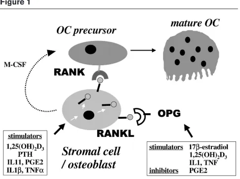
Recent studies have demonstrated that osteotropic factors and hormones such as PTH, 1,25(OH)2D3, IL-11, IL-1β, TNF-α or prostaglandin E2 upregulate RANKL expression in osteoblast/stromal cells (Fig. 1). In addition, osteoprotegerin expression is downregulated by prostaglandin E2, and is upregulated by oestrogens [21•].
RANK expression has not yet been extensively studied.
An integrated view and clinical implications
An emerging concept is that cytokines and hormonal factors that are involved in bone resorption may act by a common final pathway involving RANKL and RANK [21•].
In accordance with this concept, a recent in vivo study [39••] has shown that a recombinant chimaeric Fc fusion
form of osteoprotegerin inhibited hypercalcaemia and bone resorption induced by IL-1β, TNF-α, PTH and 1,25(OH)2D3 in mice. This convergence theory is probably not exclusive because recent studies [40,41] have suggested that the effects of TNF-αor IL-6 may involve different effectors.
Therapeutic perspectives
The concept presented above will probably lead to new therapeutic approaches in several diseases that are char-acterized by excessive bone resorption. Thus,
osteoprote-Figure 1
New members of the TNF receptor ligand family: role of RANKL (ODF, TRANCE, OPGL) and its receptor RANK in osteoclast differentiation. RANKL, a membrane-bound TNF-related factor, is expressed by osteoblast/stromal cells and is upregulated by osteotropic factors such as 1,25(OH)2D3, PTH, IL-6 or IL-11. Osteoclast (OC) precursors
[image:3.612.57.300.90.271.2]and cartilage destruction as a result of chronic synovitis. Numerous studies have pointed out the role of cytokines such as TNF-αor IL-1 in the joint destruction [43]. Recent studies suggest that RANKL mRNA is highly expressed in synovial tissues from patients with RA, but not in normal synovial tissues. This expression is detected in synovial fibroblasts, as well as in activated T cells derived from RA synovial tissues, suggesting that these cells may con-tribute to osteoclast formation at the specific sites of bone destruction in RA [44•,45•]. In addition, in rat
adjuvant-induced arthritis, RANKL is expressed on the surface of activated T cells isolated from affected rats, and may be secreted in T cell cultures. Activated T cells could there-fore directly induce osteoclastogenesis through mem-brane-bound and soluble RANKL [28]. These data suggest that RANKL may have a major pathophysiological importance in the bone and joint destruction observed in inflammatory arthritides such as RA. Activated T cells, which play a central role in the pathogenesis of RA, may (in addition to stromal cells) contribute to the osteoclast-mediated bone resorption via RANKL expression [46••].
Conclusion
Osteoclasts are multinucleated cells that are formed by fusion of osteoclast precursors from haematopoietic origin. These cells are responsible for bone resorption and osteoclast differentiation and represent an evident point of control of bone resorption. Bone resorption is closely regulated in vivo by many cellular and hormonal factors, which affect not only osteoclast activity, but also osteoclast formation. The recent discovery of new members of the TNF receptor ligand family (ODF, TNF-related induced cytokine, osteoprotegerin ligand) have emphasized the crucial role of RANKL, which is expressed by osteoblast/stromal cells, and its receptor RANK, which is expressed by osteoclast cells, in osteo-clast differentiation and activation. This system is com-pleted by osteoprotegerin, which is a secreted TNF receptor. Osteoprotegerin recognizes RANKL, and this decoy receptor blocks the interaction between RANK and RANKL. A number of osteotropic factors and hor-mones may modulate bone resorption via this common final pathway, which may represent a potential
therapeu-3. Takahashi N, Akatsu T, Udagawa N, Sasaki T, Yamaguchi A, Moseley JM, Martin TJ, Suda T: Osteoblastic cells are involved in osteoclast formation.Endocrinology1988, 123:2600–2602.
4. Quinn JM, McGee JO, Athanasou NA: Cellular and hormonal factors influencing monocyte differentiation to osteoclastic bone-resorb-ing cells.Endocrinology1994, 134:2416–2423.
5. Kurihara N, Civin C, Roodman GD: Osteotropic factor responsive-ness of highly purified populations of early and late precursors for human multinucleated cells expressing the osteoclast phenotype.
J Bone Miner Res1991, 6:257–261.
6. Matayoshi A, Brown C, DiPersio JF, Haug J, Abu-Amer Y, Liapis H, Kuestner R, Pacifici R: Human blood-mobilized hematopoietic pre-cursors differentiate into osteoclasts in the absence of stromal cells.Proc Natl Acad Sci USA1996, 93:10785–10790.
7. Roux S, Quinn J, Pichaud F, Orcel P, Chastre E, Jullienne A, De Verne-joul MC: Human cord blood monocytes undergo terminal osteo-clast differentiation in vitro in the presence of culture medium conditioned by giant cell tumor of bone.J Cell Physiol1996, 168: 489–498.
8. de Vernejoul MC, Cohen-Solal M, Orcel P: Bone cytokines. Curr Opin Rheumatol1993, 5:332–338.
9. Roodman GD: Interleukin-6: an osteotropic factor?J Bone Miner Res1992, 7:475–478.
10. Pfeilschifter J, Chenu C, Bird A, Mundy GR, Roodman GD: Inter-leukin-1 and tumor necrosis factor stimulate the formation of human osteoclastlike cells in vitro. J Bone Miner Res 1989,
4:113–118.
11. Yoshida H, Hayashi S, Kunisada T, Ogawa M, Nishikawa S, Okamura H, Sudo T, Shultz LD, Nishikawa S: The murine mutation osteopet-rosis is in the coding region of macrophage colony stimulating factor gene.Nature1990, 345:442–444.
12. Tanaka S, Takahashi N, Udagawa N, Tamura T, Akatsu T, Stanley ER, Kurokawa T, Suda T: Macrophage colony-stimulating factor is indispensable for both proliferation and differentiation of osteo-clast progenitors.J Clin Invest1993, 91:257–263.
13. Orcel P, Feuga M, Bielakoff J, de Vernejoul MC: Local bone injec-tions of LPS and M-CSF increase bone resorption by different pathways in vivo in rats.Am J Physiol1993, 264:E391–E397.
14. Ishimi Y, Abe E, Jin CH, Miyaura C, Hong MH, Oshida M, Kurosawa H, Yamaguchi Y, Tomida M, Hozumi M: Leukemia inhibitory factor/ dif-ferentiation-stimulating factor (LIF/D-Factor): regulation of its production and possible roles in bone metabolism.J Cell Physiol
1992, 152:71–78.
15. Girasole G, Passeri G, Jilka RL, Manolagas SC: Interleukin-11: a new cytokine critical for osteoclast development. J Clin Invest1994,
16. Lacey DL, Erdmann JM, Teitelbaum SL, Tan H, Ohara J, Shioi A: Inter-leukin 4, Interferon-γγand prostaglandin E impact the osteoclastic cell-forming potential of murine bone marrow macrophages.
Endocrinology1995, 136:2367–2376.
17. Oreffo RO, Bonewald L, Kukita A, Garrett IR, Seyedin SM, Rosen D, Mundy GR: Inhibitory effects of bone-derived growth factors, osteoinductive factor and transforming growth factor-ββ on iso-lated osteoclasts.Endocrinology1990, 126:3069–3075.
18. Chenu C, Pfeilschifter J, Mundy GR, Roodman GD: Tansforming growth factor ββinhibits formation of osteoclast-like cells in long-term human marrow cultures.Proc Natl Acad Sci USA1988, 85: 5683–5687.
19. Mbalaviele G, Orcel P, Bouizar Z, Julienne A, de Vernejoul MC: Trans-forming growth factor ββenhances calcitonin-induced cyclic AMP production and the number of calcitonin receptors in long term cultures of human umbilical cord blood monocytes in the pres-ence of 1,25-dihydroxycholecalciferol.J Cell Physiol 1992, 152: 486–494.
20. Orcel P, Bielakoff J, de Vernejoul MC: Effect of transforming growth factor ββon long-term human cord blood monocytes cultures.J Cell Physiol1990, 142:293–298.
21. Hofbauer LC, Khosla S, Dunstan CR, Lacey DL, Boyle WJ, Riggs BL:
• The roles of osteoprotegerin and osteoprotegerin ligand in the paracrine regulation of bone resorption.J Bone Miner Res2000,
15:2–12.
This review concerns RANKL, RANK and osteoprotegerin. Osteoclast dif-ferentiation may be determined by the relative ratio of RANKL to osteoprote-gerin in the bone marrow microenvironment. These factors mediate the effects of large numbers of upstream hormones and cytokines, suggesting a final common pathway in the regulation of osteoclastogenesis.
22. Suda T, Takahashi N, Udagawa N, Jimi E, Gillespie MT, Martin TJ:
• Modulation of osteoclast differentiation and function by the new members of the tumor necrosis factor receptor and ligand fami-lies.Endocr Rev1999, 20:345–357.
This review concerns RANKL, RANK and osteoprotegerin – three key mole-cules that regulate osteoclast recruitment and function.
23. Lacey DL, Timms E, Tan HL, Kelley MJ, Dunstan CR, Burgess T, Elliott
•• R, Colombero A, Elliott G, Scully S, Hsu H, Sullivan J, Hawkins N, Davy E, Capparelli C, Eli A, Qian YX, Kaufman S, Sarosi I, Shalhoub V, Senaldi G, Guo J, Delaney J, Boyle WJ: Osteoprotegerin ligand is a cytokine that regulates osteoclast differentiation and activation.
Cell1998, 93:165–176.
The identification and cloning of the ligand of osteoprotegerin from a murine myelomonocytic cell line is discussed. Data are presented that suggest that OPGL is an osteoclast differentiation and activation factor.
24. Yasuda H, Shima N, Nakagawa N, Yamaguchi K, Kinosaki M,
•• Mochizuki S, Tomoyasu A, Yano K, Goto M, Murakami A, Tsuda E, MorinagaT, Higashio K, Udagawa N, Takahashi N, Suda T: Osteoclast differentiation factor is a ligand for osteoprotegerin/osteoclasto-genesis-inhibitory factor and is identical to TRANCE/RANKL.Proc Natl Acad Sci USA1998, 95:3597–3602.
This paper describes the identification and cloning of the ligand of osteo-protegerin, ODF, from mouse stromal cell ST2. ODF mediates an essential signal to osteoclast progenitors for their differentiation into osteoclasts.
25. Quinn JM, Elliott J, Gillespie MT, Martin TJ: A combination of osteo-•• clast differentiation factor and macrophage-colony stimulating
factor is sufficient for both human and mouse osteoclast forma-tion in vitro.Endocrinology1998, 139:4424–4427.
This study demonstrates that murine and human osteoclast precursors, when cultured in the presence of M-CSF and a soluble form of murine ODF, form bone-resorbing osteoclasts in the absence of osteoblast/stromal cells.
26. Jimi E, Akiyama S, Tsurukai T, Okahashi N, Kobayashi K, Udagawa N, Nishihara T, Takahashi N, Suda T: Osteoclast differentiation factor acts as a multifunctional regulator in murine osteoclast differenti-ation and function.J Immunol1999, 163:434–442.
27. Kong YY, Yoshida H, Sarosi I, Tan HL, Timms E, Capparelli C, Morony
•• S, Oliveira-dos-Santos AJ, Van G, Itie A, Khoo W, Wakeham A, Dunstan CR, Lacey DL, Mak TW, Boyle WJ, Penninger JM: OPGL is a key regulator of osteoclastogenesis, lymphocyte development and lymph-node organogenesis.Nature1999, 397:315–323. Osteoprotegerin ligand-deficient mice develop severe osteopetrosis and a defect in tooth eruption, and completely lack osteoclasts as a result of an inability of osteoblasts to support osteoclastogenesis.
28. Kong YY, Feige U, Sarosi I, Bolon B, Tafuri A, Morony S, Capparelli C, Li J, Elliott R, McCabe S, Wong T, Campagnuolo G, Moran E, Bogoch ER, Van G, Nguyen LT, Ohashi PS, Lacey DL, Fish E, Boyle WJ, Pen-ninger JM: Activated T cells regulate bone loss and joint destruc-tion in adjuvant arthritis through osteoprotegerin ligand.Nature
1999, 402:304–309.
29. Nakagawa N, Kinosaki M, Yamaguchi K, Shima N, Yasuda H, Yano K,
•• Morinaga T, Higashio K: RANK is the essential signaling receptor for osteoclast differentiation factor in osteoclastogenesis.
Biochem Biophys Res Commun1998, 253:395–400.
This paper describes the cloning of ODF receptor, RANK, from a mouse macrophage-like osteoclast progenitor cell line.
30. Hsu H, Lacey DL, Dunstan CR, Solovyev I, Colombero A, Timms E, Tan HL, Elliott G, Kelley MJ, Sarosi I, Wang L, Xia XZ, Elliott R, Chiu L, Black T, Scully S, Capparelli C, Morony S, Shimamoto G, Bass MB, Boyle WJ: Tumor necrosis factor receptor family member RANK mediates osteoclast differentiation and activation induced by osteoprotegerin ligand.Proc Natl Acad Sci USA1999, 96:3540– 3545.
31. Dougall WC, Glaccum M, Charrier K, Rohrbach K, Brasel K, De Smedt T, Daro E, Smith J, Tometsko ME, Maliszewski CR, Armstrong A, Shen V, Bain S, Cosman D, Anderson D, Morrissey PJ, Peschon JJ, Schuh J: RANK is essential for osteoclast and lymph node devel-opment.Genes Dev1999, 13:2412–2424.
32. Simonet WS, Lacey DL, Dunstan CR, Kelley M, Chang MS, Luthy R,
•• NguyenHQ, Wooden S, Bennett L, Boone T, Shimamoto G, DeRose M, Elliott R, Colombero A, Tan HL, Trail G, Sullivan J, Davy E, Bucay N, Renshaw-Gegg L, Hughes TM, Hill D, Pattison W, Campbell P, Boyle WJ: Osteoprotegerin: a novel secreted protein involved in the reg-ulation of bone density.Cell1997, 89:309–319.
This paper describes the identification of osteoprotegerin, a potent inhibitor of bone resorption, from a foetal rat intestine cDNA library. In vivo, over-expression of osteoprotegerin in transgenic mice or administration of recom-binant osteoprotegerin into normal mice results in a severe osteopetrosis, secondary to a decrease in later stages of osteoclast differentiation.
33. Tsuda E, Goto M, Mochizuki S, Yano K, Kobayashi F, Morinaga T,
•• Higashio K: Isolation of a novel cytokine from human fibroblasts that specifically inhibits osteoclastogenesis.Biochem Biophys Res Commun1997, 234:137–142.
Identification of osteoclastogenesis inhibitory factor from human embryonic lung fibroblasts is described. This factor, which is identical to osteoprote-gerin, inhibits osteoclast-like cell formation stimulated through three distinct signalling pathways involving 1α,25-dihydroxyvitamin D3, PTH or IL-11.
34. Bucay N, Sarosi I, Dunstan CR, Morony S, Tarpley J, Capparelli C,
•• Scully S, Tan HL, Xu W, Lacey DL, Boyle WJ, Simonet WS: Osteo-protegerin-deficient mice develop early onset osteoporosis and arterial calcification.Genes Dev1998, 12:1260–1268.
Osteoprotegerin-deficient mice develop an osteoporosis that is character-ized by severe trabecular and cortical bone porosity, marked thinning of the parietal bones of the skull, and a high incidence of fractures. Osteoprote-gerin-deficient mice also exhibit medial calcification of the aorta and renal arteries.
35. Franzoso G, Carlson L, Xing L, Poljak L, Shores EW, Brown KD, Leonardi A, Tran T, Boyce BF, Siebenlist U: Requirement for NF-kappaB in osteoclast and B-cell development.Genes Dev1997,
11:3482–3496.
36. Iotsova V, Caamano J, Loy J, Yang Y, Lewin A, Bravo R: Osteopetro-sis in mice lacking NF-kappaB1 and NF-kappaB2.Nature Med
PTH related-protein, and 1,25(OH)2D3).
40. Hofbauer LC, Lacey DL, Dunstan CR, Spelsberg TC, Riggs BL, Khosla S: Interleukin-1beta and tumor necrosis factor-alpha, but not interleukin- 6, stimulate osteoprotegerin ligand gene expres-sion in human osteoblastic cells.Bone1999, 25:255–259.
41. Kobayashi K, Takahashi N, Jimi E, Udagawa N, Takami M, Kotake S, Nakagawa N, Kinosaki M, Yamaguchi K, Shima N, Yasuda H, Morinaga T, Higashio K, Martin TJ, Suda T: Tumor necrosis factor alpha stim-ulates osteoclast differentiation by a mechanism independent of the ODF/RANKL-RANK interaction.J Exp Med2000, 191:275–286.
42. Chikatsu N, Takeuchi Y, Tamura Y, Fukumoto S, Yano K, Tsuda E,
•• Ogata E, Fujita T: Interactions between cancer and bone marrow cells induce osteoclast differentiation factor expression and osteoclast-like cell formation in vitro. Biochem Biophys Res Commun2000, 267:632–637.
Enhanced osteoclastogenesis in the presence of cancer cells might be due to an increase in ODF (RANKL) activity. The interactions between cancer cells and mouse bone marrow cells induce ODF expression and suppress osteoprotegerin level in bone metastases, resulting in increased local bone destruction.
43. Duff GW: Cytokines and anti-cytokines. Br J Rheumatol 1993,
32(Suppl 1):15–20.
44. Gravallese EM, Manning C, Tsay A, Naito A, Pan C, Amento E,
• Goldring SR: Synovial tissue in rheumatoid arthritis is a source of osteoclast differentiation factor.Arthritis Rheum2000, 43:250–258. ODF (RANKL) is expressed in synovial tissues from RA but not in normal synovium. This expression is detected in cultured synovial fibroblasts and in activated T cells derived from RA synovial tissue.
45. Takayanagi H, Iizuka H, Juji T, Nakagawa T, Yamamoto A, Miyazaki T,
• Koshihara Y, Oda H, Nakamura K, Tanaka S: Involvement of receptor activator of nuclear factor kappaB ligand/osteoclast differentia-tion factor in osteoclastogenesis from synoviocytes in rheumatoid arthritis.Arthritis Rheum2000, 43:259–269.
RANKL is highly expressed in synovial tissues from RA, but not in normal synovium or in osteoarthritic synovium. Cultured rheumatoid synovial fibro-blasts expressed RANKL and are able to induce osteoclast differentiation, which requires cell–cell contact between synovial cells and osteoclast pre-cursors.
46. Horwood NJ, Kartsogiannis V, Quinn JM, Romas E, Martin TJ, Gillespie
•• MT: Activated T lymphocytes support osteoclast formation in vitro.
Biochem Biophys Res Commun1999, 265:144–150.