Available online on 30.08.2019 at http://jddtonline.info
Journal of Drug Delivery and Therapeutics
Open Access to Pharmaceutical and Medical Research© 2011-18, publisher and licensee JDDT, This is an Open Access article which permits unrestricted non-commercial use, provided the original work is properly cited
Open Access
Research Article
Prepare and Evaluate Mucoadhesive Formulations of Lamivudine with
Better Controlled/ Sustained Drug Release Profile
Kavita Rathore, C.K. Tyagi, Harish Pandey, Sunil Kumar Shah *
Department of Pharmaceutics, College of Pharmacy, Sri Satya Sai University of Technology & Medical Sciences, Sehore, Madhya Pradesh, India.
ABSTRACT
The aim of present study was to formulate & evaluate the mucoadhesive sustained release formulations of lamivudine and to fulfill this aim, two mucoadhesive formulations Gels and Tablets were prepared by using three different polymers: HPMC K15, poloxamer 407 & carbopol 934. Three mucoadhesive gel and nine tablet formulations were prepared and evaluated for various parameters. All three gels were able to give sustained release up to 12 hours. Tablet formulations, F1 to F5 failed to fulfill the aim. Only F6, F7, F8 & F9 formulations were selected, as all gave sustained release up to 12 hours, except F6, which gave sustained release profile only till 7 hours. From the drug release plots, it was concluded that the type of polymer and concentration of polymer have distinct effect on in vitro drug release profile and all the formulations follow first order mechanism with anomalous diffusion or non-fickian diffusion, except carbopol gel and poloxamer tablets. Carbopol gel follows zero order release rate with super case II transport and poloxamer tablets (F6) follow higuchi with non-fickian diffusion. It is concluded that mucoadhesive formulations of lamivudine can be prepared for sustaining its release. And the successful outcome of the present study also encourage for further studies to assess the ability of the mucoadhesive formulations of lamivudine in providing an effective sustained and safe therapy for AIDS.
Keywords: NDDS, ODDS, BDDS, HPMC K 15, Carbopol 934, Poloxamer 407
Article Info:Received 18 June 2019; Review Completed 19 Aug 2019; Accepted 23 Aug 2019; Available online 30 Aug 2019
Cite this article as:
Rathore K, Tyagi CK, Pandey H, Shah SK, Prepare and Evaluate Mucoadhesive Formulations of Lamivudine with Better Controlled/ Sustained Drug Release Profile, Journal of Drug Delivery and Therapeutics. 2019; 9(4-A):694-700
http://dx.doi.org/10.22270/jddt.v9i4-A.3479
*Address for Correspondence:
Dr. Sunil Kumar Shah, Department of Pharmaceutics, College of Pharmacy, Sri Satya Sai University of Technology & Medical Sciences, Sehore, Madhya Pradesh, India.
INTRODUCTION
The principle of mucoadhesive preparation is simple practical approach and is particularly useful to prolong the retention time of a dosage form in the stomach and improving the oral bioavailability of the drug1.The aim was
to develop drug delivery systems that would increase the absorption of a drug for both local and systemic effects as a result of intimate and prolonged contact at the site of absorption2-4. Among the early work on bioadhesive systems
is that of Nagai and coworkers, who showed that the treatment was improved for several administration routes when adhesive formulations were used5. For example, the
treatment of aphthae, an infection in the mouth, and the treatment of uterine cancer were improved with local delivery using mucoadhesive tablets. In addition, mucoadhesive preparations for delivery of insulin through the buccal and the nasal routes of administration were investigated6,7. The term mucoadhesion appeared in the
literature for the first time in 1977. In a medical research paper describing a clinical trial of a locally delivered anaesthetic. In the mid and late 1980s the concept of mucoadhesion became more commonly recognised9. Over
the years, mucoadhesive and bioadhesive systems have been
used for NDDS, ODDS, BDDS, vaginal, rectal and oral drug delivery10.
Nowadays, mucoadhesion in the literature, covering a wide variety of applications11. In some studies, the term
mucoadhesive formulation is used in a routine and noncritical way, e.g. different formulations and polymers have been ranked as more or less mucoadhesive by using randomly chosen methods12-16. polymers have been
developed and used with the intention of learning more about the kind of interactions that can occur between the formulation and the mucosa17. But even now, the
understanding of the phenomenon is not yet complete18. One
of the reasons for this is probably that there are so many different formulations involving a large variety of adhesion mechanisms that no single existing theory can explain them all. In our present work we are preparing two different types of mucoadhesive formulations-mucoadhesive gel and mucoadhesive tablets19-20.
MATERIALS
were procured from Loba Chemie Pvt.Ltd, Mumbai. Other excipients such as Aerosil and magnesium stearate were procured from Alfa Laboratories, Mumbai.
METHODS OF PREPARATION OF MUCOADHESIVE GELS: Preparation of Lamivudine Gel by Using HPMC K15: 2%, 4%, 6%, 8%, 10% & 12%, plane gel formulation of HPMC K15 were prepared in distilled water by simple mixing method21-23. 12%, formulation was selected on the
bases of consistency of gel. Lamivudine (150mg) was dissolved in small amount of distilled water and then incorporated in 12% HPMC gel with continuous stirring. After that, set aside the formulation for some time at room temperature24,25.
Preparation of Lamivudine Gel by Using Corbopol934:
0.5, 1% & 2%, plane gel formulation of Carbopol were prepared in distilled water. Out of these, 1% gel formulation was selected on the bases of gel consistency26,27. As on
incorporation of lamivudine (150mg), the formulation was precipitated28,29. Therefore 0.5% gel was selected to get the
desired gel formulation. And gel was prepared by simply adding the lamivudine (already dissolved in small amount of water) into 0.5% carbopol gel, with continuous stirring30. Preparation of Lamivudine gel by using POLOXAMER 407: The Pluronic F127 (Poloxamer 407) were prepared by
modification of the “Cold dispersion” method described by Schmolka. The weighed amount of poloxamer (1g) was placed in beaker and left in an oven at 110°C for 15 minutes to obtain a homogeneous liquefied mixture then 150mg lamivudine (which was already dissolved in small amount of water) added with continuous stirring31. The solution was
cooled to room temperature, & beaker was left in a refrigerator until a clear solution was obtained. The gel was formed when the solution was brought back to room temperature and stored at ambient temperature prior to use. Formulation of Mucoadhesive Tablets:
Mucoadhesive tablets of Lamivudine were made by direct compression method. Nine formulations (F1-F9) were formulated by using three different mucoadhesive polymers (HPMC K15, Carbopol 934 & Poloxamer 407). Mucoadhesive polymers were used as binder, lactose as diluents and talc as lubricant32.
The mucoadhesive tablets were prepared by mixing of drug with binder, in a pestle and mortar until homogenized. Then all other excipients were added. Mixture was passed through sieve no. 60. Finally the blend was compressed using the round concave punches (10.3mm in diameter) and dies by rotary tablet punching machine33. The tablet weight was
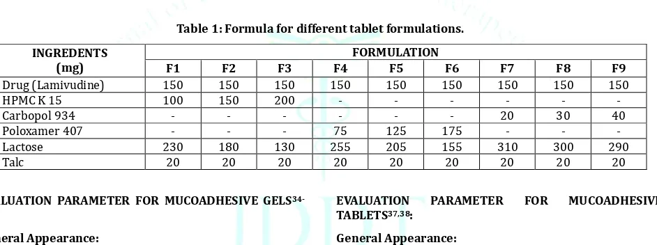
adjusted to 500mg and 75 tablets for each batch were prepared. Formula for nine batches is given in table 1.
Table 1: Formula for different tablet formulations. INGREDENTS
(mg)
FORMULATION
F1 F2 F3 F4 F5 F6 F7 F8 F9
Drug (Lamivudine) 150 150 150 150 150 150 150 150 150
HPMCK15 100 150 200 - - - -
Carbopol 934 - - - 20 30 40
Poloxamer 407 - - - 75 125 175 - - -
Lactose 230 180 130 255 205 155 310 300 290
Talc 20 20 20 20 20 20 20 20 20
EVALUATION PARAMETER FOR MUCOADHESIVE GELS
34-36:
General Appearance:
A Visual inspection was done to know the general appearance of gels. There should be no sign of grittiness and formulation must be of uniform texture.
Mucoadhesive Force:
Mucoadhesive force of all three gel formulation was determined by method and apparatus reported by Choi et al., 1998 & koffi et al, 2006. (by using goat stomach mucosa). Drug Release Profile:
Drug release profile of all three gel formulations was carried out in distilled water, by using cellophane membrane and drug release rate kinetics of the formulations was determined by fitting release result in models of data treatment as follows:
1.Log cumulative percent drug remaining versus time (first order kinetic model).
2.Cumulative percent drug release versus time (zero order kinetic model).
3.Higuchi release kinetic model. 4.Korsmeyer- peppas model.
EVALUATION PARAMETER FOR MUCOADHESIVE
TABLETS37,38: General Appearance:
General appearance was examined by visual inspection. All tablets should be of uniform size, shape, color and surface textures etc.
Weight variation:
Randomly, twenty tablets were selected and individually weighed. The average weight of tablet was calculated. Then individual weight was compared with average weight of tablets. It should be within the range ±5 for tablet more than 324mg.
Thickness:
Six tablets were selected randomly from each batch and thickness was measured by using vernical caliper.
Friability:
Twenty tablets were weighted and placed in Roche friabilator and equipment was rotated at 25 rpm for 4 minute. The tablets were taken out, dusted and reweighed. The percentage friability of tablets should be within range of ±1.
Hardness:
tester. Six tablets from each batch were tested randomly and the average reading noted.
Swelling index:
Swelling index was determined by method as descried earlier for three tablets of each batch. i.e. tablet was weighed and placed in a beaker containing 200 ml of distilled water. The tablet was gently removed at time intervals such as 1, 10 and 20mins, blotted dry and the weight determined. This was stopped once the tablets start eroding or became too gelatinous and swelling index was calculated.
Mucoadhesive Strength:
Mucoadhesive strength of tablets was measured on the modified physical balance as described earlier by using goat stomach mucosa.
Drug Content:
Take 100mg equivalent weight of crushed powder of tablets and dissolved in 1000ml of distilled water. Solution was filtered and diluted appropriately. Then, solution was analyzed by UV Spectrophotometer at λ max.
In-vitro dissolution studies and release kinetic analysis: In vitro release rate study of mucoadhesive tablet was carried out by using the Apparatus 2 i.e. Basket apparatus, using distilled water as dissolution media. And absorbance of samples was measured at λ max. To analyze the drug release rate kinetics of the formulations, the results of in-vitro release profiles were fitted to following models.
1. Log cumulative percent drug remaining versus time (first order kinetic model).
2. Cumulative percent drug release versus time (zero order kinetic model).
3. Higuchi release kinetic model. 4. Korsmeyer-peppas model.
RESULT AND DISCUSSSION
Preliminary Investigation of Drug (Lamivudine): Physical Appearance
Lamivudine was white color powder. Melting Point
Melting point of lamivudine was found to be 161°C. Solubility study
Solubility profile of lamivudine in various solvents, are given in table 2.
Table 2: Solubility profile of Lamivudine in various solvents
S. No. Solvent Solubility
1 Distilled water +
2 Ethanol +
3 Methanol +
4 Acetone -
5 Chloroform -
6 Ethyl acetate -
7 0.1N HCl +
8 0.1 N NaOH +
λ max OF LAMIVUDINE:
λ max of lamivudine was found to be 271.5 nm in distilled water.
Figure 1: Scanning of Lamivudine in UV range Standard Curve of Lamivudine
Standard calibration curve of lamivudine was determined by plotting absorbance v/s concentration on double beam U.V. spectrophotometer using λ max = 271.5 nm. Straight line was obtained after plotting concentration on X axis. It follows the beer’s law. As beer’s law is concentration dependent and on increasing the concentration from 5μg/ml to 30μg/ml, gave liner increase in absorbance39-40. The
regression equation was y = 0.0247x + 0.0093, which was further used for calculation of concentration of unknown samples. The R2 value of standard curve was 0.9978, which
signify that plot was linear. The results are shown in table 3and figure 1.
Table 3: Absorbance of Lamivudine in distilled water at λmax271.5 nm
S. No. Concentration(µg/ml) Absorbance (nm)±(SD)
1 0 0
2 5 0.116±0.002
3 10 0.238±0.001
4 15 0.334±0.003
5 20 0.493±0.002
6 25 0.613±0.002
7 30 0.738±0.001
EVALUATION PARAMETER FOR MUCOADHESIVE GELS41:
General Appearance
All three gel formulations were good texture profile. They were transparent in appearance and no sign of grittiness was observed.
Mucoadhesive Force
Mucoadhesive force of all three gel formulations were determined by using goat stomach mucosa and is given in table 4. Out of all three polymers, Carbopol showed the maximum mucoadhesive force.
Table 4: Mucoadhesive force of polymers used in gel formulations.
S. No. Polymers Mucoadhesive force
(dyne/cm²)
1 Poloxamer 407 2.2455
2 HPMC K15 2.6271
3 Carbopol 934 3.3618
Drug Release Study
Table 5: Data of Release profile of formulated gels.
Time (hr) HPMC K15 gel Cumulative % drug releasePoloxamer 407 gel Carbopol 934 gel
0 0 0 0
1 18.43±1.5 12.92±2.3 4.62±1.9
2 37.45±2.7 27.63±2.8 13.83±3.4
3 46.45±3.8 36.6±3.5 18.8±2.2
4 64.07±2.4 47.43±2.2 25.02±1.5
5 73.56±4.2 59.82±3.1 34.87±1.7
6 83.92±2.8 66.01±4.3 42.09±2.9
7 87.12±2.1 72.32±2.4 61.83±2.6
8 91.32±1.6 79.71±1.8 67.63±1.8
10 94.22±1.2 83.87±1.2 73.98±2.3
12 97.89±0.6 88.95±0.88 78.38±1.4
The above drug release data & plot show that the prepared gel formulations released drug up to 12 hours and more or less, all three gel formulations were giving sustained drug release profile. These gel formulations were further studied to know the drug release kinetics.
Drug release kinetic study: HPMC K15 gel:
Table 1.6: Data for drug release kinetic study of HPMC K15 gel
Time (hr)
Square root of
time Log time
Cumulative %
drug released Log (Mt/M∞)
Cumulative % drug remaining
to release
Log cumulative % drug remaining to
release
0 0 - 0 - 100 2
1 1 0 18.43 1.2655 81.57 1.9115
2 1.414 0.301 37.45 1.5734 62.55 1.7962
3 1.732 0.477 46.45 1.6669 53.55 1.7287
4 2 0.602 64.07 1.8066 35.93 1.5554
5 2.236 0.6989 73.56 1.8666 26.44 1.4222
6 2.449 0.778 83.92 1.9238 16.08 1.2062
7 2.645 0.845 87.12 1.9401 12.88 1.1099
8 2.828 0.903 91.32 1.9605 8.68 0.9385
10 3.162 1 94.22 1.9741 5.78 0.7619
12 3.464 1.079 97.89 1.9907 2.11 0.3242
Zero order
R² First order R² Higuchi R² Korsmeyer- peppas R² n
0.8538 0.9896 0.9598 0.9488 0.6792
On the basis of R² values of above release kinetic plots, it was determined that the HPMC gel follows first order drug release kinetic model. As R² value of first order, 0.9896 was highest among all. And in Korsmeyer- peppas plot, n= 0.6792 (i.e. 0.45<n<0.89), indicates anomalous diffusion or non-fickian diffusion. That means, release rate of the HPMC gel was controlled by the combination of both, diffusion and erosion release mechanism.
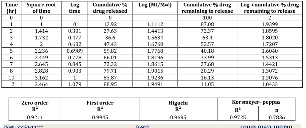
Poloxamer 407 gel
Table 1.7: Data for drug release kinetic study of poloxamer 407 gel Time
(hr) Square root of time time Log Cumulative % drug released Log (Mt/M∞) remaining to release Cumulative % drug Log cumulative % drug remaining to release
0 0 - 0 - 100 2
1 1 0 12.92 1.1112 87.08 1.9399
2 1.414 0.301 27.63 1.4413 72.37 1.8595
3 1.732 0.477 36.6 1.5634 63.4 1.8020
4 2 0.602 47.43 1.6760 52.57 1.7207
5 2.236 0.6989 59.82 1.7768 40.18 1.6040
6 2.449 0.778 66.01 1.8196 33.99 1.5313
7 2.645 0.845 72.32 1.8615 27.68 1.4421
8 2.828 0.903 79.71 1.9015 20.29 1.3072
10 3.162 1 83.87 1.9236 16.13 1.2076
12 3.464 1.079 88.95 1.9491 11.05 1.0433
Zero order
R² First order R² Higuchi R²
Korsmeyer- peppas
R² n
On the basis of R² values of above release kinetic plots, it was determined that the poloxamer gel follows first order drug release kinetic model. As R² value of first order, 0.9945 was highest among all. And in Korsmeyer- peppas plot, n= 0.7836
(i.e. 0.45<n<0.89), indicates anomalous diffusion or non-fickian diffusion. That means, release rate of the poloxamer gel was controlled by the combination of both, diffusion and erosion release mechanism.
Zero order
R² First order R² Higuchi R² Korsmeyer- peppas R² n
0.9597 0.9568 0.8883 0.981 1.1678
On the basis of R² values of above release kinetic plots, it was determined that the carbopol gel follows zero order drug release kinetic model. As R² value of zero order, 0.9597was highest among all. And in Korsmeyer- peppas plot, n= 1.1678 (i.e. higher than 0.89), indicates super case II transport. As per super Case II transport mechanism, the release mechanism was not significantly influenced by formulation variables swelling dispersed within a glassy polymer. Initially the polymer begin to swell in contact of water, .as the penetrant enters the glassy polymer , the glass transition temperature of the polymer is lowered and become rubbery show diffusion allowing relaxation of macromolecular chains and drug diffuse out from the swollen rubbery area of polymer wall (Bhowmik B.B. et al, 2009).
Evaluation Parameter for Mucoadhesive Tablets42:
GENERAL APPEARANCE
General appearance was examined by visual inspection. All tablets were good in appearance; they were white colored oval shaped tablets with smooth surface texture and no pinholes were observed.
WEIGHT VARIATION
All nine tablet batches passed the weight variation test as percentage weight variation was within the pharmacopoeia limits (±5%). Results are shown table 8.
Table 8: weight variation
BATCH CODE WEIGHT VARIATION (mg) (N=20) RESULT
F1 498±1.9 PASSED
F2 497±2.6 PASSED
F3 502±1.76 PASSED
F4 499±1.6 PASSED
F5 501±2.8 PASSED
F6 497±2.9 PASSED
F7 496±3.3 PASSED
F8 500±1.88 PASSED
F9 498±2.3 PASSED
1. THICKNESS
Thickness of all tablet batches is given in table 9. The thickness of the tablets was found in the range of 5.6– 6.1 mm.
Table 9: Thickness of tablets
BATCH CODE THICKNESS (mm) (N=6)
F1 5.8±0.18
F2 5.9±0.177
F3 6.1±0.076
F4 6±0.11
F5 5.8±0.2
F6 5.9±0.16
F7 5.8±0.084
F8 6±0.15
F9 5.9±0.23
2. FRIABILITY
The friability of all nine batches is given in table 1.10. Friability of tablets was observed in acceptable range of 0.34-0.84%. It was within the pharmacopeia limit i.e. less than 1%. That means all tablets had good mechanical strength.
Table 1.10: Friability of tablets.
BATCH CODE FRIABILITY (%)
(N=20)
F1 0.84
F2 0.76
F3 0.72
F4 0.69
F5 0.63
F6 0.56
F7 0.48
F8 0.46
F9 0.34
Hardness
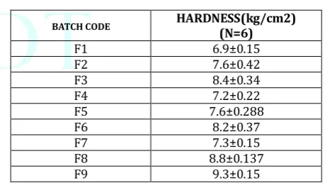
Hardness of all batches is given in table 11. Hardness of the tablets was found in the range of 6.8-9.4 kg/cm2. That was satisfactory for sustained release formulations and also indicates good mechanical strength to withstand physical and mechanical stress conditions while handling.
Table 11: Hardness of tablets.
BATCH CODE HARDNESS(kg/cm2)
(N=6)
F1 6.9±0.15
F2 7.6±0.42
F3 8.4±0.34
F4 7.2±0.22
F5 7.6±0.288
F6 8.2±0.37
F7 7.3±0.15
F8 8.8±0.137
F9 9.3±0.15
Swelling Index
Table 12: Swelling studies of tablets.
BATCH CODE
% swelling index (±SD) (N=3) Time (mins)
1 10 20
F1 10.2±2.3 18.6±3.7 32.1±4.2 F2 12.4±1.8 20±3.4 46.3±2.5 F3 15.1±2.4 24.6±2.1 54.2±3.6 F4 13.4±2.2 21.3±4.2 43.5±2.8 F5 17.7±1.5 32.3±5.1 49.6±2.3 F6 22.2±2.5 40.2±3.6 57.2±4.3 F7 27.4±1.2 75±2.6 97.7±3.8 F8 36.7±2.4 84.6±2.1 102±4.1 F9 39.2±1.7 90.3±3.4 116±3.3
Mucoadhesive studies
Mucoadhesive strength of tablets was measured on the modified physical balance as described earlier. The highest adhesion force and highest strength of the mucoadhesive bond was observed with the carbopol formulations. And it was increasing with increase in concentration of polymer.
Table 13: Mucoadhesive strength & force of tablets.
BATCH CODE
MUCOADHESIVE
STRENGTH (g) ±SD
MUCOADHESIVE FORCE
(N)
F1 17.3±1.4 0.170
F2 21.9±0.95 0.215
F3 24.6±0.74 0.241
F4 20.4±1.2 0.201
F5 23.5±1.5 0.231
F6 27.4±0.99 0.269
F7 33.2±2.2 0.326
F8 37.5±2.3 0.368
F9 42.8±1.8 0.417
CONCLUSION
The aim of present study was to formulate & evaluate the mucoadhesive sustained release formulations of lamivudine. And to fulfill this aim, two mucoadhesive formulations- gels and tablets were prepared by using three different polymers: HPMC K15, poloxamer 407 & carbopol 934. Three mucoadhesive gel and nine tablet formulations were prepared and evaluated for various parameters.
All prepared gel & tablet formulations had good physico-mechanical properties. Among all the formulations, carbopol gel and tablets showed the highest mucoadhesive force, although, each formulation had good adhesive force. All three gels were able to give sustained release up to 12 hours. Tablet formulations, F1 to F5 failed to fulfill the aim. Only F6, F7, F8 & F9 formulations were selected, as all gave sustained release up to 12 hours, except F6, which gave sustained release profile only till 7 hours. From the drug release plots, it was concluded that the type of polymer and concentration of polymer have distinct effect on in vitro drug release profile. This can further be justified with in vivo studies. And all the formulations follow first order mechanism with anomalous diffusion or non-fickian diffusion, except carbopol gel and poloxamer tablets. Carbopol gel follows zero order release rate with super case II transport and poloxamer tablets (F6) follow higuchi with non-fickian diffusion.
So from this study, it is concluded that mucoadhesive
formulations of lamivudine can be prepared for sustaining its release. And the successful outcome of the present study also encourage for further studies to assess the ability of the mucoadhesive formulations of lamivudine in providing an effective sustained and safe therapy for AIDS.
REFERENCES
1. Yadav SK, Khan G, Bonde GV, Bansal M, Mishra B. Design,
optimization and characterizations of chitosan fortified calcium alginate microspheres for the controlled delivery of dual drugs. Artif Cells Nanomed Biotechnol 2018;46:1180-93.
2. Deshmukh MT, Mohite SK. Preparation and evaluation of
mucoadhesive microsphere of fluoxetine Hcl. Int J Pharm Sci Res 2017; 8:3776-85.
3. Velmurugana S, Ali MA. Preparation and evaluation of
maraviroc mucoadhesive microspheres for gastro retentive drug delivery. Int J Pharm Pharm Sci 2015;7:208-14.
4. Abdul M, Rao AS, Martha S, Sirisha Y, Chandrika PU.
Development of a floating multiple unit controlled-release beads of zidovudine for the treatment of AIDS. JOPR J Pharm Res 2012;6:78–83.
5. Biswal I, Dinda A, Das D, Si S, Chowdary KA. Encapsulation
protocol for highly hydrophilic drug using non-biodegradable polymer. Int J Pharm Pharm Sci 2011;3:256–9.
6. Ma N, Xu L, Wang Q, Zhang X, Zhang W, Li Y, et al.
Development and evaluation of new sustained-release floating microspheres. Int J Pharm 2008;358:82–90.
7. Shah S.K., Mewada B., Tyagi C.K., Chaubey N., Pandey H.,
Formulation Development and evaluation of extended release Matrix tablet of tramadol hydrochloride using combination of Hydrophobic and hydrophilic polymer, International Journal of Pharmacy & Technology, 2017, 9 (3), 30647-30662.
8. Patel PN, Patel MM, Rathod DM, Patel JN, Modasiya MMK.
Sustain release drug delivery: a theoretical prospective. J Pharm Res 2012;5:4165–8.
9. Raffin RP, Colome LM, Schapoval EES, Pohlmann AR, Guterres
SS. Increasing sodium pantoprazole photostability by microencapsulation: Effect of the polymer and the preparation technique. Eur J Pharm Biopharm 2008;69:1014– 8.
10. Sharma H.K., Shah S.K., Kumar N., Choubey N., Tyagi C.K.,
Pandey H., Formulation and Optimization of Expandable Gastro Retentive Floating Matrix Tablet of Mosapridecitrate Using Factorial Design, IJPPR, 2018; 12 (1): 77-96.
11. El-menshawe SE, Kharshoum RM, Hamad DS, Halawa A. Effect
of biodegradable copolymers and divalent cations on the sustained release ability of propranolol hydrochloride loaded biomaterial microspheres. Int J Pharm Pharm Sci 2016;8:311-7.
12. Dasari A, Velmurugana S. Formulation and evaluation of
nevirapine mucoadhesive microspheres. Int J Pharm Pharm Sci 2015;7:342-8.
13. Vasir JK, Tambwekar K, Garg S. Bioadhesive microspheres as a
controlled drug delivery system. Int J Pharm 2003;255:13–32.
14. Chowdary KPR, Srinivas Rao S. Mucoadhesive microspheres
and microcapsules: current status. Indian J Pharm Sci 2005;67:141–50.
15. Chowdary KR, Rao YS. Mucoadhesive microspheres for
controlled drug delivery. Biol Pharm Bull 2004;27:1717–24.
16. Jain SK, Chourasia MK, Jain AK, Jain RK, Shrivastava AK.
Development and characterization of mucoadhesive microspheres bearing salbutamol for nasal delivery. Drug Delivery 2004;11:113–22.
17. George M, Abraham TE. Polyionic hydrocolloids for the
intestinal delivery of protein drugs: alginate and chitosan–a review. J Controlled Release 2006;114:1–14.
18. Patidar D, Shah S.K., Tyagi C.K., Pandey H., Controlled - Release
Effervescent Floating Tablet of Verapamil Hydrochloride: Development and Opitmization, /J. Pharm. Sci. & Res. 2016, 8(8), 732-740.
19. Kroll E, Winnik FM, Ziolo RF. In situ preparation of
nanocrystalline?-Fe2O3 in iron (II) cross-linked alginate gels. Chem Mater 1996;8:1594–6.
20. Llanes F, Ryan DH, Marchessault RH. Magnetic
21. Gowda DV, Nawaz M, Vishnu DM. Design and evaluation of carboxymethyl tamarind kernel polysaccharide (cmtkp) controlled release spheroids/pellets and investigating the influence of compression. Int J Pharm Pharm Sci 2014;6:103-9.
22. Jana S, Lakshman D, Sen KK, Basu SK. Development and
evaluation of epichlorohydrin cross-linked mucoadhesive patches of tamarind seed polysaccharide for buccal application. Int J Pharm Sci Drug Res 2010;2:193–8.
23. Jani GK, Shah DP, Prajapati VD, Jain VC. Gums and mucilages:
versatile excipients for pharmaceutical formulations. Asian J Pharm Sci 2009;4:309–23.
24. Sahoo R, Sahoo S, Nayak PL. Release behavior of anticancer
drug paclitaxel from tamarind seed polysaccharide galactoxyloglucan. Eur J Sci Res 2010;47:197–206.
25. Kulkarni D, Dwivedi AK, Sarin JPS, Singh S. Tamarind seed
polyose: a potential polysaccharide for sustained release of verapamil hydrochloride as a model drug. Indian J Pharm Sci 1997;59:1–7.
26. Patel B, Patel P, Bhosale A, Hardikar S, Mutha S, Chaulang G.
Evaluation of tamarind seed polysaccharide (TSP) as a mucoadhesive and sustained release component of nifedipinebuccoadhesive tablet and comparison with HPMC and Na CMC. Int J Pharm Tech Res 2009;1:404–10.
27. Anumolu PD, Anusha K, Mrudula Kiran A, Monika P,
Sowndarya NSKR, Sunitha G. Liquid chromatographic quantification of ternary mixture of anti-viral drugs and application to assessment of their tablet dosage form. Int J Pharm Pharm Sci 2016;8:237-40.
28. Benjamin J Eckhardt, Roy M Gulick. Drugs for HIV infection.
Infectious Diseases 2017;4:1293-308.
29. Deveswaran R, Bharath S, Furtado S, Abraham S, Basavaraj
BV, Madhavan V. Isolation and evaluation of tamarind seed polysaccharide as a natural suspending agent. Int J Pharm Biol Arch 2010;1:360-3.
30. Erik AR, Angelica RG, Raquel GR, Victor VG, Cesar PA.
Rheological properties of tamarind (Tamarindusindica L.) seed mucilage obtained by spray-drying as a novel source of hydrocolloid. Int J Biol Macromol 2018;107:817-24.
31. Shah Sunil, Shukla Dilip* , Pandey Harish, Formulation
Optimization of Polyox Based Modified Release Drug Delivery System, Journal of Drug Delivery & Therapeutics. 2019; 9(4-s):551-561
32. Gada SG, Anandkumar Y, Setty CM. Preparation and evaluation
of zidovudine mucoadhesive microspheres. Eur J Pharm Med Res 2017;4:570-6.
33. Tavakol M, Ebrahim VF, Sameereh HN. The effect of polymer
and CaCl2 concentrations on the sulfasalazine release from alginate-N, O-carboxymethyl chitosan beads. Prog Biomater 2013;2:10.
34. Obeidat WM, Price JC. Preparation and evaluation of Eudragit
S 100 microspheres as pH-sensitive release preparations for piroxicam and theophylline using the emulsion-solvent evaporation method. J Microencapsul 2006;23:195-202.
35. Sharma M, Jain K, Dev SK, Choudhury PK. Formulation and
evaluation of sodium alginate beads by emulsion gelation method. Asian J Pharm 2017;11:S101-6.
36. Maiti S, Dey P, Banik A, Biswanath S, Ray S, Kaity S. Tailoring
of locust bean gum and development of hydrogel beads for controlled oral delivery of glipizide. Drug Delivery 2010;17:288–300.
37. Verma S, Kumar V, Jyoti, Mishra DN. Formulation, evaluation
and optimization of mucoadhesive microspheres of acyclovir. Bull Pharm Res 2014;4:14-20.
38. Nagasree K, Chowdary GV, Kumar MCB, Reddy MTR,
Bhikshapathi DVRN. Design and evaluation of sodium alginate microspheres loaded with gatifloxacin. Der Pharm Lett 2016;8:361-70.
39. Pal DK, Nayak AK. Novel tamarind seed
polysaccharide-alginate mucoadhesive microspheres for oral gliclazide delivery: in vitro–in vivo evaluation. Drug Delivery 2012;19:123–31.
40. Sharma VK, Bhattacharya A. Release of metformin
hydrochloride from Ispaghula-sodium alginate beads adhered on cock intestinal mucosa. Indian J Pharm Educ Res 2008;42:365–72.
41. Nayaka AK, Saquib MHS, Beg S, Alam MI. Mucoadhesive beads
of gliclazide: design, development, and evaluation. Sci Asia 2010;36:319–25.
42. Shah S K , B. Prabhakar, Pandey S., Kushwaha S, Khan F,
Design of Extended Release Matrix Tablet of Tramadol hydrochloride Using Combination of Hydrophobic and Hydrophilic Polymer, Int. J. Pharm. Sci. Drug Res. 2017; 9(5): 214-219