P1: MBL/dat P2: M/NBL/vks QC: MBL/uks T1: MBL
August 21, 1997 10:19 Annual Reviews AR041-04
Annu. Rev. Cell Dev. Biol. 1997. 13:83–117
Copyrightc 1997 by Annual Reviews Inc. All rights reserved
MICROTUBULE POLYMERIZATION
DYNAMICS
Arshad Desai and Timothy J. Mitchison
∗Department of Biochemistry and Biophysics, and∗Department of Cellular and Molecular Pharmacology, University of California, San Francisco, California 94143; e-mail: [email protected]; [email protected]
KEY WORDS: cytoskeleton, dynamic instability, mechanism, regulation
ABSTRACT
The polymerization dynamics of microtubules are central to their biological func-tions. Polymerization dynamics allow microtubules to adopt spatial arrangements that can change rapidly in response to cellular needs and, in some cases, to per-form mechanical work. Microtubules utilize the energy of GTP hydrolysis to fuel a unique polymerization mechanism termed dynamic instability. In this review, we first describe progress toward understanding the mechanism of dynamic insta-bility of pure tubulin and then discuss the function and regulation of microtubule dynamic instability in living cells.
CONTENTS
INTRODUCTION . . . 84
MICROTUBULE STRUCTURE. . . 85
MICROTUBULE DYNAMICS IN VITRO. . . 88
Brief History . . . 88
Observation of Dynamic Instability In Vitro. . . 89
Thermodynamic Basis of Dynamic Instability. . . 91
Evidence for a Stabilizing Structure at Microtubule Ends . . . 94
The GTP CAP Model. . . 95
Structural Basis of Dynamic Instability. . . 97
Relationship of Structural and Chemical Transitions. . . 99
MICROTUBULE DYNAMICS IN VIVO. . . .100
Functions of Microtubule Dynamic Instability In Vivo. . . .100
Methodology for Analysis of Microtubule Dynamics In Vivo. . . .102
Features of Microtubule Dynamics In Vivo. . . .103
Microtubule Stabilizing Factors: MAPs. . . .105
Microtubule Destabilizing Factors . . . .107
83 1081-0706/97/1115-0083$08.00
P1: MBL/dat P2: M/NBL/vks QC: MBL/uks T1: MBL
August 21, 1997 10:19 Annual Reviews AR041-04
84 DESAI & MITCHISON
Microtubule Nucleating Factors. . . .109 EVOLUTION OF MICROTUBULE DYNAMICS. . . .110
INTRODUCTION
Microtubules (MTs) are noncovalent polymers of the protein tubulin found in all dividing eukaryotic cells and in most differentiated cell types. During cell division, a large dynamic array of MTs, called the mitotic spindle, functions to physically segregate the chromosomes and to orient the plane of cleavage. In nondividing cells, MTs organize the cytoplasm, position the nucleus and organelles, and serve as the principal structural element of flagella and cilia. MTs are physically robust polymers, with an intrinsic resistance to bending and compression. The mechanical properties of the ensemble of MTs, actin fila-ments, and intermediate filaments provide shape and strength to the cytoplasm, justifying the use of the term cytoskeleton. This term is misleading, however, in the sense that it suggests a static structure. Cytoskeletal polymers are in fact highly dynamic, capable of polymerizing, depolymerizing, and moving within the cytoplasm on a time scale of seconds to minutes.
The dynamic properties of MTs were apparent to early cytologists, who depicted mitotic spindles as compositions of linear elements whose arrangement changed rapidly with time (Wilson 1928). The first proof that spindles are composed of dynamic linear elements came with the advent of polarization microscopy, which allowed the observation of MTs in living cells (reviewed in Inou´e & Salmon 1995). This method was also used to demonstrate the importance of polymerization dynamics to MT function during mitosis (Inou´e & Sato 1967).
The subunit of a MT, a heterodimer ofα- andβ-tubulin, was first purified using its affinity for colchicine, one of the many natural product drugs that targets mitosis (Weisenberg et al 1968). Further biochemical studies led to the discovery thatβ-tubulin hydrolyzes GTP during polymerization (Weisenberg et al 1976). The energy input from GTP hydrolysis allows for nonequilibrium polymerization dynamics including dynamic instability, a behavior in which individual MT ends alternate stochastically between prolonged phases of poly-merization and depolypoly-merization (Mitchison & Kirschner 1984a). Dynamic instability appears to dominate the behavior of many types of MT arrays in living cells, but its precise mechanism and biological functions are still poorly understood.
Cytoskeletal polymer dynamics, including dynamic instability, are energet-ically expensive yet evolutionarily conserved, suggesting important biologi-cal roles. Both tubulin and actin use intrinsic nucleoside triphosphate (NTP) hydrolysis (Mitchison 1992), whereas intermediate filaments use accessory
P1: MBL/dat P2: M/NBL/vks QC: MBL/uks T1: MBL
August 21, 1997 10:19 Annual Reviews AR041-04
MICROTUBULE DYNAMICS 85
proteins, notably kinases and phosphatases (Eriksson et al 1992), to transduce chemical energy into polymer dynamics. Although we remain ignorant of many of the ways that polymerization dynamics are utilized in vivo, we can make some general remarks. At the most fundamental level, polymerization dynam-ics allow the cytoskeleton to rapidly reorganize. Were the cytoskeletal polymers to assemble to true thermodynamic equilibrium in vivo, it would be slow and difficult to change their spatial organization (Kirschner & Mitchison 1986). A second general property of MTs and actin filaments that arises from the direct coupling of polymerization dynamics to NTP hydrolysis is the potential for polymerization and depolymerization to perform mechanical work. Although precise mechanisms are poorly understood, there is good evidence that actin polymerization is harnessed to produce force in cells (Lauffenburger & Horwitz 1996), and MTs have been implicated in generating both pushing force by poly-merization and pulling force by depolypoly-merization (Inou´e & Salmon 1995).
In this review, we set the stage for a discussion of MT polymerization dy-namics by reviewing what is known about MT structure. We then focus on the mechanism of dynamic instability of pure tubulin, discussing the thermo-dynamic basis of thermo-dynamic instability and the evidence for special structures at the ends of polymerizing and depolymerizing MTs. Finally, we discuss the functions and regulation of dynamic instability in vivo.
MICROTUBULE STRUCTURE
Theα- andβ-tubulin monomers, which make up the heterodimer subunit of a MT, are≈50% identical at the amino acid level (Burns 1991), and each has a molecular mass of about 50,000. Tubulin heterodimers copurify with two moles of guanine nucleotide per moleαβdimer (Weisenberg et al 1968). During polymerization, GTP bound toβ-tubulin (at the exchangeable or E-site) is hydrolyzed (David-Pfeuty et al 1977, MacNeal & Purich 1978); the resulting E-site GDP does not exchange, whileβ-tubulin remains in the polymer. Upon depolymerization, the released tubulin subunits can exchange E-site GDP for GTP and undergo another round of polymerization.α-tubulin also binds GTP, but this GTP is bound in a non-exchangeable manner (at the N-site) and is not hydrolyzed during polymerization (Spiegelman et al 1977). In vitro, MT assembly occurs in two phases, nucleation and elongation. Our focus is on the elongation phase during which tubulin dimers add in an end-wise manner to preformed MT seeds or physiological nucleating structures such as axonemes and centrosomes (for a discussion of MT nucleation, see Fygenson et al 1994, 1995, Erickson & Stoffler 1996).
Within a MT, tubulin heterodimers are arranged in linear protofilaments (Figure 1a) that associate laterally to form 25 nm wide hollow cylindrical
P1: MBL/dat P2: M/NBL/vks QC: MBL/uks T1: MBL
August 21, 1997 10:19 Annual Reviews AR041-04
86 DESAI & MITCHISON
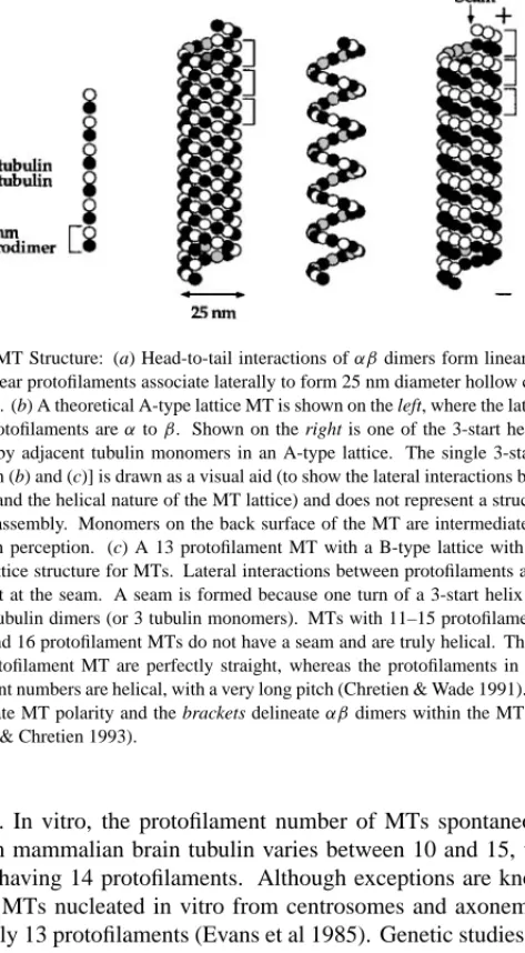
Figure 1 MT Structure: (a) Head-to-tail interactions ofαβdimers form linear protofilaments. Thirteen linear protofilaments associate laterally to form 25 nm diameter hollow cylindrical poly-mers (MTs). (b) A theoretical A-type lattice MT is shown on the left, where the lateral interactions
between protofilaments areαtoβ. Shown on the right is one of the 3-start helices that would
be formed by adjacent tubulin monomers in an A-type lattice. The single 3-start helix [on the
right in both (b) and (c)] is drawn as a visual aid (to show the lateral interactions between adjacent
monomers and the helical nature of the MT lattice) and does not represent a structural intermedi-ate of MT assembly. Monomers on the back surface of the MT are intermediintermedi-ate shades of gray to aid depth perception. (c) A 13 protofilament MT with a B-type lattice with seam (left), the
accepted lattice structure for MTs. Lateral interactions between protofilaments areαtoαandβ
toβ, except at the seam. A seam is formed because one turn of a 3-start helix results in a rise
of 1.5αβtubulin dimers (or 3 tubulin monomers). MTs with 11–15 protofilaments must have a
seam; 10 and 16 protofilament MTs do not have a seam and are truly helical. The protofilaments in a 13-protofilament MT are perfectly straight, whereas the protofilaments in MTs with other protofilament numbers are helical, with a very long pitch (Chretien & Wade 1991). Plus and minus
signs indicate MT polarity and the brackets delineateαβdimers within the MT lattice (adapted
from Wade & Chretien 1993).
polymers. In vitro, the protofilament number of MTs spontaneously assem-bled from mammalian brain tubulin varies between 10 and 15, with the vast majority having 14 protofilaments. Although exceptions are known, MTs in vivo and MTs nucleated in vitro from centrosomes and axonemes have pre-dominantly 13 protofilaments (Evans et al 1985). Genetic studies suggest that, in addition to control by a nucleating structure, protofilament number can also be controlled by specific isoforms ofβ-tubulin (Savage et al 1989, Raff et al 1997).
MTs are polar structures formed by the head-to-tail association ofαβ het-erodimers (Amos & Klug 1974). The different polymerization rates of the two ends of the MT are a consequence of this polarity; the faster growing end is
P1: MBL/dat P2: M/NBL/vks QC: MBL/uks T1: MBL
August 21, 1997 10:19 Annual Reviews AR041-04
MICROTUBULE DYNAMICS 87
referred to as the plus end and the slower growing end as the minus end (Allen & Borisy 1974). The polarity of the MT lattice is also central to the function of MT motor proteins of the kinesin (R Vale & R Fletterick, this volume) and dynein (Hyams & Lloyd 1994) families, which utilize the energy of ATP hydrolysis to move unidirectionally along the MT. After considerable controversy, a consen-sus has been reached on the orientation of theαβdimer relative to the polarity of the MT lattice. Within each protofilament,αβheterodimers are oriented with theirβ-tubulin monomer pointing toward the plus end of the MT. Therefore, β-tubulin is exposed at the plus end andα-tubulin is exposed at the minus end of the MT. Three lines of evidence support this orientation. First, GTP-coated fluorescent beads bind exclusively to MT plus ends, presumably through the E-site onβ-tubulin (Mitchison 1993). Second, the motor domain of kinesin binds primarily toβ-tubulin in the presence of AMPPNP, and ultrastructural studies of motor-decorated MT lattices have shown that the kinesin-binding tubulin monomer is at the plus end (Hirose et al 1995). Third, beads coated with an antibody specific to a peptide inα-tubulin bind to the minus ends of MTs (Fan et al 1996); the minus ends were unambiguously identified using kinesin motility assays.
Until recently, the precise nature of the lateral interactions between sub-units of adjacent protofilaments was also controversial. Two distinct lattice structures are possible: (a) an A-type lattice, in which the lateral associations between protofilaments arise from interactions betweenα andβ monomers (i.e. theαmonomers of one protofilament interact withβ monomers of adja-cent protofilaments and vice versa); and (b) a B-type lattice, in which theα andβmonomers of one protofilament associate with theαandβ monomers, respectively, of adjacent protofilaments. The lateral bonds between monomers in adjacent protofilaments in a MT lattice deviate from the horizontal with a 10◦ pitch, thereby forming a helical path that travels up the MT lattice. This path is called a 3-start helix because if you follow the path of adjacent monomers for one complete helical turn you end up three monomers above where you started, and three such parallel helices must be started to cover the entire surface of the MT lattice (Figure 1b,c). MTs were originally postulated to have an A-type lattice, where neighboring monomers in each 3-start helical path alternate be-tweenαandβwith perfect helical continuity (Figure 1b; Amos & Klug 1974). However, ultrastructural analysis of motor-decorated MTs has established the correct lattice structure as the B-type lattice with a seam (Figure 1c; Mandelkow et al 1986, Song & Mandelkow 1993, Kikkawa et al 1994). In this arrangement, the neighboring monomers within a 3-start helical path are either bothαor both βexcept at the seam, where there is a discontinuity and each 3-start helical path changes fromαtoβ or vice versa (Figure 1c). Although the MT lattice can be formally described as helical, it is now known that MTs do not assemble by
P1: MBL/dat P2: M/NBL/vks QC: MBL/uks T1: MBL
August 21, 1997 10:19 Annual Reviews AR041-04
88 DESAI & MITCHISON
a classical helical polymerization. Rather, MTs appear to grow as a sheet of interacting protofilaments that later close into a tube (discussed below).
A key missing element in the study of MT structure is an atomic resolution picture of the tubulin molecule. Stable protofilament sheets formed by tubulin in the presence of zinc ions have been utilized in electron crystallography to obtain a 6.5 ˚A structure of tubulin (Nogales et al 1995). To date, the lability of tubulin, combined with its strong tendency to aggregate or polymerize, has hindered higher resolution structural studies using X-ray crystallography.
MICROTUBULE DYNAMICS IN VITRO
Brief History
The characterization of MT dynamics in vitro began when Weisenberg demon-strated the reversible self-assembly of tubulin in buffers containing calcium chelators and GTP (Weisenberg 1972). Since then, the analysis of MT dynam-ics has passed through three discrete phases. Initially, the dynamdynam-ics of MTs were interpreted in terms of the classical polymerization theory of Oosawa (1975). Subunit exchange at polymerization steady state was thought to be lim-ited to the slow association-dissociation of tubulin dimers at MT ends. In the late 1970s and early 1980s, observation of continuous incorporation of tubulin into MTs at steady state led to the concept of treadmilling (Margolis & Wilson 1978). Treadmilling was predicted from a consideration of the consequences of nucleotide hydrolysis on the assembly of a polar polymer, and experimental evidence for treadmilling had been obtained for actin filaments (Wegner 1976). At steady state, a treadmilling polymer has constant assembly of subunits at one end, with a balanced loss of subunits at the opposite end. In 1984, a novel mechanism, termed dynamic instability, was postulated for MT dynamics based on an analysis of the length distributions of fixed MTs (Mitchison & Kirschner 1984a,b). According to this model, although a population of MTs exhibits a bulk steady state, a single MT never reaches a steady state length but persists in prolonged states of polymerization and depolymerization that interconvert infrequently. The existence of dynamic instability was confirmed by real-time analysis of single MT polymerization dynamics using dark field and DIC (dif-ferential interference contrast, or Nomarski) video microscopy (Horio & Hotani 1986, Walker et al 1988). Extensive studies since 1984 have convincingly es-tablished the phenomenon of MT dynamic instability both in vitro and in vivo, and it has come to gain acceptance as the predominant mechanism governing MT polymerization dynamics (Cassimeris et al 1987, Gelfand & Bershadsky 1991, Erickson & O’Brien 1992, Wordeman & Mitchison 1994).
Much of the work on MT dynamics in vitro over the last ten years concerns the mechanism of dynamic instability. We divide our discussion of this work into three sections. The first section describes some of the important issues raised by
P1: MBL/dat P2: M/NBL/vks QC: MBL/uks T1: MBL
August 21, 1997 10:19 Annual Reviews AR041-04
MICROTUBULE DYNAMICS 89
observation of dynamic instability of pure tubulin in vitro. The second section discusses the thermodynamic basis of dynamic instability, which is now well established. The third section concerns the precise kinetic mechanism, which is much less certain, and in this context we will discuss progress on testing the GTP cap model and analyzing the structural basis of dynamic instability.
Observation of Dynamic Instability In Vitro
Direct observation of MTs assembled from purified tubulin has led to a descrip-tion of MT dynamic instability by four parameters: the rates of polymerizadescrip-tion and depolymerization, and the frequencies of catastrophe (the transition from polymerization to depolymerization) and of rescue (the transition from depoly-merization to polydepoly-merization) (Figure 2). In the polydepoly-merization phase, GTP-tubulin subunits add to the end of a MT. During or soon after polymerization, the tubulin subunits hydrolyze their bound GTP and subsequently release the hydrolyzed phosphate (Pi). In the depolymerization phase, GDP-tubulin sub-units are released from MT ends at a very rapid rate. The central questions in the analysis of dynamic instability are how MT ends maintain prolonged states of polymerization and depolymerization and how these states interconvert.
In their pioneering study, Walker et al (1988) used DIC microscopy of sin-gle MTs to measure all four parameters of dynamic instability as a function of tubulin concentration. MT polymerization is a bimolecular reaction, dependent on free tubulin concentration, whereas MT depolymerization is a unimolecular reaction, independent of free tubulin concentration. By measuring rates of MT polymerization and depolymerization at various free tubulin concentrations, Walker et al (1988) determined the rate constants for association and dissocia-tion of GTP-tubulin at polymerizing ends and for dissociadissocia-tion of GDP-tubulin at depolymerizing ends. Their original contribution was the measurement of the frequencies of catastrophe and rescue as a function of tubulin concentra-tion. These values, and subsequent values obtained by similar analyses, must be accounted for by theoretical models attempting to explain dynamic instability. The rate constant for GTP-tubulin association with MTs is generally agreed to be in the range of 2–10 × 106M−1s−1. However, a controversy has arisen over the value for the rate constant of GTP-tubulin dissociation during the polymerization phase. There is nearly a 500-fold discrepancy in the estimates of this rate (0.1 dimers s−1versus 45 dimers s−1) in similar studies by different groups (Mitchison & Kirschner 1984a, Walker et al 1988, O’Brien et al 1990, Drechsel et al 1992, Trinczek et al 1993). Because the rate of GTP-tubulin dissociation can contribute significantly to a mechanism for dynamic instability (discussed in Walker et al 1988, Bayley et al 1994), this discrepancy needs to be resolved.
Defining the relationship between the free tubulin concentration and the transition frequencies is central to understanding the mechanism of dynamic
P1: MBL/dat P2: M/NBL/vks QC: MBL/uks T1: MBL
August 21, 1997 10:19 Annual Reviews AR041-04
90 DESAI & MITCHISON
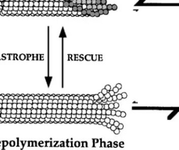
Figure 2 Microtubule dynamic instability: Dynamic instability is characterized by the coexistence of polymerizing and depolymerizing MTs. GTP-tubulin is incorporated at polymerizing MT ends,
the bound GTP is hydrolyzed during or soon after polymerization, and Piis subsequently released.
Thus the MT lattice is predominantly composed of GDP-tubulin (and is often referred to as a GDP MT in the text). Polymerizing MTs infrequently transit to the depolymerization phase (catastrophe). Depolymerization is characterized by the very rapid loss of GDP-tubulin subunits and oligomers from the MT end. Depolymerizing MTs can also infrequently transit back to the polymerization phase (rescue). The transitions in dynamic instability are measured as frequencies (e.g. catastrophe
frequency=number of catastrophes per unit time in the polymerization phase). The term frequency
is used rather than rate because it is not clear if the transitions are simple first order processes.
This representation incorporates the notions of a small GTP/GDP·Picap acting as a stabilizing
structure at polymerizing ends and different conformational configurations at polymerizing and depolymerizing ends, both of which are discussed in the text. For quantitative details on the various parameters, see Walker et al (1988) (adapted from Inou´e & Salmon 1995).
instability. Increasing the tubulin concentration, and thus the polymerization rate, results in a decrease in the catastrophe frequency, but the relationship between these two parameters is complex and poorly understood (Erickson & O’Brien 1992). In addition, there exist clear examples where catastrophe frequency is uncoupled from the polymerization rate. For example, similar catastrophe frequencies occur at MT plus ends when Mg2+is increased from 0.5 to 6 mM, despite a twofold increase in polymerization rate (O’Brien et al 1990). Catastrophes are assumed to be stochastic events with first order ki-netics; however, plus end catastrophes display non-first order kinetics, indi-cating hidden complexities in this phase transition (Odde et al 1995). The relationship between rescue frequency and the tubulin concentration is even
P1: MBL/dat P2: M/NBL/vks QC: MBL/uks T1: MBL
August 21, 1997 10:19 Annual Reviews AR041-04
MICROTUBULE DYNAMICS 91
less well understood, and it is not even clear that any significant dependency exists (O’Brien et al 1990, Walker et al 1991, Erickson & O’Brien 1992).
A surprising result of the real-time analysis of MT dynamics was the extent of minus end dynamic instability. Minus end behavior can be thought of as the dark side of MT dynamics. Dynamic instability of minus ends is probably not physiologically relevant because minus ends in cells are either capped by other proteins, for example at centrosomes, or depolymerizing when free in the cytoplasm (discussed below). However, the minus ends of MTs assembled from pure tubulin exhibit dynamic instability, which is quite similar to that of plus ends (Walker et al 1988, Erickson & O’Brien 1992). This is surprising, given their different structure and different association and dissociation rate constants. The behavior of minus ends in vitro must reflect intrinsic properties of the mechanism of dynamic instability and provides useful constraints for the development of mechanistic models.
Finally, two additional observations may be relevant to understanding dy-namic instability. First, MTs sometimes pause, where they neither polymerize nor depolymerize. Pauses are frequent in vivo (Shelden & Wadsworth 1993) and also occur in vitro with pure tubulin, although much less frequently (Walker et al 1988). Substoichiometric amounts of MT destabilizing drugs can enhance the paused state at MT plus ends, both in vitro and in vivo (Toso et al 1993, Dhamodharan et al 1995, Wilson & Jordan 1995). Second, polymerization and depolymerization rates of individual MTs exhibit significant variability (O’Brien et al 1990, Drechsel et al 1992, Gildersleeve et al 1992). This obser-vation implies that some structural feature governing rates of polymerization and depolymerization, although transient relative to the lifetime of the MT, is stably maintained over many subunit addition/loss events. This feature might be protofilament number (Chretien et al 1992) or perhaps some propagating structure at MT ends (Gildersleeve et al 1992, Chretien et al 1995).
Under special solution conditions, the transitions in MT dynamics become synchronized for the whole MT population, resulting in oscillatory polymeriza-tion cycles. Oscillatory polymerizapolymeriza-tion can be treated as a special manifestapolymeriza-tion of dynamic instability, and the mechanism for this intriguing behavior has been discussed by Mandelkow & Mandelkow (1992).
Thermodynamic Basis of Dynamic Instability
Dynamic instability is a profoundly nonequilibrium behavior and thus requires an energy source. Because the only possible source is GTP hydrolysis by β-tubulin during polymerization, we may safely state that GTP hydrolysis powers dynamic instability. One approach to understanding the role of GTP hydrolysis in dynamic instability is to ask how the free energy from the hydro-lysis of GTP (≈7.5 kcal mol−1under standard conditions, or≈12.5 kcal mol−1
P1: MBL/dat P2: M/NBL/vks QC: MBL/uks T1: MBL
August 21, 1997 10:19 Annual Reviews AR041-04
92 DESAI & MITCHISON
in vivo; Lehninger et al 1993) is partitioned among the different reactions in the kinetic cycle. To perform such an energy partitioning, the energy changes associated with polymerization and depolymerization must be determined in the absence of GTP hydrolysis. One way to ascertain this is to analyze tubulin polymerization/depolymerization with a nonhydrolyzable GTP analogue bound to its E-site.
Studies of tubulin polymerization in the presence of the classic nonhydrolyz-able GTP analogues GMPPNP and GMPPCP led to the important conclusions that polymerization does not require GTP hydrolysis and that the MT lattice is more stable with a GTP analogue bound toβ-tubulin than with GDP (Kirschner 1978, Mejillano et al 1990). However, these studies were complicated by the very weak affinity ofβ-tubulin for most GTP analogues, relative to GTP and GDP (Erickson & O’Brien 1992). Recent studies with the analogue GMPCPP, which binds relatively well toβ-tubulin, have shown that, contrary to earlier claims (Sandoval & Weber 1980), the normal P-O-P linkage between theβ andγphosphates in this analogue is resistant to hydrolysis by tubulin (Hyman et al 1992). Under standard conditions, the hydrolysis of GMPCPP in the MT lattice is negligible over the time course of most experiments. Tubulin poly-merizes normally with GMPCPP, confirming that the free energy of hydrolysis is not required for this step of the reaction. GMPCPP MTs are structurally more rigid than GDP MTs (Vale et al 1994, Mickey & Howard 1995), depoly-merize extremely slowly (≈0.1 dimers s−1versus≈1000 dimers s−1for GDP MTs), and do not exhibit dynamic instability. These properties suggest that the primary role of GTP hydrolysis is to destabilize the MT lattice by creating GDP-bound subunits that make weaker intersubunit contacts. Direct evidence for this conclusion was obtained when buffer conditions were found that trig-ger hydrolysis of GMPCPP in the MT lattice (substitution of Na+for K+and addition of glycerol); this hydrolysis destabilizes the lattice and results in rapid depolymerization (Caplow et al 1994).
By comparing tubulin polymerization and depolymerization in the presence of GMPCPP and its hydrolyzed product GMPCP, Caplow et al (1994) deter-mined how the free energy of hydrolysis of GMPCPP is partitioned in the poly-merization cycle of tubulin. Their analysis showed that the polypoly-merization of GMPCPP-tubulin is 4 kcal mol−1more favorable than that of GMPCP-tubulin. This difference in the stability of the two lattices must be derived from the hy-drolysis of GMPCPP. Because the free energy of hyhy-drolysis of GMPCPP is only −5.2 kcal mol−1, this study suggests that most of the free energy released upon GMPCPP hydrolysis is used to destabilize the MT lattice. The same general conclusion is thought to hold for GTP hydrolysis.
It is interesting to try and extend these thermodynamic conclusions from pure tubulin to the situation in living cells. Such an analysis can tell us how
P1: MBL/dat P2: M/NBL/vks QC: MBL/uks T1: MBL
August 21, 1997 10:19 Annual Reviews AR041-04
MICROTUBULE DYNAMICS 93
Figure 3 Thermodynamics of the tubulin polymerization cycle in vivo: The free energy for
GTP hydrolysis in vivo is≈−12.5 kcal mol−1. This free energy is partitioned in the tubulin
polymerization cycle as indicated. The main purpose of this figure is illustrative because several parameters needed for a complete quantitative analysis have not been measured. The free energy of polymerization (1Gpoly = −RTln (kon/koff) = −3 kcal mol−1) is obtained assuming a free
GTP-tubulin concentration of 10µM, an association rate constant of 2 × 10⁶M−1s−1and a
dissociation rate constant of 0.1 s−1(k
onrepresents the dimer association rate, which is the product of the association rate constant and the free GTP-tubulin concentration; these values result in
kon/koff = 200). The free energy of nucleotide exchange (1Gexch = −RTln (39) = −2 kcal
mol−1) is obtained by accounting for the threefold higher affinity of tubulin for GTP versus GDP
(Purich & Angelastro 1994) and an intracellular GTP/GDP ratio of 13:1 (Angelastro & Purich 1992). The dashed line, whose position depends on the free energy change accompanying hydrolysis and Pirelease on the polymerized tubulin dimer, is difficult, if not impossible, to measure directly and
is estimated here as−2.5 kcal mol−1by assuming that1Gdepol = −5 kcal mol−1(Caplow et al
1994). Maximal pushing and pulling forces can be calculated from1Gpolyand1Gdepolassuming
an average displacement of 0.61 nm for a tubulin dimer at a MT end (for discussion see Caplow et al 1994, Inou´e & Salmon 1995).
much free energy is released during polymerization and depolymerization in vivo, which puts upper bounds on the force that could be generated by motile processes driven by polymerization dynamics. We lack detailed information on several parameters, so only an approximation is possible, as shown in Figure 3. According to this estimate, polymerization could produce a pushing force of up to 35 pN/MT and depolymerization a pulling force of up to 60 pN/MT. For comparison, the stall force for a single kinesin is≈5 pN (Svoboda & Block 1994).
P1: MBL/dat P2: M/NBL/vks QC: MBL/uks T1: MBL
August 21, 1997 10:19 Annual Reviews AR041-04
94 DESAI & MITCHISON
Utilizing the free energy released during polymerization/depolymerization to move structures within the cell requires a molecular interface that can cou-ple MT dynamics to movement. For pushing force, this interface can be a simple barrier; MT polymerization inside synthetic vesicles has been observed to deform the vesicle membrane (Hotani & Miyamoto 1990, Elbaum et al 1996). A more complex coupling interface of unknown molecular composi-tion, termed TAC (Tip Attachment Complex), was implied by observations of MT polymerization-driven extension of membrane tubules in Xenopus extracts (Waterman-Storer et al 1995). Intuitively, it seems more difficult to couple MT depolymerization to movement because the coupling interface would have to hold on to a depolymerizing end. However, recent studies have shown that pure kinesins coupled to beads can remain attached to depolymerizing MTs (Lombillo et al 1995). Although the precise mechanism is not understood, the resulting minus end-directed motility is not dependent on ATP or on the inherent directionality of the kinesin. Therefore, in addition to being motile ATPases, kinesins can also act as coupling factors to depolymerizing MTs. The biological importance of MT polymerization dynamics is highlighted dur-ing chromosome movement in mitosis. Defindur-ing the mechanisms by which chromosome movement is tightly coupled to MT dynamics remains one of the greatest challenges in the study of MT dynamics (for a detailed discussion, see Inou´e & Salmon 1995).
Although we can conclude from thermodynamic analysis of dynamic insta-bility that GTP hydrolysis weakens the MT lattice, and we can estimate the capacity of MT dynamics to perform mechanical work, we can not tell much about the detailed mechanism of dynamic instability. Most importantly, we do not know how a MT persists for many minutes in a polymerizing state or how this state decays infrequently when a catastrophe occurs. To address these issues, we need to analyze the kinetic processes underlying dynamic instability.
Evidence for a Stabilizing Structure at Microtubule Ends
GTP hydrolysis is known to occur very rapidly during polymerization, and from thermodynamic analysis we know that GDP-tubulin makes a very unstable lattice. So how can tubulin polymerize at all? A fundamental idea underlying all recent studies of MT dynamics is that polymerizing MTs are stabilized by some special structure at their ends. This structure was originally postulated to be a cap of GTP-tubulin (Mitchison & Kirschner 1984a). Below we examine the evidence for such a cap, but first, what is the evidence that any stabilizing structure exists?
The most direct way to test for a special stabilizing structure at polymerizing ends is to cut a polymerizing MT and determine the behavior of the newly ex-posed ends. In the original dynamic instability paper, severing MTs by shearing
P1: MBL/dat P2: M/NBL/vks QC: MBL/uks T1: MBL
August 21, 1997 10:19 Annual Reviews AR041-04
MICROTUBULE DYNAMICS 95
was found to promote rapid depolymerization (Mitchison & Kirschner 1984a); this conclusion was confirmed by more quantitative studies using inelastic light scattering (Keates & Hallett 1988). A more elegant approach involves using microscopy to directly observe the effect of severing an individual MT. Salmon and coworkers (Walker et al 1989; PT Tran, RA Walker & ED Salmon, personal communication) have characterized the behavior of MTs severed by a UV mi-crobeam or a fine glass needle. As predicted, if a special stabilizing structure is required at polymerizing ends, the newly exposed plus ends were unstable and rapidly depolymerized. Surprisingly, newly exposed minus ends were sta-ble and immediately resumed polymerization. This behavior may suggest that minus ends do not require a stabilizing structure or that rescue is very efficient at minus ends. Alternatively, the stability of minus ends could be explained by the existence of an intermediate between polymerization and depolymerization (see below for details).
The MT-cutting experiments provide compelling evidence that polymerizing MT plus ends are stabilized by a special structure located near or at their ends (how close to the end is far from clear). What is this structure? Below we discuss evidence that it is a part of the MT lattice differentiated either by different chemistry, for example the presence of GTP, or by a different structure, for example a flatter, sheet-like lattice. These two views are not exclusive, and we conclude with a discussion about their integration.
The GTP CAP Model
Soluble tubulin has a very slow rate of GTP hydrolysis (David-Pfeuty et al 1977, Caplow & Shanks 1990); this rate increases tremendously when tubulin subunits are incorporated into a MT. Thus by analogy with signaling GTPases, tubulin can be thought of as its own GAP (GTPase Activating Protein), acceler-ating hydrolysis by the formation of intersubunit contacts during polymeriza-tion. The original model for dynamic instability proposed that polymerizing MTs are stabilized by a cap of subunits in which GTP hydrolysis has not yet occurred (Mitchison & Kirschner 1984a). The infrequent loss of such a GTP cap would result in a catastrophe, whereas the reacquisition of such a cap by a depolymerizing end would result in a rescue. This model was based on early observations of a relatively long kinetic lag between tubulin polymerization and GTP hydrolysis (Carlier & Pantaloni 1981).
A stabilizing cap at polymerizing MT ends could be composed of either GTP-tubulin or GDP·Pi–tubulin subunits. In proteins where nucleotide hydrolysis drives a conformational change, the step correlating with the conformational change is often not hydrolysis but phosphate (Pi) release (Vale 1996). In actin polymerization, where NTP hydrolysis also accompanies polymerization, a conformational change that weakens the polymer is thought to occur upon Pi
P1: MBL/dat P2: M/NBL/vks QC: MBL/uks T1: MBL
August 21, 1997 10:19 Annual Reviews AR041-04
96 DESAI & MITCHISON
release (Carlier 1989, 1991). If this were also true for tubulin, we might expect a GDP·PiMT lattice to be as stable as a GTP lattice. MTs appear to be stabilized by the phosphate analogue BeFx(Carlier et al 1989); however, unlike actin, addition of high concentrations of phosphate does not stabilize MTs (Caplow et al 1989, Trinczek et al 1993). Therefore, whether a GDP·Pilattice is stable remains to be resolved.
Whether one proposes a stabilizing cap of GTP-tubulin or of GDP·Pi-tubulin, the key question is the same: At rates of MT polymerization relevant to dynamic instability, does a polymerizing end accumulate a run of GTP (or GDP·Pi) sub-units sufficient to stabilize it against rapid depolymerization? Experimentally, this question resolves into two issues: (a) How long is the lag between poly-merization and GTP hydrolysis/Pirelease (or how tight/loose is the coupling between polymerization and GTP hydrolysis/Pirelease?); and (b) how many subunits at a MT end need to be bound to GTP or GDP·Pito stabilize it?
Contrary to earlier observations, several recent studies indicate that there is little, if any, lag between polymerization and GTP hydrolysis (discussed in de-tail by Caplow 1992, Erickson & O’Brien 1992). Therefore, can we conclude there is no GTP cap under dynamic instability conditions? Unfortunately, we do not conclusively know the answer. A stabilizing GTP cap may be as small as one layer of subunits, thereby escaping detection in the assays employed to date (Erickson & O’Brien 1992, Bayley et al 1994, Caplow & Shanks 1996, Flyvbjerg et al 1996). An additional complication is that MT plus ends have exposed E-sites on the terminalβ-tubulin subunits (Mitchison 1993) whose nucleotide state may affect the behavior of MT ends (Caplow & Shanks 1995). Recently, more progress has been made in determining the lag between poly-merization and Pirelease. Studies using relatively low time resolution (5–20 s) filtration methods presented contradictory evidence regarding the existence of a significant lag between polymerization and Pirelease (Melki et al 1990, Stewart et al 1990, Caplow 1992). This issue was reexamined by simultaneous moni-toring of polymerization and Pirelease using an enzyme-linked spectrophoto-metric assay for detecting free Pi(Melki et al 1996). Convincing evidence for a lag between assembly and Pirelease was obtained for rapid taxol-driven poly-merization. The extent of such a lag under conditions of dynamic instability remains to be determined. Nevertheless, these data support a model in which polymerizing ends are stabilized by a cap of predominantly GDP·Pi-tubulin subunits.
Two alternate approaches have been taken to measure the size of a GTP (or GDP-Pi) cap required to stabilize a polymerizing MT. The first involves esti-mating the kinetic lifetime of the cap by rapidly diluting polymerizing MTs and measuring the time lag before the MTs undergo a catastrophe. Observations on individual MTs in a microscopic flow cell (Walker et al 1991) showed that
P1: MBL/dat P2: M/NBL/vks QC: MBL/uks T1: MBL
August 21, 1997 10:19 Annual Reviews AR041-04
MICROTUBULE DYNAMICS 97
polymerizing MTs transit very rapidly (within 1–4 s) upon dilution, suggest-ing that the cap size is fairly small, less than 100 subunits. This analysis also showed that cap size is independent of the polymerization rate (within a tenfold range). These results are inconsistent, within the explored range of tubulin con-centrations, with models predicting larger caps at faster polymerization rates. Dilution of bulk populations of brain MTs has yielded similar conclusions (Voter et al 1991). The second method used to estimate the minimal cap size is to define the smallest number of GMPCPP-tubulin subunits required to sta-bilize a GDP MT to depolymerization by dilution. Using statistical analysis of fixed MTs and direct fluorescence measurements, Drechsel & Kirschner (1994) concluded that as few as 22±11 GMPCPP subunits—one to three layers of the lattice—were sufficient to stabilize a MT. Using real-time analysis, Caplow & Shanks (1996) obtained a similar conclusion; they provided further evidence for stabilization by a single layer of GMPCPP subunits, by analyzing MTs composed of a mixture of GMPCPP- and GDP-tubulin. Such mixed lattice MTs exhibit a mean lifetime of a subunit at a MT end (reciprocal of the de-polymerization rate) that is proportional to the 13th or 14th power of the mole fraction of GMPCPP in the mixed lattice. This remarkable proportionality sug-gests that a single monolayer of tubulin-GMPCPP subunits (and by extension tubulin-GTP/GDP·Pisubunits) is both necessary and sufficient to stabilize a MT. Whether this is indeed the case in MTs undergoing dynamic instability remains a daunting challenge for future studies.
Structural Basis of Dynamic Instability
Since electron microscopy (EM) was first used to study tubulin polymerization, there have been clues suggesting an intimate relationship between MT dynam-ics and the geometry of the intersubunit bonds in the MT lattice (reviewed in Kirschner 1978). Within MTs, protofilaments are relatively straight, whereas the depolymerization products of MTs are often highly curved protofilament oligomers (Kirschner 1978, Mandelkow & Mandelkow 1985, Mandelkow et al 1991, Tran et al 1997). Cryoelectron microscopy (cryoEM) makes it feasible to trap kinetic intermediates by rapid freezing and image them directly with-out staining (Mandelkow & Mandelkow 1986, Wade & Chretien 1993). Us-ing cryoEM and X-ray scatterUs-ing to analyze polymerizUs-ing and depolymerizUs-ing MTs, Mandelkow et al (1985, 1988, 1991) hypothesized that the predominant driving force for MT depolymerization is the curling up of protofilaments. Recent cryoEM studies have confirmed the existence of curved oligomers at the ends of depolymerizing MTs (Figure 4a). Divalent cations stabilize these curved protofilament oligomers, thereby promoting protofilament peeling and increasing the depolymerization rate, which results in ram’s horn-type struc-tures at the depolymerizing MT ends (Tran et al 1997).
P1: MBL/dat P2: M/NBL/vks QC: MBL/uks T1: MBL
August 21, 1997 10:19 Annual Reviews AR041-04
98 DESAI & MITCHISON
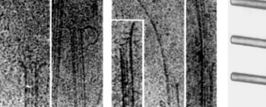
Figure 4 Cryoelectron microscopy of (a) depolymerizing and (b) polymerizing microtubule ends: In cryoEM images, the body of the MT is delineated by two thick edges. Between these thick edges, discrete lines can be seen running along the length of the MT. These lines arise from the superposition of protofilaments on opposite sides of the MT cylinder, and their number and long range periodicities can be quantitatively interpreted to determine the protofilament number and other structural parameters of the MT lattice (Chretien & Wade 1991). Note the curvature of protofilament oligomers at depolymerizing MT ends in (a) and the presence of sheets at ends of polymerizing MTs in (b). The sheets tend to orient perpendicular to the surface and often appear as a single thick line (the projection image of multiple protofilaments). (c) Diagrammatic representation of the structure of polymerizing (top) and depolymerizing (bottom) MT ends with a hypothetical structural mechanism for catastrophe. Catastrophe is postulated to occur as a consequence of sheet closure catching up to a MT end (middle) (see text for details) [images in (a) and (b) reprinted from Chretien et al 1995].
CryoEM studies have also led to a structural hypothesis for the mechanism of catastrophe. Chretien et al (1995) analyzed MTs nucleated by centrosomes and observed striking long protofilament sheets at the plus ends of polymerizing MTs (Figure 4b). This observation demonstrates that polymerization occurs primarily by extension of protofilament sheets as opposed to helical subunit addition. A similar conclusion had been reached in earlier negative stain EM studies (Erickson 1974, Kirschner et al 1975, Detrich & Jordan 1986, Simon & Salmon 1990), and protofilament sheets have also been observed at MT ends in vivo (McIntosh et al 1985). These protofilament sheets eventually close to form the cylindrical body of the MT, presumably along the seam in the lattice (Figure 1c), although this is not known for certain. Chretien et al (1995) hypoth-esize that sheet closure occurs at a variable rate, with the consequence of sheet closure catching up to the polymerizing end being a catastrophe (Figure 4c). Thus the sheets may represent a structural cap that stabilize a polymerizing MT. This suggestion is intriguing, and it seems plausible that sheet closure induces catastrophe by some physical mechanism. At present, however, the evidence that protofilament sheets stabilize polymerizing ends is only a correlation. A
P1: MBL/dat P2: M/NBL/vks QC: MBL/uks T1: MBL
August 21, 1997 10:19 Annual Reviews AR041-04
MICROTUBULE DYNAMICS 99
careful comparison of sheets at plus and minus ends of MTs, and under condi-tions that appear to uncouple catastrophe frequency from polymerization rate (discussed above), may shed further light on the relationship of sheet closure to the mechanism of catastrophe.
Relationship of Structural and Chemical Transitions
How does the structural view of dynamic instability mesh with the chemical transitions that occur as a consequence of GTP hydrolysis? The effect of GTP hydrolysis on MT structure has been analyzed by comparison of GMPCPP and GDP MTs using cryoEM. The lattice structure of GMPCPP and GDP MTs appears to be very similar. Using optical diffraction and lattice accommodation theory (Chretien & Wade 1991), Hyman et al (1995) showed that the inter-monomer spacing along the protofilament decreases from 4.2 nm in GMPCPP MTs to 4.05 nm in GDP MTs. More significantly, the curvature of protofilament oligomers at depolymerizing ends of GDP MTs is twofold greater than that of similar oligomers at ends of GMPCPP MTs (the latter could be induced to de-polymerize at a reasonable rate using calcium; T Muller-Reichert, D Chretien, F Severin & AA Hyman, personal communication). These results provide quan-titative support for a model in which GTP hydrolysis causes tubulin to enter a curved conformation, destabilizing the MT lattice (Melki et al 1989). Because GDP-tubulin is prevented from adopting the fully curved conformation while in the lattice (presumably by specific lattice interactions), the energy of GTP hydrolysis is stored in the lattice as mechanical strain. This strain is released when the GDP-tubulin subunits are exposed at MT ends and provides the driving force for the rapid depolymerization phase of dynamic instability. This struc-tural explanation for how the free energy of GTP hydrolysis destabilizes the MT lattice is incorporated in the representation of dynamic instability in Figure 2.
Although these studies define the structural differences between GTP-like and GDP MT lattices, they do not relate the kinetics of structural changes to the relative rates of polymerization, hydrolysis and Pirelease. Ideally, we would like to understand the cause-effect relationships between chemical and structural changes at MT ends. Chretien et al (1995) hypothesize that closure of protofilament sheets triggers GTP hydrolysis. In our opinion, the change in the rate of GTP hydrolysis between soluble tubulin and tubulin in the MT lattice is so large that it seems unlikely to be triggered by a structural change as subtle as a change in the curvature of the protofilament sheet. Furthermore, hydrolysis is known to occur in flat tubulin sheets formed in the presence of zinc ions (Melki & Carlier 1993), in taxol-induced oligomers that are also often sheet-like (Melki et al 1996), and upon interaction of tubulin dimers with MT ends at concentrations too low to support MT assembly (Caplow & Shanks 1990). The identification of cause-effect relationships between chemical and
P1: MBL/dat P2: M/NBL/vks QC: MBL/uks T1: MBL
August 21, 1997 10:19 Annual Reviews AR041-04
100 DESAI & MITCHISON
structural transitions will require determining where GTP and GDP·Piare still present in the lattice of the growing MT end. This information most likely will come from refinement of the kinetic data and correlation with structural analysis. The more ambitious idea of directly visualizing the bound nucleotide in the lattice is attractive, but it is not clear what technology could achieve this. The striking structural and kinetic differences between polymerizing and de-polymerizing ends highlight the central mystery of dynamic instability—the interconversion of polymerizing and depolymerizing ends. The current two-state model suggests that interconversion can be explained by stochastic loss or reacquisition of a stabilizing cap. However, pauses in MT dynamics and non-first order kinetics for catastrophes (discussed above) have provided sug-gestive evidence for an intermediate state between polymerization and depoly-merization. To explain the disparate stability of plus and minus ends in cutting experiments, a three-state model has been proposed recently that postulates the existence of a kinetic intermediate between polymerization and depolymeriza-tion (PT Tran, RA Walker & ED Salmon, personal communicadepolymeriza-tion). Because of the polarity of the MT lattice, such an intermediate could be significantly different at plus and minus ends. This model has important implications for the mechanisms of the transitions of dynamic instability. Although this three-state model is derived purely from kinetic analysis, it is tempting to speculate that a closed-tube state, which presumably exists as a structural intermediate between polymerizing ends with sheets and depolymerizing ends with peeling GDP-tubulin oligomers (Figure 4c), may represent a structural correlate of such a kinetic intermediate.
MICROTUBULE DYNAMICS IN VIVO
Functions of Microtubule Dynamic Instability In Vivo
As discussed in the introduction, polymerization dynamics facilitate spatial or-ganization and rapid remodeling of the cytoskeleton. But what are the specific in vivo benefits of dynamic instability? Several biological functions have been proposed (Kirschner & Mitchison 1986). One simple idea is that dynamic in-stability allows newly formed regions of cytoplasm to rapidly fill with MTs which, in turn, facilitate recruitment of membrane systems using MT motor proteins. Such colonization of newly extruded cytoplasm with MTs has been visualized during motility of growth cones (Tanaka & Kirschner 1991).
A more interesting idea for the function of dynamic instability is that it allows MTs to search three-dimensional space more effectively than equilibrium polymerization, thereby enabling MTs to find specific target sites within the cell (the search-capture model). This idea has been conceptually verified by quantitative modeling (Holy & Leibler 1994). The search-capture model was initially formulated to account for a difficult targeting problem—the capture of
P1: MBL/dat P2: M/NBL/vks QC: MBL/uks T1: MBL
August 21, 1997 10:19 Annual Reviews AR041-04
MICROTUBULE DYNAMICS 101
MTs by the kinetochore region of chromosomes early in mitosis. Plus ends of centrosome-nucleated dynamically unstable MTs were hypothesized to probe through the cytoplasm, searching for binding sites on kinetochores that could capture them. This process has been visualized in living newt lung cells (Hayden et al 1990) and is thought to account for the initial attachment of outlying chromosomes to the spindle.
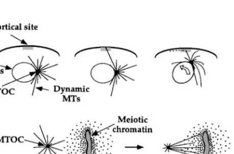
The search-capture model is particularly attractive as a conceptual basis for understanding the generation of asymmetric MT arrays. In several biological processes, one can hypothesize that a single MT nucleated by the MT organiz-ing center (MTOC) is initially captured by a specialized cortical site, leadorganiz-ing to subsequent movement of the MTOC towards the capture site and forma-tion of a multi-MT attachment (Figure 5a). Captured MTs may be stabilized
Figure 5 Mechanisms for generating asymmetric microtubule distributions: (a) Search-capture mediated by dynamic instability. Search-capture has been implicated in attachment of the meiotic spindle to a specific cortical site in marine eggs prior to polar body formation (Lutz et al 1988); in asymmetric cell division in nematode embryos (Hyman 1989); in positioning of the budding yeast spindle to the mother-bud neck prior to anaphase (Yeh et al 1995); and in the reorientation of the interphase centrosome toward the site of cell-cell interaction in cytotoxic and helper lymphocytes (Kupfer & Singer 1989). (b) Local regulation of MT dynamics: As shown here, chromatin in meiotic spindles produces a gradient of MT stabilization, presumably through local post-translational reg-ulation of MAPs and/or catastrophe factors (Karsenti et al 1984, Zhang & Nicklas 1995, Dogterom et al 1996). (c) Movement of pre-existing MTs through the cytoplasm: The example depicted here occurs during growth cone pathfinding (Tanaka & Kirschner 1995); another example is movement mediated by MT motors during mitotic spindle assembly (Gaglio et al 1996, Heald et al 1996).
P1: MBL/dat P2: M/NBL/vks QC: MBL/uks T1: MBL
August 21, 1997 10:19 Annual Reviews AR041-04
102 DESAI & MITCHISON
subsequently or differentiated by post-translational modifications ofα-tubulin (Bulinski & Gundersen 1991). Search-capture, when first proposed in 1986, was thought to be the dominant mechanism for inducing asymmetry in the MT cytoskeleton. Since then it has become clear that several alternative processes are also important. Two such processes are local regulation of factors control-ling MT dynamics (Figure 5b) and movement of pre-existing MTs through the cytoplasm (Figure 5c). In general, dissecting the mechanisms by which com-plex asymmetric arrays of MTs are assembled remains a fascinating research goal.
Although the precise biological functions of dynamic instability continue to be explored, a considerable amount is known about the behavior of MTs in cells. In the remainder of this review we focus on MTs in vivo by first describing some of the methodology used to analyze MT dynamics, then describing the nature and regulation of MT dynamics in vivo, and finally discussing specific regulatory molecules that modulate MT dynamics.
Methodology for Analysis of Microtubule Dynamics In Vivo
Most of our information on MT dynamics in vivo has come from optical mi-croscopy. Single MTs can be visualized using DIC microscopy in thin, outlying regions of certain cells (Cassimeris et al 1988). This type of imaging results in minimal light-induced damage, but unfortunately its use is restricted to ex-tremely flat cells. More generally applicable is fluorescence imaging of individ-ual MTs (Sammak & Borisy 1988, Schulze & Kirschner 1988). Photodamage is a greater concern with this method, but it can be limited in certain cases by oxygen-scavenging systems (Tanaka & Kirschner 1991, Waterman-Storer et al 1993). To date, most observations have been performed on cells microinjected with rhodamine-labeled tubulin. However, the advent of green fluorescent pro-tein (GFP) fusions will extend these observations to a wider range of cell types, including yeast cells (Stearns 1995). All imaging methods suffer problems in more interior regions of the cell and in MT-rich structures such as the mitotic spindle, where high MT density makes visualization of single MTs difficult. Overall, single MT imaging has shown that dynamic instability occurs in cells, and values for the different parameters have been obtained (Cassimeris et al 1988, Hayden et al 1990, Shelden & Wadsworth 1993).
Live cell measurements have been supplemented by fluorescence observa-tions in crude extracts, notably from Xenopus eggs where spindle assembly and function can also be followed. Extract work suffers the disadvantage that true physiological rates may not be observed but has the great advantage that the system can be perturbed in specific ways, either by control of cell-cycle state (Belmont et al 1990, Verde et al 1992) or by removal of specific proteins with antibodies (Walczak et al 1996). Video-enhanced DIC microscopy can be
P1: MBL/dat P2: M/NBL/vks QC: MBL/uks T1: MBL
August 21, 1997 10:19 Annual Reviews AR041-04
MICROTUBULE DYNAMICS 103
used in cell extracts clarified by centrifugation (Gliksman et al 1992, Parsons & Salmon 1997). Although clarified extracts are less physiological than crude extracts, they represent a useful starting point for the biochemical purification of proteins that influence MT dynamics.
Fluorescence perturbation techniques, notably photobleaching and photoac-tivation of fluorescence, have been used to observe the average rate of turnover of MTs in a small region of the cell. These assays led to the discoveries that MT dynamics are regulated in the cell cycle (Salmon et al 1984, Saxton et al 1984), that different regions of the spindle turn over at different rates (Zhai et al 1995), and that MTs flux polewards in spindles (Mitchison 1989). Photobleaching is probably the more convenient technique, but it has been criticized because of the likelihood of photodamage artifacts. Photobleaching of GFP-tagged pro-teins, including tubulin, may turn out to be less toxic and artifact-prone than bleaching of conventional fluorophores (Cole et al 1996), presumably because the fluorophore in GFP is contained within a protein capsule (Ormo et al 1996). Simpler assays measuring the amount of tubulin partitioning into detergent-soluble (αβdimers) and detergent-insoluble (polymer) fractions have also been informative. The amount of tubulin in each fraction has been determined by at least two methods: quantitative immunoassay (Solomon 1986, Liao et al 1995) and fluorescence microscopy (Zhai & Borisy 1994). This type of simple assay has been used to analyze the fraction of tubulin polymerized at different stages of the cell cycle (Zhai & Borisy 1994) and in response to expression of proteins regulating MT stability (Marklund et al 1996). Finally, we should mention an interesting biochemical technique for analyzing turnover in vivo that relies on incorporation of radioactive GTP into the polymer fraction (Purich & Angelastro 1994).
A promising new approach to understanding the role of GTP hydrolysis by β-tubulin in dynamic instability, as well as the biological role of MT dynam-ics, is mutational analysis of yeastβ-tubulin (summarized in Burns & Farrell 1996). Farrell and coworkers have characterized the in vivo phenotypes and in vitro properties ofβ-tubulin mutations (Davis et al 1994, Sage et al 1995). In addition to defining the residues important for GTP binding and hydrolysis, this approach has demonstrated that mutationally altered GTP hydrolysis byβ -tubulin strongly affects MT function in vivo and MT dynamic instability in vitro.
Features of Microtubule Dynamics In Vivo
How do the parameters of MT dynamic instability in vivo compare with those measured for pure tubulin? MTs in vivo differ from pure tubulin primar-ily in their rapid polymerization rates and their high transition frequencies (Cassimeris 1993). The polymerization rate of tubulin in vivo is about five-to tenfold higher than that of a similar concentration of pure tubulin. Despite
P1: MBL/dat P2: M/NBL/vks QC: MBL/uks T1: MBL
August 21, 1997 10:19 Annual Reviews AR041-04
104 DESAI & MITCHISON
these rapid polymerization rates, MTs in vivo exhibit a high frequency of catas-trophe. If we use the relationship between polymerization rate and catastrophe frequency for pure tubulin as a reference, at the polymerization rates observed in vivo we would expect a near-zero frequency of catastrophe. This apparent para-dox can be resolved if distinct mechanisms exist to promote polymerization and to induce catastrophes in vivo. In order to elucidate these mechanisms, many efforts have been directed at identifying and characterizing cellular factors that modulate dynamic instability.
MT dynamics in vivo change extensively in response to regulatory signals, providing further support for the existence of mechanisms for modulating MT dynamics. A well-studied example of intracellular regulation of MT dynamics is the interphase-mitosis transition. MTs in interphase tissue culture cells turn over with a half-life of greater than 5–10 min, whereas MTs in mitosis turn over with a half-life of 30s−1 min (reviewed in McNally 1996). MT stability can also change significantly (over two to three orders of magnitude) as a conse-quence of cellular differentiation (Bulinski & Gundersen 1991). Differentiation of both neuronal (Baas et al 1991) and epithelial (Bre et al 1990) cells is cor-related with an increase in MT stability. MTs are also likely to be regulated by signal transduction pathways, but relatively little is known in this area. Stimula-tion of macrophages with phorbol esters, which presumably trigger endogenous signaling pathways, causes a rapid increase in both the total polymer level and number of MTs (Robinson & Vandre 1995). Dissecting the regulation of MT dy-namics by signaling pathways will be an important area of research in the future. In the cases where the parameters of dynamic instability have been mea-sured, the transition frequencies appear to be the primary targets of regula-tory molecules. Regulating the transition frequencies is attractive because MT lengths and overall dynamic behavior are very sensitive to changes in these parameters (Verde et al 1992, Gliksman et al 1993). For example, in Xenopus extracts induced to enter a mitosis-like state by the addition of cyclin, a five-to tenfold increase in the catastrophe frequency, without significant changes in other parameters of dynamic instability, results in the transformation of relatively stable arrays of long MTs into very dynamic arrays of short MTs (Belmont et al 1990, Verde et al 1992). In urchin eggs treated with phosphatase inhibitors, a decrease in the rescue frequency has been shown to promote a sim-ilar transformation (Gliksman et al 1992). Thus the increased turnover of MTs in mitosis appears to be driven primarily by changes in catastrophe and rescue frequencies. However, the extent to which it depends on regulation of catastro-phe versus rescue is controversial and may vary between different cell types. Studies on regulation of MT dynamics in Xenopus extracts were performed in the presence of protein synthesis inhibitors, implying that rapid regulation of MT dynamics can be completely post-translational; however, transcriptional
P1: MBL/dat P2: M/NBL/vks QC: MBL/uks T1: MBL
August 21, 1997 10:19 Annual Reviews AR041-04
MICROTUBULE DYNAMICS 105
regulation is also important in many cases, as in the induction of MAP synthesis during neuronal differentiation (Drubin et al 1985).
In order to account for the dynamics of MTs in cells, much effort has been directed toward identifying proteins that regulate MT dynamics and under-standing how these proteins are themselves regulated. Below we discuss some of these factors with an emphasis on how they modulate the polymerization dynamics of MTs.
Microtubule Stabilizing Factors: MAPs
Until recently, all of the proteins known to regulate MT polymerization were MAPs (Microtubule Associated Proteins); MAPs are proteins that bind in a nucleotide-insensitive manner to the MT lattice. A great deal of characteri-zation has been performed on what we term classical MAPs: MAP1, MAP2, and tau in neurons, and MAP4 in non-neuronal cells, and detailed summaries have been recently presented (Hyams & Lloyd 1994). These proteins bind to, stabilize, and promote the assembly of MTs. Neuronal MAPs weakly in-crease the polymerization rate of pure tubulin, strongly suppress catastrophes, and promote rescues (Drechsel et al 1992, Pryer et al 1992, Trinczek et al 1995). The net effect of these changes is to reduce the turnover rate and in-crease the fraction of tubulin in polymer at steady state. Neuronal MAPs are thought to exert their effects primarily by binding to the MT lattice in such a way as to cross link adjacent tubulin subunits. Consistent with this idea, the tubulin-binding sites in neuronal MAPs often consist of repeated motifs (Lewis et al 1988), and neuronal MAPs saturate their binding sites on MTs at ratios of 1:4–10 (MAP:tubulin). These cross links have the effect of suppressing subunit dissociation and, perhaps, also inhibiting protofilament peeling, thus inhibiting catastrophe and promoting rescue. Neuronal MAPs also promote polymerization, but whether this involves increasing the association rate or decreasing the dissociation rate of GTP-tubulin subunits at MT ends remains controversial (Drechsel et al 1992, Pryer et al 1992, Trinczek et al 1995). MAP4 is evolutionarily conserved from Drosophila to humans and is found in both neuronal and non-neuronal cell types. MAP4 promotes MT assembly in vitro, although, unlike the neuronal MAPs, it does so by strongly enhancing the rescue frequency without decreasing the catastrophe frequency (Ookata et al 1995). Surprisingly, genetic as well as biochemical disruption of MAP4 function failed to show a significant phenotype, perhaps due to functional overlap with other MAPs (Pereira et al 1992, Wang et al 1996).
XMAP215 was identified in Xenopus eggs (Gard & Kirschner 1987), and recently a human homologue has been identified (Charrasse et al 1996). XMAP215 affects MT dynamics in a very different manner than conventional MAPs (Vasquez et al 1994). XMAP215 strongly increases the polymerization
P1: MBL/dat P2: M/NBL/vks QC: MBL/uks T1: MBL
August 21, 1997 10:19 Annual Reviews AR041-04
106 DESAI & MITCHISON
rate of pure tubulin, but only at MT plus ends. XMAP215 also increases the rate of rapid depolymerization and decreases the rescue frequency, thereby in-creasing MT turnover. The ability of XMAP215 to differentially affect the two ends of a MT and to promote assembly while also increasing turnover suggests a novel mechanism of action. Furthermore, XMAP215 may play an important role in vivo, where rapid polymerization is also matched by rapid turnover. Because XMAP215 decreases the rescue rate, it must bind to the MT lattice in a manner that differs from classic MAPs. This mode of MT-binding by XMAP215 is supported by its lower saturation stoichiometry (1:20 XMAP215:tubulin) relative to classic MAPs (1:4–10 MAP:tubulin). Investi-gation of the novel mechanism of action of XMAP215 and its interaction with MT lattices deserves future study.
In addition to the MAPs discussed above, many MT-binding proteins have been isolated in diverse systems (Kreis & Vale 1993, Maccioni & Cambiazo 1995). SDS-PAGE of MAP preparations, particularly from non-neuronal cell types, demonstrates a large number of bands of which relatively few have been identified (e.g. Andersen et al 1994). Therefore, it is not clear if most of the MAPs controlling MT dynamics are known, or if we are only at the tip of the iceberg.
MAP REGULATION Most identified MAPs are known to be regulated by
phos-phorylation, and in all cases the more phosphorylated forms are inhibited in their ability to stabilize MTs (Drechsel et al 1992, Trinczek et al 1995). The binding of MAPs to MTs is predominantly electrostatic, involving the highly acidic C-terminal domains of bothα- and β-tubulin (Rodionov et al 1990). Phosphorylation inhibits MAP function by reducing the affinity of the MAP for the MT lattice, presumably by weakening this electrostatic interaction, al-though exceptions to this rule are known (Ookata et al 1995). The inactivation of MAPs by phosphorylation reduces the frequency of rescue and is one mech-anism by which MT turnover can be increased in vivo (McNally 1996). In addition to phosphorylation, alternative mechanisms may exist for regulating MAP-MT interactions as demonstrated by the recent characterization of map-modulin (Ultizer et al 1997), a protein capable of modulating the binding of multiple MAPs to MTs in vitro.
Kinases that phosphorylate a given MAP in vivo have not been rigorously identified in most cases, although many kinases are known to phosphorylate MAPs in vitro. Extensive phosphorylation site analysis has been performed for classic MAPs (summarized in Hyams & Lloyd 1994), especially tau, which has been implicated in Alzheimer’s disease. Biochemical fractionation of brain extracts for an activity capable of phosphorylating a specific region in the tau MT-binding domain resulted in the identification of a novel protein kinase,
P1: MBL/dat P2: M/NBL/vks QC: MBL/uks T1: MBL
August 21, 1997 10:19 Annual Reviews AR041-04
MICROTUBULE DYNAMICS 107
p110MARK, which can phosphorylate and inactivate multiple MAPs in vitro and cause reorganization of the MT cytoskeleton when expressed in tissue culture cells (Drewes et al 1996). p110MARKshows sequence similarity to a protein kinase required for the asymmetric division of the one-cell nematode embryo and also to protein kinases found in budding yeast. These homologies suggest exciting new avenues for the analysis of MAP regulation in diverse biolog-ical processes. In addition to the conservation of a protein kinase probably important for regulating MAPs, proteins that exhibit motifs characteristic of the MT-binding sites in neuronal MAPs have been found in both nematodes and budding yeast (Irminger-Finger et al 1996, McDermott et al 1996). Along with providing the ability to utilize genetic systems to analyze the function and regulation of MAPs, such evolutionary conservation indicates the fundamental importance of MAPs to the MT cytoskeleton.
Microtubule Destabilizing Factors
The high frequency of catastrophe of MTs in vivo compared with pure tubulin suggests the existence of factors that induce catastrophes. Such factors would destabilize MTs and oppose the action of MAPs, reducing net assembly and in-creasing turnover. Regulation of catastrophe factor activity may underly rapid changes in MT dynamics in vivo, as during the interphase-mitosis transition (McNally 1996). In addition to their potential importance in regulation of dy-namics, study of such factors may help elucidate the mechanism of catastrophe, which is still unclear from pure tubulin work. Recently three types of molecules have been identified that destabilize MTs but appear to do so using different mechanisms.
Op18/stathmin is a small heat-stable protein that was first identified because it is abundant, highly induced in some tumor cells, and is a substrate for a number of protein kinases (Sobel 1991). Op18 is probably ubiquitous in vertebrate cells but is not present in budding yeast in a form recognizable by sequence. Op18 was purified as a MT destabilizing factor from calf thymus with a MT polymer-ization inhibition assay (Belmont & Mitchison 1996). The ability of Op18 to destabilize MTs was confirmed by increased MT polymerization upon its im-munodepletion from Xenopus extracts and a decrease in the fraction of tubulin in polymer upon its overexpression in human tissue culture cells (Figure 6 top; Marklund et al 1996). Interestingly, Op18 is regulated negatively by phospho-rylation; it is highly phosphorylated in mitotic cells, thus presumably inactive, suggesting that its function is more important in interphase cells (Marklund et al 1996). One exciting possibility is that Op18 plays a role in signal transduction to the MT cytoskeleton. The mechanism by which Op18 destabilizes MTs is unclear. Op18 binds to theαβ tubulin dimer (the binding affinity has yet to be determined), and preliminary experiments suggest that it acts primarily to