Functional Connectivity with the Default
Mode Network Is Altered in Fibromyalgia
Patients
Nicholas Fallon1*, Yee Chiu2, Turo Nurmikko3,4, Andrej Stancak1
1Department of Psychological Sciences, Institute of Psychology, Health, and Society, University of Liverpool, Liverpool, United Kingdom,2Wirral University Teaching Hospital NHS Foundation Trust, Wirral, United Kingdom,3Pain Research Institute, Institute of Ageing and Chronic Disease, University of Liverpool, Liverpool, United Kingdom,4The Walton Centre NHS Foundation Trust, Liverpool, United Kingdom
Abstract
Fibromyalgia syndrome (FMS) patients show altered connectivity with the network main-taining ongoing resting brain activity, known as the default mode network (DMN). The con-nectivity patterns of DMN with the rest of the brain in FMS patients are poorly understood. This study employed seed-based functional connectivity analysis to investigate resting-state functional connectivity with DMN structures in FMS. Sixteen female FMS patients and 15 age-matched, healthy control subjects underwent T2-weighted resting-state MRI scan-ning and functional connectivity analyses using DMN network seed regions. FMS patients demonstrated alterations to connectivity between DMN structures and anterior midcingulate cortex, right parahippocampal gyrus, left superior parietal lobule and left inferior temporal gyrus. Correlation analysis showed that reduced functional connectivity between the DMN and the right parahippocampal gyrus was associated with longer duration of symptoms in FMS patients, whereas augmented connectivity between the anterior midcingulate and pos-terior cingulate cortices was associated with tenderness and depression scores. Our find-ings demonstrate alterations to functional connectivity between DMN regions and a variety of regions which are important for pain, cognitive and emotional processing in FMS patients, and which may contribute to the development or maintenance of chronic symptoms in FMS.
Introduction
Fibromyalgia syndrome is characterised by widespread chronic pain, cognitive and affective disturbances and fatigue. The pathophysiology of FMS is unknown with evidence for both cen-tral and peripheral contributions [1], however, ongoing chronic pain was previously shown to cause reorganisation of the default mode network (DMN) in the brain [2], and resting-state functional connectivity changes are prevalent in FMS (reviewed in [3]). Furthermore, func-tional connectivity alterations can reflect the transition from acute to chronic pain [4]. Altered central connectivity could contribute to the development and maintenance of fibromyalgia or
a11111
OPEN ACCESS
Citation:Fallon N, Chiu Y, Nurmikko T, Stancak A (2016) Functional Connectivity with the Default Mode Network Is Altered in Fibromyalgia Patients. PLoS ONE 11(7): e0159198. doi:10.1371/journal. pone.0159198
Editor:Satoru Hayasaka, University of Texas at Austin, UNITED STATES
Received:January 20, 2016
Accepted:June 28, 2016
Published:July 21, 2016
Copyright:© 2016 Fallon et al. This is an open access article distributed under the terms of the
Creative Commons Attribution License, which permits unrestricted use, distribution, and reproduction in any medium, provided the original author and source are credited.
Data Availability Statement:Data are available from the University of Liverpool Research Ethics Committee for researchers who meet the criteria for access to confidential data. The point of contact for the University Central Ethics board is 'Mr Matthew Billington, Research Integrity and Governance Officer, Central Research Ethics Support,
[email protected]. Researchers should make contact via this email address and quote the study name and authors. This request will then be referred to the departmental ethics board within the Institute of Psychology, Health and Society who will make the decision regarding access on a case by case basis.
reflect predominant symptoms in patients, and improved understanding of the mechanisms underlying FMS symptoms could aid development of therapeutic and diagnostic interventions.
The DMN comprises of regions which demonstrate coherent activation patterns [5,6] and functional connectivity [7] when the brain switches between rest and task states. DMN regions include precuneus, posterior cingulate cortex (PCC), bilateral inferior parietal cortices and medial prefrontal cortices (mPFC) [8–10]. Functional connectivity with the DMN network can be quantitatively evaluated using correlational analysis of resting-state fMRI data, utilising either a hypothesis led seed based approach with known DMN regions of interest [8,11,12], or data-driven independent component analysis (ICA) to identify resting-state networks [13]. ICA of FMS patients has indicated enhanced intrinsic connectivity between the DMN and insula cortices [14], which was shown to subside following a non-pharmacological intervention [15]. Seed based research demonstrated augmented connections between somatosensory corti-ces and DMN in FMS patients [16], and altered connectivity between pain processing struc-tures such as anterior cingulate cortex (ACC) and insula cortex, or mPFC with PCC [17]. Similarly, augmented mPFC-PCC connections have been reported in other chronic pain states such as temporomandibular disorder [18], and ACC-PCC connectivity was enhanced in chronic back pain patients [19]. Research utilising seeds located in insular cortices revealed increased functional connectivity with cingulate cortex in FMS patients [20], although another study did not identify augmented insula-DMN connectivity, but indicated that insula-somato-sensory cortex connectivity was affected by FMS [21].
Disruption of the DMN could have significant implications for cognitive and behavioural impairments in chronic pain [2], and recent studies have indicated that various successful ther-apeutic interventions in FMS patients correspond with normalisation of a wide range of rest-ing-state functional connectivity alterations [15,21–23]. Therefore, functional connectivity analysis of resting-state networks in FMS patients not only has potential to enhance our under-standing of mechanisms underlying symptoms, but could also be used to improve clinical interventions or diagnosis. Unfortunately, it appears that alterations to DMN connectivity pat-terns vary across different studies of FMS patients, which reflects the variability seen in struc-tural brain changes (reviewed in [24]). As brain imaging studies typically employ small cohorts of patients, further data is needed to allow for the possibility of a future meta-analytic evalua-tion of the strongest and most relevant funcevalua-tional connectivity changes in FMS patients.
We utilised seed-based functional connectivity analysis to evaluate resting-state functional connectivity with DMN structures selected from meta-analyses [9], and clinical measures including duration of symptoms (years), manual tender point scores (MTPS, [25]), and Beck Depression Inventory (BDI, [26]) scores. We hypothesised that the DMN would show func-tional connectivity alterations in FMS patients with structures relevant to FMS symptoms such as insula and cingulate cortices for pain processing, somatosensory cortices, or pre-frontal/lim-bic regions important for cognitive and affective processing.
Methods
Participants
Sixteen female patients (age 38.5 ± 8.45 years, mean ± SD) took part in the study. Mean dura-tion of symptoms was 9.13 ± 6.80 years, and mean time since diagnosis was 2.88 ± 1.34 years (mean ± SD). All patients fulfilled ACR criteria for diagnosis with fibromyalgia on the day of scanning [25]. The recruitment, medication and symptom profiles for the patient population are reported in our previous morphological report [27]. Five patients were using no medica-tions for management of their FMS and the remaining 11 patients were either using permissi-ble doses of common medications with minimal central nervous efficacy or withdrew from Pending this approval we will send the data to
interested researchers.
Funding:This work was supported by the Pain Relief Foundation (www.painrelieffoundation.org.uk). The funders had no role in study design, data collection and analysis, decision to publish, or preparation of the manuscript.
Competing Interests:The authors have declared that no competing interests exist.
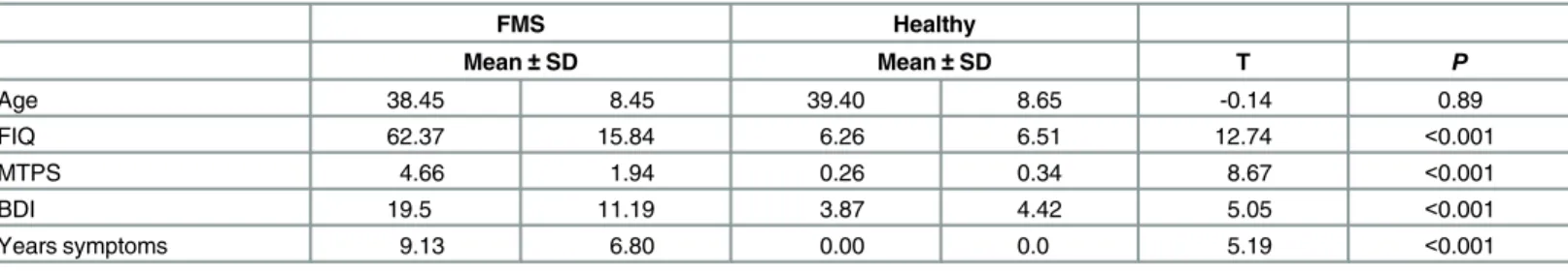
non-permitted medications, such as co-codamol, for at least 3 days prior to recordings. Informed written consent was obtained from all participants in accordance with the Declara-tion of Helsinki and the study was approved by the Research Ethics Committee of the Univer-sity of Liverpool and the Research Governance Committee of two NHS Foundation Trust hospitals; the Walton Centre, Liverpool, United Kingdom, and Wirral University Teaching Hospital, Wirral, United Kingdom. Fifteen age-matched female controls (age 39.40 ± 8.65 years, mean ± SD) were recruited through campus advertisement. Volunteers taking regular medication, currently diagnosed with any disease or disorder or demonstrating a history of major disease, alcohol/drug abuse or serious head or brain injury were excluded. Patients and volunteers were compensated for time and travel expenses.Table 1shows demographic and clinical data for healthy and patient groups, and the results of T-tests to compare mean scores on clinical measures for each group.
MRI data acquisition
Magnetic resonance images were acquired using a whole-body 3 tesla Siemens Trio MRI imag-ing system (Siemens, Magnetom, Erlangen, Germany) and an 8-channel head coil. Restimag-ing- Resting-state fMRI data were acquired using a T2-weighted sequence (32 axial slices, 0.7mm spacing, TR = 2.0 s, TE = 30 ms, flip angle = 90°, field of view = 192mm, voxel size = 3× 3 ×3.5 mm). During a 20 minute resting-state fMRI acquisition period (600 scans), participants were asked to remain awake with their eyes closed. Fifteen auditory stimuli (a one second beep tone) were delivered via headphones at pseudorandom intervals (every 60–90 s, mean onset 75 s). Partici-pants were instructed to respond to the auditory stimulus by pressing a button on a button box placed in their right hand. Stimulus-response epochs were later defined as blocks and excluded from the analysis to leave interleaved resting-state data.
Pre-processing and data analysis
Spatial pre-processing was performed in SPM8 (Welcome Trust Centre for Neuroimaging, University College London, United Kingdom) running in Matlab v.7.13 (The Mathworks Inc, USA). Functional volumes underwent realignment, slice-timing correction, normalisation to Montreal Neuroloigical Institute (MNI) space using the normalised EPI template image in SPM and spatial smoothing (8mm full width half maximum Gaussian kernel filter). Motion parameters from realignment were evaluated and a motion artefact threshold
(translation>3mm, rotation>1 degree) was employed for exclusion. No participants dis-played gross movements to require exclusion. Noise correction was performed using the ana-tomical component-based noise correction (aCompCor) method [28] implemented in the Functional Connectivity Toolbox (CONN, [11]) in SPM8. During pre-processing, high-resolu-tion T1-weighted anatomical volumes for each subject were segmented into grey matter, white
Table 1. FMS patient and healthy control clinical and demographic data FIQ = Fibromyalgia Impact Questionnaire; MTPS = Manual Tender Point Scale; BDI = Beck Depression Inventory; SD = Standard deviation.
FMS Healthy Mean±SD Mean±SD T P Age 38.45 8.45 39.40 8.65 -0.14 0.89 FIQ 62.37 15.84 6.26 6.51 12.74 <0.001 MTPS 4.66 1.94 0.26 0.34 8.67 <0.001 BDI 19.5 11.19 3.87 4.42 5.05 <0.001 Years symptoms 9.13 6.80 0.00 0.0 5.19 <0.001 doi:10.1371/journal.pone.0159198.t001
matter and cerebrospinal fluid and normalised to MNI space. BOLD signals from cerebral white matter and ventricles were removed using principal component analysis (PCA) of the multivariate BOLD signal within each these masks [11]. The residual BOLD data was previ-ously shown to benefit from improved specificity, sensitivity and validity for subsequent func-tional connectivity analyses [11,29]. Sound conditions, representing the 15 second period beginning 1 second before onset of a sound stimuli/response epoch and 14 seconds post sti-muli, were defined as blocks and removed from the data so as to only investigate the remaining interleaved resting-state data. This method was previously shown to provide appropriate data for resting-state network analysis [30]. Finally, BOLD data was bandpass filtered (0.008–0.09 Hz) to reduce low-frequency drift and noise effects.
Seed regions of interest
ROI seeds, consisting of 10mm diameter spheres, were defined in posterior cingulate cortex (PCC), precuneus (pC), medial prefrontal gyrus (MPFG), bilateral Inferior parietal lobules (L/ R.IPL), right middle temporal gyrus (R.MTG) and ventral anterior cingulate cortex (vACC). These ROIs were centred on co-ordinates from a meta-analysis of DMN fMRI studies [9], and were generated using MarsBaR software (MRC Cognition and Brain Sciences Unit, Cambridge, United Kingdom) in SPM8.Table 2shows the anatomical location, Brodmann area and MNI co-ordinates for each of the seed regions utilised. The BOLD time series extracted for each seed region was the average for all voxels making up the 10mm diameter sphere.
Seed-to-voxel analysis
Individual correlation maps throughout the whole brain were generated in the CONN
toolbox by extracting the mean resting-state BOLD time course from each seed ROI and calcu-lating correlation coefficients with the BOLD timecourse of each voxel throughout the whole brain. The resulting coefficients were converted to normally distributed scores using Fisher's transformation to give maps of voxelwise functional connectivity for each seed ROI in the DMN for each subject. The value of each voxel throughout the whole brain represents the rela-tive degree of functional connectivity with each seed [31]. These maps were subsequently used for second-level analysis of relative functional connectivity using a two-sided independentt -test, implemented in the CONN toolbox, to investigate differences in seed-to-voxel connectiv-ity between groups. As in previous studies [32,33], voxelwise statistics throughout the whole brain were performed at an uncorrected level (P<0.001) before false discovery rate (FDR) cor-rection [34] was applied at the cluster level (P<0.05).
Table 2. Seed regions of interest selected for the default mode network; the seed location, hemisphere, Brodmann area and MNI (x,y,z) co-ordi-nates are shown network (adapted from [9]).
Hemisphere Region BA x y z
Left Precuneus (pC) 7 −4 −58 44
Left Posterior cingulate (PCC) 31 −4 −52 22
Right Ventral anterior cingulate (vACC) 32 2 32 −8
Right Inferior parietal lobule (R.IPL) 40 52 −28 24
Left Medial prefrontal cortex (mPFC) 9 −2 50 18
Right Middle temporal gyrus (R.MTG) 39 46 −66 16
Left Middle frontal gyrus (L.MFG) 8 −26 16 44
Left Inferior parietal lobule (L.IPL) 40 −56 −36 28
Left Middle temporal gyrus (L.MTG) 39 −42 −66 18
ROI-ROI Analyses
To examine whether functional connectivity between DMN structures differs in FMS, the aver-age BOLD time series for all voxels in each seed ROI were normalised using Fisher’s transfor-mation and correlations performed with all other seeds in the DMN network in a 9×9 correlation matrix. The resulting correlation coefficients for each participant were then com-pared using a two-sided independent samplest-test to evaluate between-group differences in ROI-ROI connectivity for each seed. Results were thresholded at P<0.05 and FDR correction was applied to correct for multiple tests required.
Correlation analysis
Fischer transformed correlation coefficients for clusters of voxels showing a significant group difference in connectivity with DMN structures in seed-to-voxel analyses were extracted for each subject. One-tailed Pearson’s correlation analysis was performed to investigate the rela-tionship between relative functional connectivity with seed ROIs, and clinical measures includ-ing duration of symptoms (years), tenderness scores measured on the day of scanninclud-ing (MTPS), and Depression scores (BDI). As FMS patients demonstrated significantly higher scores on all clinical variables, with negligible scores evident in the healthy group, correlation analysis with clinical data was only performed within the patient population and a threshold (P<0.05) was employed to determine significance.
Results
Seed-to-voxel analysis
FMS patients, relative to healthy control participants, demonstrated significant connectivity differences between DMN seeds located in PCC, L.MFG and R.IPL and a variety of cortical regions.Table 3shows the T-maxima of the clusters demonstrating altered connectivity with DMN structures, as well as MNI co-ordinates, P values (FDR corrected) and the size of each cluster in contiguous voxels.
FMS patients exhibited reduced functional connectivity, relative to healthy controls, between the PCC seed in the DMN and the right parahippocampal gyrus (t(29) = -7.8, P = 0.011), and right inferior temporal gyrus (t(29) =−5.6, P = 0.011). FMS patients group also demonstrated enhanced connectivity, compared to healthy participants, between the right IPL seed and right hippocampal formation, (t(29) = 6.91, P = 0.034), the left MFG and left posterior parietal cortex (t(29) = 4.92, P = 0.024) and between the PCC seed and left anterior midcingulate cortex (aMCC,t(29) = 6.18, P = 0.034).Fig 1shows the locations of DMN seeds (Fig 1A), as well as the locations of clusters of voxels demonstrating altered functional connectivity to DMN seeds in
Table 3. Seed-to-voxel analysis, brain regions showing alterations to functional connectivity with DMN seeds in FMS patients, relative to healthy control subjects.Cluster location, MNI co-ordinates (x,y,z) and T maxima (cluster-level FDR corrected) are shown,k= number of contiguous voxels. PCC = posterior cingulate cortex; L.MFG = left middle frontal gyrus; R.IPL = right inferior parietal lobule; ITG = inferior temporal gyrus;
PHG = parahippocampal gyrus; aMCC = anterior midcingulate cortex; Hi = hippocampal formation; SPL = superior parietal lobule.
Seed Contrast Cluster MNI [mm] k T P
PCC HC>FMS ITG 60–30–12 283 -7.8 0.011 HC>FMS PHG 14, -12–24 264 -5.6 0.011 FMS>HC aMCC -16, 18, 24 157 6.18 0.034 R.IPL FMS>HC Hi 26–14–14 335 6.91 0.034 L.MFG FMS>HC SPL -32–62 50 294 4.92 0.024 doi:10.1371/journal.pone.0159198.t003
the FMS patient group relative to healthy control group, and histograms illustrating relative functional connectivity between seed regions and significant clusters (Fig 1B–1D).
ROI-to-ROI analysis
ROI-ROI functional connectivity was compared between FMS patients and healthy control group using two-sided independentt-test analysis implemented in the CONN toolbox. There were no group differences in functional connectivity evident between any of the allocated DMN seed ROIs (P>0.05).
Fig 1. Seed–to-voxel analysis. A.The locations of all 9 DMN seeds as specified by [9]. The red sphere indicates the location of the PCC seed, green is the left MFG and blue for right IPL seed.B.Functional connectivity with the PCC seed. Red-yellow colour indicates a cluster showing increased connectivity with PCC in FMS patients relative to healthy participants; blue-white colour indicates clusters demonstrating reduced connectivity.Top right panel;mean Fischer transformed correlation coefficients indicating relative functional connectivity with PCC seed for each significant cluster in FMS and healthy groups.C.Functional connectivity with the left MFG seed.Middle right panel;mean Fischer transformed correlation coefficients indicating relative connectivity between left MFG seed and significant clusters in FMS and healthy control groups.D.Functional connectivity with the right IPL seed.Bottom right panel;mean Fischer transformed correlation coefficients indicating relative connectivity between right IPL seed and significant clusters in FMS and healthy control groups. doi:10.1371/journal.pone.0159198.g001
Correlation analysis
Pearson’s correlation analysis was performed between functional connectivity coefficients and clinical measures in the FMS patient group. The correlation coefficients for functional connec-tivity between PCC and aMCC, which demonstrated augmented connecconnec-tivity in FMS, posi-tively correlated with clinical measures including MTPS (r(14) = 0.43, P = 0.049) and BDI scores (r(14) = 0.52, P = 0.019), suggesting that the increased connectivity between these regions in FMS patients may relate to pain and affective symptoms.Fig 2A and 2Bshows the scatterplots of correlation coefficients representing relative functional connectivity between PCC−aMCC and MTPS/BDI scores respectively. The connectivity between the cluster located in the right parahippocampal gyrus and the PCC seed, which demonstrated reduced connectiv-ity in FMS patients relative to healthy control group, negatively correlated with the duration of symptoms in FMS patients (r =−0.50, p = 0.049). The degree of disruption to functional con-nectivity between PCC and parahippocampal gyrus was associated with longer symptom dura-tion but not with other clinical measures relating to pain or affective disturbance.Fig 2Cshows the scatterplot of correlation coefficients representing relative functional connectivity between PCC and parahippocampal gyrus and duration of symptoms in the FMS patient group.
Discussion
FMS patients demonstrated increased functional connectivity between the DMN seed in the PCC and the aMCC which corresponds closely with the primary loci of anatomical brain alter-ations reported in a recent meta-analysis of studies of FMS patients [35]. Furthermore, the degree of augmented connectivity seen in patients positively correlated with widespread ten-derness indexed by MTPS scores, and depression scores as measured by the BDI questionnaire, which suggests that increased connectivity between these regions could have relevance for the development and/or maintenance of predominant FMS symptoms. According to a recent meta-analyses, the aMCC is amongst the most commonly seen pain processing regions in both clinical and experimental pain neuroimaging studies [36–38]. This region has also been shown to play an important role in processing of tonic ongoing pain [39], and recent evidence points towards the aMCC sub-serving the relationship between pain and action selection [40–42], or approach/avoidance functions relevant for pain and decision-making (reviewed in [38]). Therefore, augmented connectivity between PCC and aMCC in FMS patients during rest may reflect the additional burden of decision-making and processing of appropriate action
responses in the presence of chronic pain. The PCC in the DMN also showed reduced connec-tivity with right inferior temporal gyrus in FMS patients. This finding corresponds with reduced connectivity seen between these regions identified in patients with diabetic neuropathy which was proposed to indicate a reorganisation of resting brain networks contributing to spontaneous pain [43].
Reduced functional connectivity between the DMN seed in PCC and the right parahippo-campal gyrus was also evident in the FMS patient group. The degree of disruption to connectiv-ity correlated with the duration of symptoms in FMS patients but not tenderness scores or affective disturbance. This correlation is potentially indicative of a time-dependent alteration, although this cannot be specifically attributed to FMS symptoms. The right IPL structure in the DMN demonstrated increased connectivity with the right hippocampal formation in FMS patients relative to the healthy control group. Morphological changes in the hippocampal for-mation have previously been identified in FMS [44,45], and an animal model previously linked hippocampal neurogenesis to various chronic pain syndromes [46].
Augmented connectivity with the hippocampal formation was recently identified in long-term, but not early-stage, chronic back pain patients, and this reorganisation could represent
Fig 2. Correlation analysis. A.Scatter plot showing MTPS scores and Fischer transformed connectivity correlation coefficients representing relative functional connectivity between the aMCC and PCC and in FMS patient group. The linear regression line is also shown.B.Scatter plot showing BDI scores and relative functional connectivity between the aMCC and PCC in FMS patient group.C.Scatter plot showing duration of symptoms (in years) and relative functional connectivity between PCC and right parahippocampal gyrus in FMS patient group.
the transition from acute to chronic pain [4]. A comparison of early and late stage chronic back pain patients revealed that long-term chronic pain caused a shift towards augmented acti-vation patterns in brain regions related to cognitive-affective processing (including hippocam-pal formation), whereas early-stage pain was linked to activations in accepted pain processing regions [47]. Apkarian et al. [48] recently proposed that the development and maintenance of chronic pain could be contingent on the interaction between hippocampal learning mecha-nisms and nociceptive signals. As such the augmented hippocampal connectivity seen in the present study could be of relevance for the development of chronic symptoms in FMS. The hip-pocampal formation was also previously included as an additional structure of the DMN [49,50], and functional connectivity analysis of this seed region demonstrates strong correla-tions with DMN activity [51]. This overlap points to a link between DMN and networks under-lying cognitive function and memory [49,52] which are often impaired in FMS [53–55].
FMS patients also demonstrated increased functional connectivity between the left IPL structure in the DMN and the left superior parietal lobule. As this region is adjacent to the pari-etal activation patterns seen in the DMN, the augmented connectivity is likely to reflect an expansion of the DMN to incorporate this area in FMS patients, perhaps as a result of ongoing pain. Hyperperfusion was previously seen in the left superior parietal lobule in FMS patients at rest [56] and similar augmented DMN connectivity with this region predicted clinical pain fol-lowing movements in chronic back pain patients [19].
Due to the heterogeneity of FMS populations, it was proposed that predominant symptom profiles within a patient group can influence findings [57], which may relate to the pattern of connectivity differences in the present study, and indeed the diversity of findings in the litera-ture. Our patient population were predominantly taking minimal medications suitable for a short withdrawal or not taking medication at all [27], which could affect the severity or symp-tom profile of patients taking part. Therefore, patient heterogeneity can be considered as a limi-tation of this method for investigation of FMS, although this would also suggest that functional connectivity could be sensitive enough to evaluate specific symptom profiles which would be advantageous. Future studies should consider sub-grouping patients based on symptom pro-files, or longitudinal studies could relate the prevalence or dynamics of particular symptoms to specific functional connectivity alterations. As with all cross-sectional research, it is not possi-ble to infer causality for any of the alterations seen in the present study, and future longitudinal research is needed to consider the direction and progression of the relationship between rest-ing-state alterations and symptoms in FMS. Furthermore, although we utilised MTPS exami-nations to consider tenderness on the day of testing, a specific measure of clinical pain levels, e.g., McGill Pain Questionnaire [58] would also represent a beneficial addition in future.
Our findings suggest that FMS alters DMN connectivity with brain regions such as aMCC, hippocampal formation and inferior temporal gyrus which have implications for pain and developing chronicity, action selection, decision making, and affective/cognitive disturbances in FMS. The locations of structures exhibiting altered functional connectivity were either anatomically or functionally linked to the DMN suggesting a re-organisation of resting-state networks in FMS patients. These alterations could reflect predisposing central factors for the development of FMS, or alternatively, the consequences of ongoing symptoms on resting brain activity. Furthermore, connectivity increases between PCC and aMCC were correlated with clinical measures in patients, whereas reduced connectivity with parahippocampal gyrus was associated with longer symptom duration in FMS patients which may reflect ongoing time-dependent reorganisation or an indication of developing chronicity. Our results indicate that FMS symptoms are likely to influence functional connectivity with the DMN, and the specific nature of connectivity alterations could be of importance for understanding specific mecha-nisms underlying FMS. However, the pattern of alterations may vary as a result of the
heterogeneous symptom profile of the patient cohort or fluctuations in levels of ongoing pain and other symptoms.
Author Contributions
Conceived and designed the experiments: AS TN NF. Performed the experiments: NF. Ana-lyzed the data: NF. Contributed reagents/materials/analysis tools: AS NF. Wrote the paper: NF YC TN AS. Provided appropriate fibromyalgia patients and clinical supervision: TN YC.
References
1. Clauw DJ (2014) Fibromyalgia: a clinical review. JAMA 311: 1547–1555. doi:10.1001/jama.2014.3266 PMID:24737367
2. Baliki MN, Geha PY, Apkarian AV, Chialvo DR (2008) Beyond feeling: chronic pain hurts the brain, dis-rupting the default-mode network dynamics. J Neurosci 28: 1398–1403. 28/6/1398 [pii] doi:10.1523/ JNEUROSCI.4123-07.2008PMID:18256259
3. Napadow V, Harris RE (2014) What has functional connectivity and chemical neuroimaging in fibromy-algia taught us about the mechanisms and management of 'centralized' pain? Arthritis Res Ther 16: 425. PMID:25606591
4. Mutso AA, Petre B, Huang LJ, Baliki MN, Torbey S, Herrmann KM, et al. (2014) Reorganization of hip-pocampal functional connectivity with transition to chronic back pain. J Neurophysiol 111: 1065–1076. doi:10.1152/jn.00611.2013PMID:24335219
5. Raichle ME, MacLeod AM, Snyder AZ, Powers WJ, Gusnard DA, Shulman GL (2001) A default mode of brain function. Proc Natl Acad Sci U S A 98: 676–682. doi:10.1073/pnas.98.2.676PMID:11209064
6. Raichle ME, Snyder AZ (2007) A default mode of brain function: a brief history of an evolving idea. Neu-roimage 37: 1083–1090. doi:10.1016/j.neuroimage.2007.02.041PMID:17719799
7. Greicius MD, Krasnow B, Reiss AL, Menon V (2003) Functional connectivity in the resting brain: a net-work analysis of the default mode hypothesis. Proc Natl Acad Sci USA 100: 253–258. doi:10.1073/ pnas.0135058100PMID:12506194
8. Fox MD, Snyder AZ, Vincent JL, Corbetta M, Van Essen DC, Raichle ME (2005) The human brain is intrinsically organized into dynamic, anticorrelated functional networks. Proc Natl Acad Sci USA 102: 9673–9678. doi:10.1073/pnas.0504136102PMID:15976020
9. Laird AR, Eickhoff SB, Li K, Robin DA, Glahn DC, Fox PT (2009) Investigating the functional heteroge-neity of the default mode network using coordinate-based meta-analytic modeling. J Neurosci 29: 14496–14505. doi:10.1523/JNEUROSCI.4004-09.2009PMID:19923283
10. Whitfield-Gabrieli S, Ford JM (2012) Default mode network activity and connectivity in psychopathol-ogy. Annu Rev Clin Psychol 8: 49–76. doi:10.1146/annurev-clinpsy-032511-143049PMID:22224834
11. Whitfield-Gabrieli S, Nieto-Castanon A (2012) Conn: a functional connectivity toolbox for correlated and anticorrelated brain networks. Brain Connect 2: 125–141. doi:10.1089/brain.2012.0073PMID: 22642651
12. Biswal B, Yetkin FZ, Haughton VM, Hyde JS (1995) Functional connectivity in the motor cortex of rest-ing human brain usrest-ing echo-planar MRI. Magn Reson Med 34: 537–541. PMID:8524021
13. Beckmann CF, DeLuca M, Devlin JT, Smith SM (2005) Investigations into resting-state connectivity using independent component analysis. Philos Trans R Soc Lond B Biol Sci 360: 1001–1013. doi:10. 1098/rstb.2005.1634PMID:16087444
14. Napadow V, LaCount L, Park K, As-Sanie S, Clauw DJ, Harris RE (2010) Intrinsic brain connectivity in fibromyalgia is associated with chronic pain intensity. Arthritis Rheum 62: 2545–2555. doi:10.1002/art. 27497PMID:20506181
15. Napadow V, Kim J, Clauw DJ, Harris RE (2012) Decreased intrinsic brain connectivity is associated with reduced clinical pain in fibromyalgia. Arthritis Rheum 64: 2398–2403. doi:10.1002/art.34412 PMID:22294427
16. Pujol J, Macia D, Garcia-Fontanals A, Blanco-Hinojo L, Lopez-Sola M, Garcia-Blanco S, et al. (2014) The contribution of sensory system functional connectivity reduction to clinical pain in fibromyalgia. Pain 155: 1492–1503. doi:10.1016/j.pain.2014.04.028PMID:24792477
17. Cifre I, Sitges C, Fraiman D, Munoz MA, Balenzuela P, Gonzalez-Roldan A, et al. (2012) Disrupted functional connectivity of the pain network in fibromyalgia. Psychosom Med 74: 55–62. doi:10.1097/ PSY.0b013e3182408f04PMID:22210242
18. Kucyi A, Moayedi M, Weissman-Fogel I, Goldberg MB, Freeman BV, Tenenbaum HC, et al. (2014) Enhanced medial prefrontal-default mode network functional connectivity in chronic pain and its associ-ation with pain ruminassoci-ation. J Neurosci 34: 3969–3975. doi:10.1523/JNEUROSCI.5055-13.2014PMID: 24623774
19. Loggia ML, Kim J, Gollub RL, Vangel MG, Kirsch I, Kong J, et al. (2013) Default mode network connec-tivity encodes clinical pain: An arterial spin labeling study. Pain 154: 24–33. doi:10.1016/j.pain.2012. 07.029PMID:23111164
20. Ichesco E, Schmidt-Wilcke T, Bhavsar R, Clauw DJ, Peltier SJ, Kim J, et al. (2014) Altered resting state connectivity of the insular cortex in individuals with fibromyalgia. J Pain 15: 815–826 e811. doi:10. 1016/j.jpain.2014.04.007PMID:24815079
21. Flodin P, Martinsen S, Mannerkorpi K, Lofgren M, Bileviciute-Ljungar I, Kosek E, et al. (2015) Normali-zation of aberrant resting state functional connectivity in fibromyalgia patients following a three month physical exercise therapy. Neuroimage Clin 9: 134–139. doi:10.1016/j.nicl.2015.08.004PMID: 26413476
22. Garza-Villarreal EA, Jiang Z, Vuust P, Alcauter S, Vase L, Pasaye EH, et al. (2015) Music reduces pain and increases resting state fMRI BOLD signal amplitude in the left angular gyrus in fibromyalgia patients. Front Psychol 6: 1051. doi:10.3389/fpsyg.2015.01051PMID:26257695
23. Schmidt-Wilcke T, Ichesco E, Hampson JP, Kairys A, Peltier S, Harte S, et al. (2014) Resting state con-nectivity correlates with drug and placebo response in fibromyalgia patients. Neuroimage Clin 6: 252– 261. doi:10.1016/j.nicl.2014.09.007PMID:25379438
24. Cagnie B, Coppieters I, Denecker S, Six J, Danneels L, Meeus M (2014) Central sensitization in fibro-myalgia? A systematic review on structural and functional brain MRI. Semin Arthritis Rheum 44: 68– 75. doi:10.1016/j.semarthrit.2014.01.001PMID:24508406
25. Wolfe F, Smythe HA, Yunus MB, Bennett RM, Bombardier C, Goldenberg DL, et al. (1990) The Ameri-can College of Rheumatology 1990 Criteria for the Classification of Fibromyalgia. Report of the Multi-center Criteria Committee. Arthritis Rheum 33: 160–172. PMID:2306288
26. Beck AT, Ward CH, Mendelson M, Mock J, Erbaugh J (1961) An inventory for measuring depression. Arch Gen Psych 4: 561–571.
27. Fallon N, Alghamdi J, Chiu Y, Sluming V, Nurmikko T, Stancak A (2013) Structural alterations in brain-stem of fibromyalgia syndrome patients correlate with sensitivity to mechanical pressure. Neuroimage Clin 3: 163–170. doi:10.1016/j.nicl.2013.07.011PMID:24179860
28. Behzadi Y, Restom K, Liau J, Liu TT (2007) A component based noise correction method (CompCor) for BOLD and perfusion based fMRI. Neuroimage 37: 90–101. doi:10.1016/j.neuroimage.2007.04.042 PMID:17560126
29. Shehzad Z, Kelly AM, Reiss PT, Gee DG, Gotimer K, Uddin LQ, et al. (2009) The resting brain: uncon-strained yet reliable. Cereb Cortex 19: 2209–2229. doi:10.1093/cercor/bhn256PMID:19221144
30. Fair DA, Schlaggar BL, Cohen AL, Miezin FM, Dosenbach NU, Wenger KK, et al. (2007) A method for using blocked and event-related fMRI data to study "resting state" functional connectivity. Neuroimage 35: 396–405. doi:10.1016/j.neuroimage.2006.11.051PMID:17239622
31. Whitfield-Gabrieli S, Moran JM, Nieto-Castanon A, Triantafyllou C, Saxe R, Gabrieli JD (2011) Associa-tions and dissociaAssocia-tions between default and self-reference networks in the human brain. Neuroimage 55: 225–232. doi:10.1016/j.neuroimage.2010.11.048PMID:21111832
32. Woodward ND, Rogers B, Heckers S (2011) Functional resting-state networks are differentially affected in schizophrenia. Schizophr Res 130: 86–93. doi:10.1016/j.schres.2011.03.010PMID:21458238
33. Ichesco E, Quintero A, Clauw DJ, Peltier S, Sundgren PM, Gerstner GE, et al. (2012) Altered functional connectivity between the insula and the cingulate cortex in patients with temporomandibular disorder: a pilot study. Headache 52: 441–454. doi:10.1111/j.1526-4610.2011.01998.xPMID:21929661
34. Benjamini Y, Hochberg Y (1995) Controlling the False Discovery Rate—a Practical and Powerful Approach to Multiple Testing. J Roy Stat Soc B Met 57: 289–300.
35. Dehghan M, Schmidt-Wilcke T, Pfleiderer B, Eickhoff SB, Petzke F, Harris RE, et al. (2016) Coordi-nate-based (ALE) meta-analysis of brain activation in patients with fibromyalgia. Hum Brain Mapp. doi: 10.1002/hbm.23132
36. Duerden EG, Albanese MC (2013) Localization of pain-related brain activation: a meta-analysis of neu-roimaging data. Hum Brain Mapp 34: 109–149. doi:10.1002/hbm.21416PMID:22131304
37. Shackman AJ, Salomons TV, Slagter HA, Fox AS, Winter JJ, Davidson RJ (2011) The integration of negative affect, pain and cognitive control in the cingulate cortex. Nat Rev Neurosci 12: 154–167. doi: 10.1038/nrn2994PMID:21331082
38. Vogt BA (2016) Midcingulate cortex: Structure, connections, homologies, functions and diseases. J Chem Neuroanat 74: 28–46. doi:10.1016/j.jchemneu.2016.01.010PMID:26993424
39. Owen DG, Clarke CF, Bureau Y, Ganapathy S, Prato FS, St Lawrence KS (2012) Measuring the neural response to continuous intramuscular infusion of hypertonic saline by perfusion MRI. J Magn Reson Imaging 35: 669–677. doi:10.1002/jmri.22814PMID:21953816
40. Perini I, Bergstrand S, Morrison I (2013) Where pain meets action in the human brain. J Neurosci 33: 15930–15939. doi:10.1523/JNEUROSCI.3135-12.2013PMID:24089498
41. Misra G, Coombes SA (2015) Neuroimaging Evidence of Motor Control and Pain Processing in the Human Midcingulate Cortex. Cereb Cortex 25: 1906–1919. doi:10.1093/cercor/bhu001PMID: 24464941
42. Schmidt-Wilcke T, Kairys A, Ichesco E, Fernandez-Sanchez ML, Barjola P, Heitzeg M, et al. (2014) Changes in clinical pain in fibromyalgia patients correlate with changes in brain activation in the cingu-late cortex in a response inhibition task. Pain Med 15: 1346–1358. doi:10.1111/pme.12460PMID: 24995850
43. Cauda F, Sacco K, Duca S, Cocito D, D'Agata F, Geminiani GC, et al. (2009) Altered resting state in diabetic neuropathic pain. PLoS One 4: e4542. doi:10.1371/journal.pone.0004542PMID:19229326
44. Schmidt-Wilcke T, Luerding R, Weigand T, Jurgens T, Schuierer G, Leinisch E, et al. (2007) Striatal grey matter increase in patients suffering from fibromyalgia—a voxel-based morphometry study. Pain 132 Suppl 1: S109–116. S0304-3959(07)00267-9 [pii] doi:10.1016/j.pain.2007.05.010PMID: 17587497
45. Lutz J, Jager L, de Quervain D, Krauseneck T, Padberg F, Wichnalek M, et al. (2008) White and gray matter abnormalities in the brain of patients with fibromyalgia: a diffusion-tensor and volumetric imaging study. Arthritis Rheum 58: 3960–3969. doi:10.1002/art.24070PMID:19035484
46. Mutso AA, Radzicki D, Baliki MN, Huang LJ, Banisadr G, Centeno MV, et al. (2012) Abnormalities in Hippocampal Functioning with Persistent Pain. Journal of Neuroscience 32: 5747–5756. doi:10.1523/ Jneurosci.0587-12.2012PMID:22539837
47. Hashmi JA, Baliki MN, Huang L, Baria AT, Torbey S, Hermann KM, et al. (2013) Shape shifting pain: chronification of back pain shifts brain representation from nociceptive to emotional circuits. Brain 136: 2751–2768. doi:10.1093/brain/awt211PMID:23983029
48. Apkarian AV, Mutso AA, Centeno MV, Kan L, Wu M, Levinstein M, et al. (2015) Role of adult hippocam-pal neurogenesis in persistent pain. Pain. doi:10.1097/j.pain.0000000000000332
49. Buckner RL, Andrews-Hanna JR, Schacter DL (2008) The brain's default network: anatomy, function, and relevance to disease. Ann N Y Acad Sci 1124: 1–38. doi:10.1196/annals.1440.011PMID: 18400922
50. Greicius MD, Srivastava G, Reiss AL, Menon V (2004) Default-mode network activity distinguishes Alz-heimer's disease from healthy aging: evidence from functional MRI. Proc Natl Acad Sci USA 101: 4637–4642. doi:10.1073/pnas.0308627101PMID:15070770
51. Vincent JL, Snyder AZ, Fox MD, Shannon BJ, Andrews JR, Raichle ME, et al. (2006) Coherent sponta-neous activity identifies a hippocampal-parietal memory network. J Neurophysiol 96: 3517–3531. doi: 10.1152/jn.00048.2006PMID:16899645
52. Spreng RN, Mar RA, Kim AS (2009) The common neural basis of autobiographical memory, prospec-tion, navigaprospec-tion, theory of mind, and the default mode: a quantitative meta-analysis. J Cogn Neurosci 21: 489–510. doi:10.1162/jocn.2008.21029PMID:18510452
53. Park DC, Glass JM, Minear M, Crofford LJ (2001) Cognitive function in fibromyalgia patients. Arthritis Rheum 44: 2125–2133. doi:10.1002/1529-0131(200109)44:9<2125::AID-ART365>3.0.CO;2-1PMID: 11592377
54. Leavitt F, Katz RS (2006) Distraction as a key determinant of impaired memory in patients with fibromy-algia. J Rheumatol 33: 127–132. PMID:16395760
55. Dick BD, Verrier MJ, Harker KT, Rashiq S (2008) Disruption of cognitive function in fibromyalgia syn-drome. Pain 139: 610–616. doi:10.1016/j.pain.2008.06.017PMID:18691816
56. Usui C, Hatta K, Doi N, Nakanishi A, Nakamura H, Nishioka K, et al. (2010) Brain perfusion in fibromyal-gia patients and its differences between responders and poor responders to gabapentin. Arthritis Res Ther 12: R64. doi:10.1186/ar2980PMID:20374641
57. May A (2011) Structural brain imaging: a window into chronic pain. Neuroscientist 17: 209–220. doi: 10.1177/1073858410396220PMID:21489967
![Table 2. Seed regions of interest selected for the default mode network; the seed location, hemisphere, Brodmann area and MNI (x,y,z) co-ordi- co-ordi-nates are shown network (adapted from [9]).](https://thumb-us.123doks.com/thumbv2/123dok_us/9745956.2856401/4.918.54.865.848.1039/regions-selected-default-network-location-hemisphere-brodmann-network.webp)

![Fig 1. Seed –to-voxel analysis. A. The locations of all 9 DMN seeds as specified by [ 9]](https://thumb-us.123doks.com/thumbv2/123dok_us/9745956.2856401/6.918.217.865.122.701/fig-seed-voxel-analysis-locations-dmn-seeds-specified.webp)

