3-
O
-Ethyl-
L-ascorbic acid
Shu Jin* and Xiaoqin Miao
Nanjing Research Institute for Comprehensive Utilization of Wild Plants, Jiang-wangmiaojie 4#, Nanjing 210042, People’s Republic of China
Correspondence e-mail: [email protected]
Received 27 March 2008; accepted 11 April 2008
Key indicators: single-crystal X-ray study;T= 293 K; mean(C–C) = 0.005 A˚; Rfactor = 0.040;wRfactor = 0.138; data-to-parameter ratio = 7.5.
In the crystal structure of the title compound, C8H12O6,
molecules are linked to each other by O—H O hydrogen bonding.
Related literature
For general background, see: Nihroet al.(1992); Satohet al.
(1994).
Experimental Crystal data
C8H12O6 Mr= 204.18
Orthorhombic,P212121
a= 4.6690 (9) A˚
b= 11.939 (2) A˚
c= 16.794 (3) A˚
V= 936.2 (3) A˚3
Z= 4
MoKradiation
= 0.13 mm1 T= 293 (2) K 0.200.200.10 mm
Data collection
Enraf–Nonius CAD-4 diffractometer
Absorption correction: none 1973 measured reflections 1024 independent reflections
882 reflections withI> 2(I)
Rint= 0.028
3 standard reflections every 200 reflections intensity decay: none
Refinement
R[F2> 2(F2)] = 0.040 wR(F2) = 0.138
S= 1.00 1024 reflections 137 parameters 1 restraint
H atoms treated by a mixture of independent and constrained refinement
max= 0.18 e A˚
3
min=0.26 e A˚
3
Table 1
Hydrogen-bond geometry (A˚ ,).
D—H A D—H H A D A D—H A
O2—H2A O5i
0.83 (3) 2.06 (3) 2.873 (3) 168 (5) O5—H5A O3ii
0.91 (5) 1.90 (4) 2.748 (4) 154 (4) O6—H6A O6iii
0.87 (5) 1.87 (5) 2.715 (4) 163 (4)
Symmetry codes: (i) x;y1 2;zþ
3
2; (ii) x;yþ 1 2;zþ
3 2; (iii)
x1 2;yþ
3 2;zþ1.
Data collection: CAD-4 Software (Enraf–Nonius, 1989); cell
refinement: CAD-4 Software; data reduction: XCAD4 (Harms &
Wocadlo, 1995); program(s) used to solve structure: SHELXTL
(Sheldrick, 2008); program(s) used to refine structure:SHELXTL;
molecular graphics:SHELXTL; software used to prepare material
for publication:SHELXTL.
Supplementary data and figures for this paper are available from the IUCr electronic archives (Reference: XU2411).
References
Enraf–Nonius (1989).CAD-4 Software. Enraf–Nonius, Delft, The Nether-lands.
Harms, K. & Wocadlo, S. (1995).XCAD4. University of Marburg, Germany. Nihro, Y., Sogawa, S. & Izumi, A. (1992).J. Med. Chem.35, 1618–1623. Satoh, T., Niino, Y. & Matsumoto, H. (1994). Jpn Patent JP6228557. Sheldrick, G. M. (2008).Acta Cryst.A64, 112–122.
Acta Crystallographica Section E Structure Reports
Online
supporting information
Acta Cryst. (2008). E64, o860 [doi:10.1107/S1600536808009963]
3-
O
-Ethyl-
L-ascorbic acid
Shu Jin and Xiaoqin Miao
S1. Comment
L-Ascorbic acid has been widely employed as an antioxidant for stabilization of nutrients. However, the low lipophilicity
of it and its susceptibility to thermal and oxidative degradation restricts its field of application and has raised considerable
interest in the study of ascorbic acid derivatives with increased lipophilicity and stability. The title compound is one of
the lipophilic ascorbic acid derivatives, which exhibit antioxidative properties (Nihro et al.,1992) and can be used as
antioxidant in food (Satoh et al., 1994). As part of our ongoing study on ascorbic acid derivatives, we report here the
crystal structure of the title compound (Fig. 1).
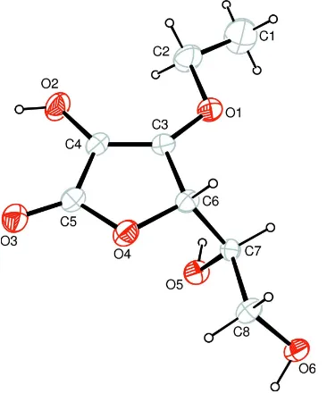
The geometrical parameters of the compound are normal. The C3—C4 bond distance of 1.332 (5) Å and O3—C5 bond
distance of 1.215 (4) Å indicate typical C═C and O═C double bonds. Molecules are linked to each other by O—H···O
hydrogen bonding (Table 1).
S2. Experimental
0.1 mol 5,6-O,O-Isopropylidene L-ascorbic acid was dissolved in 100 ml DMSO at room temperature, and 0.12 mol
NaHCO3 was added with stirring. After the addition of 0.1 mol ethyl bromide, the mixture was stirred at 313 K for 6 h.
The solvent was distilled of at 333 K under reduced pressure. The residue was dissolved in 50 ml water and extracted five
times with ethyl acetate (100 ml/time). The collected organic phase was dried over Na2SO4 and the solvent was
evaporated at reduced pressure. 100 ml 0.1 M HCl was added to the residue, refluxed for 15 min and then the solvent was
evaporated at reduced pressure. The residue was dissolved in ethyl acetate; single crystals were obtained by slow
evaporation of the ethyl acetate solution.
S3. Refinement
Hydroxyl H atoms were located in a difference Fourier map and positional parameters were refined, Uiso(H) = 1.5Ueq(O).
Other H atoms were positioned geometrically with C—H = 0.96–0.98 Å and refined using a riding model with Uiso(H) =
Figure 1
The molecular structure of (I), with atom labels and 30% probability displacement ellipsoids for non-H atoms.
3-O-Ethyl-L-ascorbic acid
Crystal data
C8H12O6
Mr = 204.18
Orthorhombic, P212121 Hall symbol: P 2ac 2ab
a = 4.6690 (9) Å
b = 11.939 (2) Å
c = 16.794 (3) Å
V = 936.2 (3) Å3
Z = 4
F(000) = 432
Dx = 1.449 Mg m−3 Melting point: 385 K
Mo Kα radiation, λ = 0.71073 Å Cell parameters from 25 reflections
θ = 10–13°
µ = 0.13 mm−1
Data collection
Enraf–Nonius CAD-4 diffractometer
Radiation source: fine-focus sealed tube Graphite monochromator
ω/2θ scans
1973 measured reflections 1024 independent reflections 882 reflections with I > 2σ(I)
Rint = 0.028
θmax = 25.2°, θmin = 2.1°
h = 0→5
k = 0→14
l = −20→20
3 standard reflections every 200 reflections intensity decay: none
Refinement
Refinement on F2 Least-squares matrix: full
R[F2 > 2σ(F2)] = 0.040
wR(F2) = 0.138
S = 1.00 1024 reflections 137 parameters 1 restraint
Primary atom site location: structure-invariant direct methods
Secondary atom site location: difference Fourier map
Hydrogen site location: inferred from neighbouring sites
H atoms treated by a mixture of independent and constrained refinement
w = 1/[σ2(F
o2) + (0.1P)2 + 1.3P] where P = (Fo2 + 2Fc2)/3 (Δ/σ)max < 0.001
Δρmax = 0.18 e Å−3 Δρmin = −0.26 e Å−3
Extinction correction: SHELXL, Fc*=kFc[1+0.001xFc2λ3/sin(2θ)]-1/4 Extinction coefficient: 0.121 (14)
Special details
Experimental. 1H NMR (500 MHz, CDCl3): δ1.39 (3H, t), 3.86 (2H, m), 3.96 (1H, m), 4.54 (2H, q), 4.71 (1H, d) p.p.m.
Geometry. All e.s.d.'s (except the e.s.d. in the dihedral angle between two l.s. planes) are estimated using the full covariance matrix. The cell e.s.d.'s are taken into account individually in the estimation of e.s.d.'s in distances, angles and torsion angles; correlations between e.s.d.'s in cell parameters are only used when they are defined by crystal symmetry. An approximate (isotropic) treatment of cell e.s.d.'s is used for estimating e.s.d.'s involving l.s. planes.
Refinement. Refinement of F2 against ALL reflections. The weighted R-factor wR and goodness of fit S are based on F2, conventional R-factors R are based on F, with F set to zero for negative F2. The threshold expression of F2 > σ(F2) is used only for calculating R-factors(gt) etc. and is not relevant to the choice of reflections for refinement. R-factors based on F2 are statistically about twice as large as those based on F, and R- factors based on ALL data will be even larger.
Fractional atomic coordinates and isotropic or equivalent isotropic displacement parameters (Å2)
x y z Uiso*/Ueq
O1 0.5474 (6) 0.5579 (2) 0.82156 (13) 0.0513 (7) C1 0.6281 (17) 0.6421 (5) 0.9472 (3) 0.0896 (18)
H1A 0.5996 0.6352 1.0036 0.134*
H1B 0.5371 0.7093 0.9285 0.134*
H1C 0.8295 0.6455 0.9359 0.134*
O2 0.1035 (7) 0.3667 (2) 0.85756 (15) 0.0563 (8)
H2A 0.027 (12) 0.305 (2) 0.851 (3) 0.084*
C2 0.5046 (14) 0.5463 (4) 0.90741 (19) 0.0664 (14)
H2B 0.3016 0.5418 0.9192 0.080*
H2C 0.5953 0.4782 0.9262 0.080*
C5 0.1347 (8) 0.3595 (3) 0.7110 (2) 0.0419 (8) O5 0.0863 (5) 0.64292 (19) 0.67658 (15) 0.0419 (6)
H5A 0.088 (11) 0.671 (3) 0.727 (3) 0.063*
O6 0.3693 (6) 0.7318 (2) 0.53918 (14) 0.0466 (7)
H6A 0.213 (11) 0.729 (4) 0.511 (3) 0.070*
C6 0.4518 (7) 0.4996 (2) 0.68745 (17) 0.0355 (8)
H6B 0.6523 0.4838 0.6744 0.043*
C7 0.3734 (7) 0.6151 (3) 0.65672 (17) 0.0329 (7)
H7A 0.5026 0.6705 0.6804 0.039*
C8 0.4004 (9) 0.6198 (3) 0.56722 (18) 0.0427 (9)
H8A 0.5861 0.5910 0.5514 0.051*
H8B 0.2544 0.5729 0.5432 0.051*
Atomic displacement parameters (Å2)
U11 U22 U33 U12 U13 U23
O1 0.0599 (17) 0.0583 (14) 0.0356 (11) −0.0134 (14) −0.0067 (13) 0.0078 (11) C1 0.122 (5) 0.091 (3) 0.056 (3) −0.020 (4) 0.007 (3) −0.013 (2) O2 0.0731 (19) 0.0566 (14) 0.0391 (12) −0.0154 (17) 0.0083 (14) 0.0088 (12) C2 0.096 (4) 0.068 (2) 0.0350 (16) −0.016 (3) −0.005 (2) 0.0046 (17) C3 0.0394 (17) 0.0395 (15) 0.0365 (15) 0.0018 (17) −0.0037 (15) 0.0049 (13) O3 0.0678 (17) 0.0441 (13) 0.0544 (14) −0.0087 (15) −0.0043 (15) 0.0031 (11) C4 0.0452 (19) 0.0366 (16) 0.0385 (16) 0.0016 (15) 0.0020 (16) 0.0092 (13) O4 0.0605 (16) 0.0383 (12) 0.0364 (11) −0.0028 (12) 0.0032 (12) 0.0020 (9) C5 0.0473 (19) 0.0352 (15) 0.0432 (18) 0.0034 (18) 0.0011 (17) 0.0056 (14) O5 0.0367 (13) 0.0485 (12) 0.0406 (12) 0.0064 (12) 0.0060 (11) 0.0020 (10) O6 0.0455 (14) 0.0504 (13) 0.0440 (13) −0.0009 (13) −0.0044 (12) 0.0163 (11) C6 0.0328 (16) 0.0358 (15) 0.0378 (15) 0.0055 (15) 0.0032 (14) 0.0039 (13) C7 0.0293 (15) 0.0367 (15) 0.0326 (15) −0.0029 (14) 0.0023 (13) 0.0023 (12) C8 0.054 (2) 0.0394 (16) 0.0345 (16) 0.0045 (19) 0.0078 (17) 0.0044 (14)
Geometric parameters (Å, º)
O1—C3 1.332 (4) C4—C5 1.456 (5)
O1—C2 1.462 (4) O4—C5 1.353 (4)
C1—C2 1.444 (7) O4—C6 1.432 (4)
C1—H1A 0.9600 O5—C7 1.421 (4)
C1—H1B 0.9600 O5—H5A 0.91 (4)
C1—H1C 0.9600 O6—C8 1.425 (4)
O2—C4 1.356 (4) O6—H6A 0.87 (5)
O2—H2A 0.83 (3) C6—C7 1.516 (4)
C2—H2B 0.9700 C6—H6B 0.9800
C2—H2C 0.9700 C7—C8 1.510 (4)
C3—C4 1.332 (5) C7—H7A 0.9800
C3—C6 1.500 (4) C8—H8A 0.9700
O3—C5 1.215 (4) C8—H8B 0.9700
C2—C1—H1A 109.5 O4—C5—C4 109.9 (3)
C2—C1—H1B 109.5 C7—O5—H5A 107 (3)
H1A—C1—H1B 109.5 C8—O6—H6A 103 (4)
C2—C1—H1C 109.5 O4—C6—C3 104.2 (2)
H1A—C1—H1C 109.5 O4—C6—C7 111.0 (3)
H1B—C1—H1C 109.5 C3—C6—C7 113.8 (3)
C4—O2—H2A 110 (4) O4—C6—H6B 109.2
C1—C2—O1 109.1 (4) C3—C6—H6B 109.2
C1—C2—H2B 109.9 C7—C6—H6B 109.2
O1—C2—H2B 109.9 O5—C7—C8 107.7 (3)
C1—C2—H2C 109.9 O5—C7—C6 111.1 (3)
O1—C2—H2C 109.9 C8—C7—C6 110.7 (3)
H2B—C2—H2C 108.3 O5—C7—H7A 109.1
O1—C3—C4 134.8 (3) C8—C7—H7A 109.1
O1—C3—C6 115.7 (3) C6—C7—H7A 109.1
C4—C3—C6 109.5 (3) O6—C8—C7 110.8 (3)
C3—C4—O2 129.9 (3) O6—C8—H8A 109.5
C3—C4—C5 107.4 (3) C7—C8—H8A 109.5
O2—C4—C5 122.6 (3) O6—C8—H8B 109.5
C5—O4—C6 109.1 (2) C7—C8—H8B 109.5
O3—C5—O4 121.1 (3) H8A—C8—H8B 108.1
C3—O1—C2—C1 −169.9 (4) C5—O4—C6—C3 1.1 (3)
C2—O1—C3—C4 3.1 (6) C5—O4—C6—C7 −121.7 (3)
C2—O1—C3—C6 179.3 (3) O1—C3—C6—O4 −178.1 (3)
O1—C3—C4—O2 1.1 (6) C4—C3—C6—O4 −0.9 (4)
C6—C3—C4—O2 −175.3 (3) O1—C3—C6—C7 −57.1 (4)
O1—C3—C4—C5 176.8 (4) C4—C3—C6—C7 120.0 (3)
C6—C3—C4—C5 0.4 (4) O4—C6—C7—O5 61.3 (3)
C6—O4—C5—O3 178.3 (3) C3—C6—C7—O5 −55.8 (4)
C6—O4—C5—C4 −1.0 (4) O4—C6—C7—C8 −58.3 (4)
C3—C4—C5—O3 −178.9 (4) C3—C6—C7—C8 −175.4 (3)
O2—C4—C5—O3 −2.8 (6) O5—C7—C8—O6 67.1 (4)
C3—C4—C5—O4 0.3 (4) C6—C7—C8—O6 −171.2 (3)
O2—C4—C5—O4 176.4 (3)
Hydrogen-bond geometry (Å, º)
D—H···A D—H H···A D···A D—H···A
O2—H2A···O5i 0.83 (3) 2.06 (3) 2.873 (3) 168 (5) O5—H5A···O3ii 0.91 (5) 1.90 (4) 2.748 (4) 154 (4) O6—H6A···O6iii 0.87 (5) 1.87 (5) 2.715 (4) 163 (4)