3-(4-Methoxybenzoyl)propionic acid
Sajid Ali,aNasim Hassan Rama,aGhulam Qadeera* and Ales Ruzickab
a
Department of Chemistry, Quaid-i-Azam University, Islamabad 45320, Pakistan, andbDepartment of General and Inorganic Chemistry, Faculty of Chemical Technology, University of Pardubice, Nam. Cs. Legii’ 565, 53210 Pardubice, Czech Republic
Correspondence e-mail: [email protected]
Received 18 October 2008; accepted 22 October 2008
Key indicators: single-crystal X-ray study;T= 150 K; mean(C–C) = 0.002 A˚; Rfactor = 0.048;wRfactor = 0.112; data-to-parameter ratio = 16.4.
In the crystal of the title compound, C11H12O4, inversion
dimers arise from pairs of intermolecular O—H O hydrogen bonds and C—H O bonds further consolidate the packing. There is also a C—H contact between the benzene ring and the methylene group.
Related literature
For general background, see: Hashemet al.(2007); Husainet al.(2005). For bond-length data, see: Allenet al.(1987).
Experimental
Crystal data
C11H12O4
Mr= 208.21 Monoclinic,P21=c a= 5.0511 (3) A˚
b= 10.0219 (7) A˚
c= 20.0840 (12) A˚ = 90.107 (6)
V= 1016.67 (11) A˚3
Z= 4
MoKradiation = 0.10 mm1
T= 150 (1) K 0.200.180.13 mm
Data collection
Bruker–Nonius KappaCCD area-detector diffractometer Absorption correction: integration
(Coppens, 1970)
Tmin= 0.979,Tmax= 0.987
8320 measured reflections 2236 independent reflections 1662 reflections withI> 2(I)
Rint= 0.048
Refinement
R[F2> 2(F2)] = 0.048
wR(F2) = 0.112
S= 1.13 2236 reflections
136 parameters
H-atom parameters constrained
max= 0.18 e A˚
3
min=0.20 e A˚
3
Table 1
Hydrogen-bond geometry (A˚ ,).
D—H A D—H H A D A D—H A
O2—H2 O1i 0.82 1.81 2.628 (3) 173
C6—H6 O3ii
0.93 2.34 3.247 (3) 164
C11—H11B O4iii
0.96 2.60 3.328 (3) 133
C3—H3B Cg1iv 0.97 2.74 3.591 (3) 146
Symmetry codes: (i)x;y;z; (ii)x;y1
2;zþ12; (iii)xþ2;y;zþ1; (iv) xþ1;y;z.Cg1 is the centroid of the phenyl ring.
Data collection: COLLECT (Hooft, 1998); cell refinement:
COLLECTandDENZO(Otwinowski & Minor, 1997); data reduc-tion:COLLECTandDENZO; program(s) used to solve structure:
SIR92(Altomareet al., 1994); program(s) used to refine structure:
SHELXL97(Sheldrick, 2008); molecular graphics:PLATON(Spek, 2003); software used to prepare material for publication:
SHELXL97.
The authors gratefully acknowledge funds from the Higher Education Commission, Islamabad, Pakistan.
Supplementary data and figures for this paper are available from the IUCr electronic archives (Reference: HK2556).
References
Allen, F. H., Kennard, O., Watson, D. G., Brammer, L., Orpen, A. G. & Taylor, R. (1987).J. Chem. Soc. Perkin Trans. 2, pp. S1–19.
Altomare, A., Cascarano, G., Giacovazzo, C., Guagliardi, A., Burla, M. C., Polidori, G. & Camalli, M. (1994).J. Appl. Cryst.27, 435.
Coppens, P. (1970).Crystallographic Computing, edited by F. R. Ahmed, S. R. Hall & C. P. Huber, pp. 255–270. Copenhagen: Munksgaard.
Hashem, A. I., Youssef, A. S. A., Kandeel, K. A. & Abou-Elmangd, W. S. I. (2007).Eur. J. Med. Chem.42, 934–939.
Hooft, R. W. W. (1998).COLLECT. Nonius BV, Delft, The Netherlands. Husain, A., Khan, M. S. Y., Hasan, S. M. & Alam, M. M. (2005).Eur. J. Med.
Chem.40, 1394–1404.
Otwinowski, Z. & Minor, W. (1997). Methods in Enzimology, Vol. 276,
Macromolecular Crystallography, Part A, edited by C. W. Carter Jr & R. M. Sweet, pp. 307–326. New York: Academic Press.
Sheldrick, G. M. (2008).Acta Cryst.A64, 112–122. Spek, A. L. (2003).J. Appl. Cryst.36, 7–13.
Acta Crystallographica Section E
Structure Reports
Online
supporting information
Acta Cryst. (2008). E64, o2197 [doi:10.1107/S1600536808034508]
3-(4-Methoxybenzoyl)propionic acid
Sajid Ali, Nasim Hassan Rama, Ghulam Qadeer and Ales Ruzicka
S1. Comment
Benzoyl propionic acids are important intermediates in heterocyclic chemistry and have been used for the synthesis of
various biologically active five -membered heterocyles such as butenolides, pyrrolones (Husain et al., 2005), oxadiazoles
and triazoles (Hashem et al., 2007). In view of the versatility of these compounds, we synthesized the title compound and
reported herein its crystal structure.
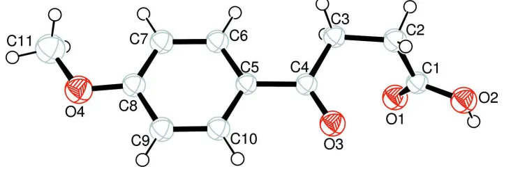
In the title compound (Fig. 1), the bond lengths (Allen et al., 1987) and angles are within normal ranges. O3, O4, C2,
C3 and C4 atoms are 0.067 (3), -0.003 (3), -0.163 (4), -0.013 (3) and 0.016 (3) Å away from the phenyl plane,
respectively.
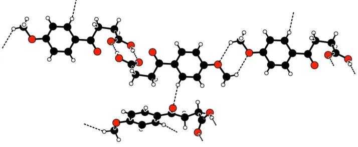
In the crystal structure, intermolecular O-H···O and C-H···O hydrogen bonds (Table 1) link the molecules (Fig. 2), in
which they may be effective in the stabilization of the structure. There also exist a C—H···π contact (Table 1) between the
phenyl ring and the methylene group.
S2. Experimental
The title compound was synthesized by the condensation of succinic anhydride (2 g, 20 mmol) with anisol (10 ml) in the
presence of alumium chloride (6 g, 42 mmol). The reaction mixture was refluxed for 4 h. After completion of the
reaction, excess solvent (anisol) was removed by steam distillation. The resultant solid product was purified by dissolving
it in sodium hydroxide solution (5%, w/v), filtering followed by addition of hydrochloric acid. The obtained solid mass
was filtered, washed with cold water, dried and crystallized from methanol (yield; 55%, m.p. 419-420 K)
S3. Refinement
H atoms were positioned geometrically, with O-H = 0.82 Å (for OH) and C-H = 0.93, 0.97 and 0.96 Å for aromatic,
[image:2.610.125.496.544.672.2]methylene and methyl H, respectively, and constrained to ride on their parent atoms with Uiso(H) = 1.2Ueq(C,O).
Figure 1
Figure 2
[image:3.610.56.492.76.721.2]A partial packing diagram. Hydrogen bonds are shown as dashed lines.
Figure 3
The formation of the title compound.
3-(4-Methoxybenzoyl)propionic acid
Crystal data
C11H12O4
Mr = 208.21 Monoclinic, P21/c
Hall symbol: -P 2ybc
a = 5.0511 (3) Å
b = 10.0219 (7) Å
c = 20.0840 (12) Å
β = 90.107 (6)°
V = 1016.67 (11) Å3
Z = 4
F(000) = 440
Dx = 1.360 Mg m−3
Melting point: 419(1) K Mo Kα radiation, λ = 0.71073 Å Cell parameters from 8408 reflections
θ = 1–27.5°
µ = 0.10 mm−1
T = 150 K Block, colorless 0.20 × 0.18 × 0.13 mm
Data collection
Bruker–Nonius KappaCCD area-detector diffractometer
Radiation source: fine-focus sealed tube Graphite monochromator
Detector resolution: 9.091 pixels mm-1
φ and ω scans
Absorption correction: integration (Coppens, 1970)
Tmin = 0.979, Tmax = 0.987
8320 measured reflections 2236 independent reflections 1662 reflections with I > 2σ(I)
Rint = 0.048
θmax = 27.5°, θmin = 2.0°
h = −6→6
k = −13→13
Refinement
Refinement on F2
Least-squares matrix: full
R[F2 > 2σ(F2)] = 0.048
wR(F2) = 0.112
S = 1.13 2236 reflections 136 parameters 0 restraints
Primary atom site location: structure-invariant direct methods
Secondary atom site location: difference Fourier map
Hydrogen site location: inferred from neighbouring sites
H-atom parameters constrained
w = 1/[σ2(F
o2) + (0.0363P)2 + 0.36P]
where P = (Fo2 + 2Fc2)/3
(Δ/σ)max < 0.001
Δρmax = 0.18 e Å−3
Δρmin = −0.20 e Å−3
Special details
Geometry. All e.s.d.'s (except the e.s.d. in the dihedral angle between two l.s. planes) are estimated using the full covariance matrix. The cell e.s.d.'s are taken into account individually in the estimation of e.s.d.'s in distances, angles and torsion angles; correlations between e.s.d.'s in cell parameters are only used when they are defined by crystal symmetry. An approximate (isotropic) treatment of cell e.s.d.'s is used for estimating e.s.d.'s involving l.s. planes.
Refinement. Refinement of F2 against ALL reflections. The weighted R-factor wR and goodness of fit S are based on F2,
conventional R-factors R are based on F, with F set to zero for negative F2. The threshold expression of F2 > σ(F2) is used
only for calculating R-factors(gt) etc. and is not relevant to the choice of reflections for refinement. R-factors based on F2
are statistically about twice as large as those based on F, and R- factors based on ALL data will be even larger.
Fractional atomic coordinates and isotropic or equivalent isotropic displacement parameters (Å2)
x y z Uiso*/Ueq
O1 0.0482 (3) −0.08173 (13) 0.07156 (6) 0.0440 (4)
O2 −0.2693 (3) 0.06464 (14) 0.04647 (6) 0.0443 (4)
H2 −0.1915 0.0654 0.0107 0.053*
O3 0.1138 (2) 0.13027 (12) 0.19230 (6) 0.0398 (3)
O4 0.7706 (3) −0.03563 (14) 0.44558 (7) 0.0477 (4)
C1 −0.1507 (3) −0.01793 (17) 0.08665 (8) 0.0323 (4)
C2 −0.2850 (3) −0.03301 (19) 0.15227 (8) 0.0355 (4)
H2A −0.4267 −0.0977 0.1478 0.043*
H2B −0.3645 0.0517 0.1644 0.043*
C3 −0.1025 (3) −0.07690 (17) 0.20779 (8) 0.0317 (4)
H3A −0.0091 −0.1569 0.1941 0.038*
H3B −0.2075 −0.0989 0.2467 0.038*
C4 0.0958 (3) 0.02952 (16) 0.22605 (8) 0.0297 (4)
C5 0.2686 (3) 0.00865 (16) 0.28458 (8) 0.0287 (4)
C6 0.2583 (3) −0.10711 (17) 0.32209 (8) 0.0333 (4)
H6 0.1367 −0.1730 0.3108 0.040*
C7 0.4243 (3) −0.12673 (18) 0.37606 (9) 0.0363 (4)
H7 0.4169 −0.2059 0.4002 0.044*
C8 0.6011 (3) −0.02784 (18) 0.39356 (8) 0.0343 (4)
C9 0.6134 (4) 0.09014 (18) 0.35680 (9) 0.0365 (4)
H9 0.7319 0.1570 0.3688 0.044*
C10 0.4502 (3) 0.10717 (17) 0.30291 (9) 0.0339 (4)
H10 0.4605 0.1855 0.2782 0.041*
C11 0.7813 (5) −0.1587 (2) 0.48107 (11) 0.0561 (6)
H11B 0.9151 −0.1535 0.5150 0.067*
H11C 0.8233 −0.2297 0.4508 0.067*
Atomic displacement parameters (Å2)
U11 U22 U33 U12 U13 U23
O1 0.0500 (8) 0.0510 (8) 0.0311 (7) 0.0157 (7) 0.0057 (6) −0.0004 (6)
O2 0.0502 (8) 0.0558 (8) 0.0268 (6) 0.0147 (7) 0.0026 (6) 0.0017 (6)
O3 0.0442 (7) 0.0337 (7) 0.0413 (7) 0.0016 (6) −0.0026 (6) 0.0066 (6)
O4 0.0528 (8) 0.0466 (8) 0.0436 (8) −0.0063 (7) −0.0184 (6) 0.0049 (6)
C1 0.0358 (9) 0.0341 (9) 0.0268 (8) −0.0008 (8) −0.0027 (7) −0.0039 (7)
C2 0.0317 (8) 0.0441 (10) 0.0307 (9) −0.0015 (8) 0.0018 (7) −0.0002 (8)
C3 0.0323 (8) 0.0360 (9) 0.0267 (8) 0.0005 (7) 0.0022 (7) −0.0010 (7)
C4 0.0303 (8) 0.0295 (9) 0.0293 (8) 0.0062 (7) 0.0066 (7) −0.0007 (7)
C5 0.0285 (8) 0.0288 (8) 0.0288 (8) 0.0017 (7) 0.0039 (6) −0.0024 (7)
C6 0.0355 (9) 0.0306 (9) 0.0338 (9) −0.0057 (7) 0.0001 (7) −0.0005 (7)
C7 0.0408 (9) 0.0334 (9) 0.0348 (9) −0.0025 (8) −0.0015 (8) 0.0046 (7)
C8 0.0343 (9) 0.0381 (10) 0.0304 (9) 0.0015 (8) −0.0025 (7) −0.0023 (7)
C9 0.0372 (9) 0.0320 (9) 0.0402 (10) −0.0068 (8) −0.0040 (8) −0.0032 (8)
C10 0.0364 (9) 0.0288 (9) 0.0364 (9) 0.0001 (7) 0.0031 (7) 0.0006 (7)
C11 0.0659 (14) 0.0541 (13) 0.0481 (12) −0.0033 (11) −0.0225 (10) 0.0106 (10)
Geometric parameters (Å, º)
O1—C1 1.230 (2) C5—C6 1.384 (2)
O2—C1 1.301 (2) C5—C10 1.396 (2)
O2—H2 0.8201 C6—C7 1.383 (2)
O3—C4 1.220 (2) C6—H6 0.9300
O4—C8 1.352 (2) C7—H7 0.9301
O4—C11 1.425 (2) C8—C7 1.379 (2)
C1—C2 1.491 (2) C8—C9 1.395 (2)
C2—C3 1.511 (2) C9—H9 0.9300
C2—H2A 0.9700 C10—C9 1.370 (2)
C2—H2B 0.9699 C10—H10 0.9299
C3—H3A 0.9700 C11—H11A 0.9600
C3—H3B 0.9701 C11—H11B 0.9600
C4—C3 1.508 (2) C11—H11C 0.9600
C5—C4 1.478 (2)
C1—O2—H2 109.2 C10—C5—C4 119.76 (15)
C8—O4—C11 117.38 (15) C7—C6—C5 121.48 (16)
O1—C1—O2 123.60 (15) C7—C6—H6 119.3
O1—C1—C2 122.58 (16) C5—C6—H6 119.2
O2—C1—C2 113.77 (15) C8—C7—C6 119.26 (16)
C1—C2—C3 113.83 (14) C8—C7—H7 120.4
C1—C2—H2A 108.8 C6—C7—H7 120.3
C3—C2—H2A 108.8 O4—C8—C7 124.36 (17)
C3—C2—H2B 108.7 C7—C8—C9 120.24 (16)
H2A—C2—H2B 107.6 C10—C9—C8 119.74 (16)
C4—C3—C2 112.19 (15) C10—C9—H9 120.1
C4—C3—H3A 109.3 C8—C9—H9 120.2
C2—C3—H3A 109.2 C9—C10—C5 120.94 (16)
C4—C3—H3B 109.2 C9—C10—H10 119.5
C2—C3—H3B 109.1 C5—C10—H10 119.6
H3A—C3—H3B 107.9 O4—C11—H11A 109.5
O3—C4—C5 120.98 (15) O4—C11—H11B 109.5
O3—C4—C3 120.04 (15) H11A—C11—H11B 109.5
C5—C4—C3 118.98 (14) O4—C11—H11C 109.4
C6—C5—C10 118.33 (15) H11A—C11—H11C 109.5
C6—C5—C4 121.91 (15) H11B—C11—H11C 109.5
Hydrogen-bond geometry (Å, º)
D—H···A D—H H···A D···A D—H···A
O2—H2···O1i 0.82 1.81 2.628 (3) 173
C6—H6···O3ii 0.93 2.34 3.247 (3) 164
C11—H11B···O4iii 0.96 2.60 3.328 (3) 133
C3—H3B···Cg1iv 0.97 2.74 3.591 (3) 146