A Telepathology Model Using JPEG Algorithm for
Histological Image Compression
Boniface K. Alese, Adebowale J. Adelakun, Otasowie Iyare, Oloruntoba S. Ogundele
Abstract—The specific objectives of the research were to examine the efficacy of Discrete Cosine Transform (DCT) as an image compression algorithm using the Joint Photographic Expert Group (JPEG) standard; and determine the suitability or otherwise of application of DCT on images required for Collaborative Pathological diagnosis. Seven histological images acquired with the use of Digital microscope from the University of Alberta, Canada were sent by e-mail by the Lead Pathologist to the researcher in Nigeria. These were compressed using GNU Image Processing software (GIMP, a third party freeware) at 25% and 50% factor and presented randomly to researchers. The result showed that JPEG is efficient in compression of medical images because the images compressed at 25% factor were reduced from between 22% - 32%. The ones compressed at 50% factor were reduced between 15% - 20% which revealed that though the JPEG algorithm does not reduce images literarily based on the factor of reduction, size reduction is still significant. The conclusion therefore is that the DCT is an efficient image compression algorithm and that using JPEG for telepathology systems is not only suitable but desirable because of the various practical usefulness to providing cost effective pathology services to the rural areas of the developing world.
Index Terms—Telepathology, compression algorithm, confidence level, histological images, discrete cosine transform
I. INTRODUCTION
he challenges of health care delivery in Africa are enormous and the larger aspect of the populace lives in the remotest part where the socio-economic conditions and means of communication are undeveloped [1]. It is also a known fact that pathological services are almost non-existent in these areas making correct diagnosis of diseases very difficult. Tanzania and Nigeria have approximately one clinical pathologist available per 3 million people while in Zambia, the entire country has only one surgical pathologist for a population of 12 million [2].
In [5], the authors identified various factors which has affected the deployment of pathology services in sub-Sahara Africa. Among which are the scarcity of clinical specialty even where pathological laboratory exists thereby limiting the level of care hospitals can give to patients; Delays and low quality of services which has made pathology test limited to clinicians;
Manuscript received March 12, 2014; revised April 3, 2014.
B. K. Alese is with the Federal University of Technology, Akure, Nigeria, e-mail: [email protected], +2348034540465
A.J. AAdelakun is with the Federal University of Technology, Akure, Nigeria, e-mail:
O. Iyare is with the Federal University of Technology, Akure, Nigeria, e-mail: [email protected], +2347033513174
O. S. Ogundele is with the Central Bank of Nigeria, e-mail: [email protected], +2348037752527
General laboratory quality standards including quality control and quality assurance which was described as non-existent or sub-optimal. In [3] and [4], it was further stated that other pervasive problems for pathology in sub-Saharan Africa are related to limited opportunities for basic training in pathology and lack of books, CDs and other educational materials for continuing improvement which has resulted in lack of operational competency with little backup.
In view of these challenges it is clear that there exists an unmet need for pathological services in the nations of Africa. It is also crucial that innovative interventions address these needs to improve the quality of laboratory services. In [5] it was described as fortuitous the fact that the world has witnessed the phenomenal advances in technology that incorporates new methods in communications and information technology into medical practices. This has largely solved the problem of technical requirement for telepathology [6].
Telepathology, which [7] described as the use of custom software and high speed internet connections to transmit images across distances for diagnosis and consultation, was pioneered by Ronald Weinstein, a cancer cell biologist and pathologist in the mid-1980s; He demonstrated that pathologists viewing images on video monitors can achieve essentially the same diagnostic accuracy as those using convectional light microscopy [7].
Telepathology is of immense benefit to Clinicians and patients in various ways among which are real time consultations. Physicians in community services (rural areas) can consult experts on different cases to better understand the diseases process in their patients. Patients can also have access to a broader array of pathologists who can consult on their diagnosis.
Telepathology has the potential of allowing pathologists from around the world to consult with each other as if their offices were only a few steps away [7].
In all, telepathology allows having the right pathologists make the right decision regarding patient’s diagnosis without regard to where the Patient or the Pathologist is. According to [7] telepathology involves the use of custom software and high speed internet connections to transmit images across distances for diagnosis and consultation. The sizes of images required for telepathology is huge so there is the need for compression. There are several methods of image compression today which fall into two general categories: loosless or loosy image compressions [8].
The Joint Photographic Expert Group (JPEG) process is a widely used form of loosy image compression that centers on the Discrete Cosine transform (DCT). The DCT works by separating images into parts of differing frequencies. During a step called quantization, where part of compression actually occurs, the less important frequencies are discarded, hence the term loosy. Then only the most important frequencies that remain are used to retrieve the image in the decompression process. As a result reconstructed images contain some distortion; which could be adjusted during the compression stage [8].
Furthermore, “A discrete cosine transform (DCT) expresses a sequence of finitely many data points in terms of a sum of cosine functions oscillating at different frequencies. DCTs are important to numerous applications in science and engineering, from lossy compression of audio and images (where small high-frequency components can be discarded), to spectral methods for the numerical solution of partial differential equations” [9]
This work is set to examine the efficacy of Discrete Cosine Transform (DCT) as an image compression algorithm using the Joint Photographic Expert Group (JPEG) standard; and also to determine the suitability or otherwise of application of DCT on images required for Collaborative Pathological diagnosis.
II. EXISTING WORKS ON COMPRESSIONS TECHNOLOGY
The authors in [10] stated that numerous studies have investigated aspects of image quality assessment of compressed medical images [11], [12] but that majority has focused on gray-level images generated for radiology, Computerised Tomography (CT) and Magnetic resonance (MR) applications. This cannot be divorced from the fact that radiology is the dominant application domain for medical imaging technology. [13] identified that significant amount of literature is available on the topic of compression of digital radiology images.
He further observed that a review of several looseless compression schemes such as Differential Pulse Code Modulation (DPCM), Hierarchical Interpolation (HINT), Difference Pyramids (DP) e.t.c. and loosy compression schemes such as Vector Quantisation (VQ) and algorithms based on Discrete Cosine Transform (DCT) and Discrete Wavelet Transform (DWT) etc. which may be used for digital radiology images is provided in [12] and [14]. He noted that [15] investigated the utility of JPEG-LES and JPEG 2000 for compression of digital radiology images representing several different image modalities such as Computed Tomography (CT), Magnetic Resonance (MR), and Ultrasound (US) etc. and concluded that both the
compression schemes offer similar or better performance than JPEG and recommended their inclusion in the DICOM standard. [16] cited by [13] also compared the performance of the JPEG and JPEG 2000 algorithm for digital mammograms in an attempt to provide more evidence to support adoption of JPEG 2000 for medical images.
In [17] also cited by [13] suggested the use of content based compression of images for static telepathology. These authors were reported to have proposed using “content based” approach which combines visually loosless and loosy compression techniques by judiciously applying either in the appropriate context across an image so as to maintain diagnostically important information while still mazimising the possible compression.
In [10], it was identified that many studies have been published on telepathology but only few have actually compared noncompressed images with compressed images using s structure study design. These authors actually studied the effect of image compression on telepathology and concluded that JPEG compression of images does not negatively affect the accuracy and confidence level of diagnosis. The author in [18] had earlier concluded that loosy compression holds promise for diagnostic telepathology. [19], proposed the use of JPEG 2000 image compression algorithm for developing a virtual slide telepathology system by using some useful features such as scalability and JPEG 2000 Interactive Protocol (JPIP) offered by the JPEG 2000 algorithm.
In the review of [13] it was noted that these few publications addressing the issue of image compression for telepathology or virtual microscopy and most of the published trials reported in [20], verifying the diagnostic accuracy of telepathology have tacitly used JPEG or JPEG 2000 algorithm for compression without an in-depth analysis of the diagnostic accuracy and communication efficiency. In [13] it was stated that whole slide images are extremely large and that storage and transmission of these images in uncompressed form is impractical. This has prompted the use of image compression algorithms. The author in [10] was of the opinion that efficient methods of image transmission, especially by compression, are of great interest to the scientific community, since high-quality digital images may attain sizes of 1 megabyte or greater.
JPEG compression adversely affect the quality of images and the accuracy of diagnosis.
III. JPEG AND THE CONCEPT OF DIGITAL TRANSFORM OF IMAGES
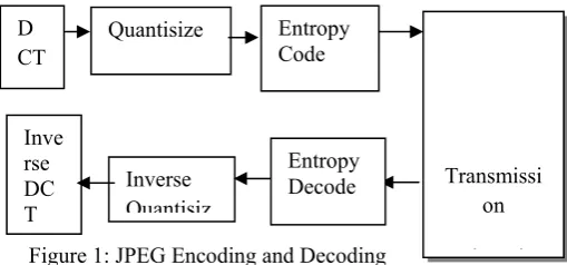
The JPEG standard was introduced by the joint ISO/CCITT committee known as the JPEG (Joint Photographic Experts Group). [22] cited in [13] said JPEG supports four modes of operation: sequential, progressive, lossless and hierarchical encoding. The baseline sequential codec implements the Discrete Cosine Transform (DCT). [8] in their general overview of JPEG methods described the itemized process required for the Discrete Cosine Transform of images in steps as follows:
Step 1: The image is broken into blocks of 8x8 blocks of pixels.
Step II: Working from left to right, top to bottom, the DCT is applied to each block.
Step III: Each block is compressed through quantisation. Step IV: The array of compressed blocks that constitute the image is stored in a drastically reduced amount of space. Step V: When desired the image is reconstructed through decompression: a process that uses the Inverse Cosine Transform (IDCT).
The process is further explained in Figure 1 and DCT equation.
[image:3.595.41.296.376.495.2]
Figure 1: JPEG Encoding and Decoding
The DCT Equation: The DCT equation (1) computes the i,jth entry of the DCT of an image.
D i, j
√ C i C j ∑ . ∑ p x, y cos Cos
(1)
√
0 (2)
P (x,y) is the x, yth element if the image represented by the
matrix p. N is the size of the block that the DCT is done on. The equation calculates one entry (i, jth) of the transformed
image from the pixel values of the original image matrix. For the standard 8x8 block that JPEG compression uses, N equals 8 and x and y range from 0 to 7. Therefore D (i,j) would be as in Equation (2.3).
D i, j
C i C j ∑ . ∑ p x, y Cos Cos
(3)
The DCT uses cosine functions the resulting matrix depends on the horizontal, diagonal and vertical frequencies. Therefore an image black with a lot of change in frequency has a very random looking resulting matrix, while an image matrix of just one color, has a resulting matrix of a large value for the first element and zeroes for other elements [8].
IV. THE PROPOSED MODEL
The proposed model is a web based application consisting flat files where digital images and result of diagnosis (consultations) are archived, after been loaded via a user interface, an inference engine (Analyser), and a report generator.
A. Mathematical Model for Image Assessment and
Comparison
The images submitted for second opinion on it’s suitability for diagnosis from participating pathologist were considered and reports were generated through the inference mechanism and the report generator.
The Degree of confidence D, which is a Percentage of agreement, is given by:
∑ / (4)
If D ≥ 90%, the (Ai) opinion is accepted and otherwise it is
rejected.
Table 1: The result of Images compressed at 25%.
Ca ses
Orig inal Size of Ima ges
Size of Images Compr essed @ 25% factor
Perce ntage Reduc tion (%)
No of respon ses(n)
No of Assess ors in Agree ment with Owne r (Ai)
Degre e of Confid ence (D%)
1 2.04
Mb
63.6Kb 32.08 10
0
2 1.96
Mb
65.1Kb 30.11 10
0
3 2.12
Mb
81.8Kb 25.92 10
0 00
4 2.30
Mb
86.3Kb 26.65 10
0 00
5 1.65
Mb
55.8Kb 29.57 10
0 00
6 1.64
Mb
57.3Kb 28.62 10
0 00
7 2.07
Mb
93.3Kb 22.19 10
0 00
Inverse Quantisiz D
CT
Quantisize Entropy Code
Inve rse DC T
Entropy Decode
Transmissi on
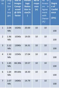
Table 2: The result of Images compressed at 50%
The Opinion of Assessors which agrees with owners opinion is denoted by Ai where i ranges from 1,2 …. n. ‘n’
is the total number of participants.
B. The conceptual and Architectural Model for the
Telepathology system
The model for the proposed telepathology system was on the description of Second Opinion Pathology as earlier discussed. This is constituted of a telepathology system where a referring pathologist can easily request a second opinion from a specialist or consulting pathologist located at a remote facility. This would have the potential of significantly improving the turn-around time by reducing the time spent in sending the glass slides or tissue block to the specialist by post. [23] demonstrated that high concordance rates ranging from 99% to 100% were achieved for clinically significant telepathology and conventional light microscopy diagnosis of 2,200 consecutive surgical pathology cases while using a hybrid dynamic/store-and-forward telepathology system. The model would help realize the assertion by [13] that telepathology can be used to support an isolated pathologist for second or expert consultation or even completely transfer diagnostic work to a remote facility.
C. The Histopathological images
Biopsies were taken from real life subjects with renal disorders. The tissues were taken fresh and fixed in 10% formal saline (formalin). They were processed as per histopathology protocols and subsequently cut into thin session and placed on glass slides and stained with various histopathology stains. The micrograph were taken with digital microscope with installed camera by the Lead pathologist at the University of Alberta, Canada and sent via e-mail to the researcher in Nigeria.
There were seven cases in all which came in sizes between 1.6MB – 2.3MB as indicated in Table 4.2 and Table 4.3. The images were later compressed using a third party software GNU Image Manipulation Program (GIMP) which uses JPEG standard for compression. The factor of compression was first set at 25% and later at 50% the results are as shown in Table 4.2 and Table 4.3 respectively.
D. Histopathological Images Compressed at 50% and
25%
The result of the images compressed at 50% and at 25% is shown in Tables 1 and 2 respectively.
E. Result of Images compressed at 25% factor
Table 1 shows that the image compressed at 25% factor with significant reduction ranging from between 22% - 32% reduction in size of the compressed images compared to the original images. Cases 1 and Case 2 assessed had 90% degree of agreement while Cases 2, 3, 4, 5 and 6 had 100%.
[image:4.595.43.293.90.463.2]F. Result of Images compressed at 50% factor
Table 2 shows the result of the compressed images at 50% reduction factor with significant reduction in size of the compressed images compared to the original ranging between 14.7% -20%. The ten cases assessed had 100% degree of agreement.
V. DISCUSSIONS
Table 1 shows that there were 10 respondents who participated in examining each of the seven cases presented. Case 1 and Case 2 had 9 of the participating Pathologist agree with the owner on the suitability of the compressed image for diagnosis that is 90% degree of confidence. In Cases 3, 4, 5, 6, and 7 all the 10 participating Pathologists agree with the owner’s opinion that is 100% degree of confidence.
Table 2 shows that there were 10 participating Pathologist and all the 10 agreed with the Owner’s opinion on the compressed images of Cases 1, 2, 3, 4, 5, 6 and 7 thereby giving a 100% degree of confidence in all the cases examined.
Table 1 also shows that the images after compression at 25% factor were reduced from between 22% - 32% while Table 4.3 shows that the images after compression using the 50% factor were reduced between 15% - 20%. These results show that the JPEG algorithm does not reduce images Cas
es Origi
nal Size of Ima ges
Size of
Images
Compr essed
@ 50%
factor
Percen tage
Reduc tion in
size(% )
No of
respo nses
(n)
No of Assess ors in Agree ment with
Owner
(Ai)
Degre e of
Confid ence
(D%)
1 2.04 Mb
102Kb 20.00 10 10
100
2 1.96 Mb
103Kb 19.03 10 10
100
3 2.12 Mb
130Kb 16.31 10 10
100
4 2.30 Mb
135Kb 17.04 10 10
100
5 1.65 Mb
84.3Kb 19.57 10 10
100
6 1.64 Mb
89.6Kb 18.30 10 10
100
7 2.07 Mb
140Kb 14.79 10 10
literarily based on the factor of reduction but the size reduction is significant.
The research question whether the Discrete Cosine Transform (DCT) algorithm implemented by JPEG is suitable for telepathology has been clearly answered in the affirmative. Since from our findings we have seen that the compression of images was significant that is 22%-32% for images compressed at 25% and 15%- 20% for images compressed at 50%.
This reduction in sizes will significantly affect the speed and time of uploading the images for diagnostic purposes thereby aiding collaborative pathological diagnosis.
It is also discovered from the responses of the very experienced Pathologists involved in the study that the compressed images do not have much difference in terms of diagnostic value from the original images.
The proposed Telepathology model will be efficient in offering collaborative pathological diagnosis both at small, medium and large scale basis. Since it is an online system, gaining the confidence of the professionals in the field and provision of legal framework would enhance the implementation.
VI. CONCLUSION
In conclusion it is clear from the findings that the Discrete Cosine Transform (DCT) as an image compression algorithm using JPEG is efficient. It is also established that JPEG is a suitable compression algorithm for implementation of telepathology, since it is established that the images after compression are still suitable for pathological diagnosis from the responses of participating pathologists. It was found that the participating pathologist agree with 90% - 100% degree of confidence with the owner’s opinion that the compressed images still had diagnostic lesion representation which justifies the finding of [10] and also confirmed the assertion of [8] as the findings show that the images compressed at 50% have higher degree of confidence and as such would be more suitable for use in the system.
In other words, the efficacy of Discrete Cosine Transform (DCT) as an image compression algorithm using the Joint Photographic Expert Group (JPEG) standard has been examined and found to be suitable for compression of images required for Collaborative Pathological diagnosis as set out in the objectives of this study.
REFERENCES
[1] INCTR(2012),
http://inctr.ctisinc.com:9000/sites/InCTR/Education/INCTR%2010th %20Anniversary%20Meeting%202010/Program%20Reports/INCTR %20Pathology%20Program%20The%20role%20of%20Telepatholog y%20in%20Improving%20Pathology%20Services%20Nina%20Hur witz.pdf
[2] Ali Lotfizadeh (2012), “Telepathology to aid diagnosis in the developing world”, http://www.patexia.com/feed/telepathology-to-aid-diagnosis-in-the-developing-world-2341 last visited 20th December, 2012.
[3] Ahmad, M. (2005), Laboratory Medicine in Developing Countries: Need for Immediate Improvement. The Intenational Network of Cancer Treatment and Research INCTR Newsletter.
URL:www.inctr.org/publications/2005_v05_n04_s03.html
[4] Ahmad, Z., Qureshi, A. & Khurshid, A. (2009).The practice of histopathology in a developing country: difficulties and challenges;
plus a discussion on the terrible disease burden we carry. J Clin
Pathol 62:97-101
[5] Malami S.A (2011). Recent Advances in Telepathology in the Developing World, Advances in Telemedicine: Applications in Various Medical Disciplines and Geographical Regions, Prof. Georgi Graschew (Ed.), ISBN: 978-953-307-161-9, InTech, Available from: http://www.intechopen.com/books/advances-intelemedicine- applications-in-various-medical-disciplines-and-geographical-regions/recent-advances-intelepathology-in-the-developing-world [6] Wells, C. A. and Sowter, C., “Telepathology: a diagnostic tool for the
millennium" Journal of Pathology, vol. 191, pp. 1-7, May 2000. [7] Scott P. Edwards (2005), “Telepathogy Services”, web document,
Baystate Health Website, Posted April, 7, 2005; Last visited December, 2008.
[8] Ken Cabeen and Peter Gent (2008), “Image Compression and the discrete Cosine Transform”, Math 45, College of the Redwoods, dct.pdf, last visited August, 2008.
[9] Wikipedia (2012),
http://en.wikipedia.org/wiki/Discrete_cosine_transform, visited 30th September.
[10] Marcelo, A., Fontelo, P., Farolan, M., and Cualing, H.(2000), “Effect of image compression on telepathology," Archives of Pathology and Laboratory Medicine, vol. 124, no. 11, pp 1653-6.
[11] Cosman, P. C., Gray, R. M., and Olshen, R. A.(1994), “Evaluating quality of compressed medical images: Snr, subjective rating, and diagnostic accuracy," Proceedings of the IEEE, vol. 82, no. 6, pp. 919-932.
[12] Wong, S., Zaremba, L., Gooden, D., and Huang, H. K(1995), “Radiologic image compression-a review," Proceedings of the IEEE, vol. 83, no. 2, pp. 194-219, 1995.
[13] Sourabh Khire (2009), “Time-sensitive communication of digital images, with application in Telepathology”, Masters Thesis in Electrical and Computer Engineering, Georgia Institute of Technology.
[14] Menegaz, G., “Trends in Medical image compression," Current Medical Imaging Reviews, vol. 2, pp. 165{185, May 2006.
[15] Clunie, D.A. (2000), “Looseless compreshension of grayscale medical images – effectiveness of traditional and state of the art approaches,” in Effectiveness of Traditional and State of the Art Approaches, SPIE Medical Imaging, pp. 74-84
[16] Wanigasekara, N. R., Ding, S., Yan, Z., and Zeng, Y. (2002), “Quality evaluation for jpeg 2000 based medical image compression," in Proceedings of the Second Joint EMBS/BMES Conference, 2002.
[17] Varga J.M, Ducksbury, P. G., and Callagy, G. (2002), “Application of content-based image compression to telepathology," in Proc. SPIE, Vol. 4681, Medical Imaging 2002: Visualization, Image-Guided Procedures, and Display.
[18] Foran, D.J, Meer, P.P., Papathomas, T., and Marsic, I.(1997), “Compression guidelines for diagnostic telepathology,” IEEE Transaction on Information Technology in Biomedicine, vol. 1, no. 1 pp. 55-60.
[19] Ortiz, J. G., Ruiz, V., and Garca, I.(2007), “Virtual slide telepathology systems with peg2000," in Proceedings of the 29th Annual Conference of the IEEE EMBS, pp. 880{883, August 2007. [20] Cross, S.S., Dennis, T., and Start, R.D.(2002), “Telepathology:
current status and future prospects in diagnostic histopathology”, Histopathology, vol. 41, no. 2 pp 91-109.
[21] Williams, S. M., Carter, A. B., and Jayant, N. S.(2008), “Diagnostically lossless compression of pathology image slides," Advancing Practice Instruction and Innovation through Informatics Conference, Pittsburgh, 2008
[22] Wallace, G. K.(1992), “The jpeg still picture compression standard," IEEE Transactions on Consumer Electronics, vol. 38, no. 1, pp. xviii-xxxiv, 1992.
[23] Dunn, B.E., Choi, H., Almargo, U.A., Recla, D.L., Krupinski, E.A, and Weistein R.S (1999), “Routine surgical telepathology in the department of veterans affairs: experiment-related improvement in pathologist performance in 2200 cases,” Telemedicine Journal, vol. 5. No. 4. Pp. 323-337.
[24] Malami, S. A. & Iliyasu, Y. (2008a). Local Audit of Diagnostic Surgical Pathology as a Tool