The effectiveness of packing post incision and drainage among patients
with skin abscesses in improved wound healing and reduced recurrence.
By
Natalie Ford, PA-S, MPH
A Capstone Paper submitted to the faculty of the University of North Carolina at Chapel Hill
in partial fulfillment of the requirements for the degree of Master of Health Sciences
in the Physician Assistant Program
Chapel Hill
December 2017
________________________________
Name and title of First Reader
______________________
Date
________________________________
Name and title of Second Reader
______________________
BACKGROUND Introduction
A skin abscess is an accumulation of pus beneath the skin and is among one of the most
common skin and soft tissue infections. Skin abscesses can occur in anyone including healthy
patients with no comorbidities. They can occur anywhere on the body, but are common in
friction-prone areas such as the buttocks, breast and groin.1 Patients present with tender nodules with fluctuance, induration, and erythema.2 Definitive management for abscesses is incision and drainage, with or without the placement of packing material.
The purpose of this article is to review the necessity of wound packing in the healing
outcomes and recurrence of infection after incision and drainage compared to no packing in
patients with a skin abscess. Evaluation of this topic has the potential to improve patient care
while reducing overall health care costs.
First, we will discuss the epidemiology, pathophysiology, diagnosis and management
options for skin abscess. Then, we will review the most recent literature to answer the
aforementioned question at hand.
Epidemiology
The epidemiology of skin abscess is unclear due to underreporting or patients
inconsistently seeking treatment. However, evidence reveals that the greatest incidence occurs
in individuals ages 18-44, African Americans, and males.3 Taira et. al found that the rate of emergency department abscess visits increased more rapidly than the overall rate of ED visits
with the rate of skin abscesses more than doubling within a decade from 1.2 million in 1996 to
Those most at risk for skin and soft tissue infections include, but are not limited to the
very young and elderly, diabetics, the immunocompromised, the obese, and those with any
recent water exposure. In addition, groups of humans in close living parameters are at an
increased risk including long term facility residents, military personnel and the incarcerated. 3 Many organisms may be found in a skin abscess. Staphylococcus aureus, a commensal
and pathogenic bacterium, is the leading cause for skin and soft tissue infections with
approximately 80% of skin infections related to S. aureus.5S. aureus is second after Clostridium difficile for leading health-care associated infections in the United States.6 Increasing resistance
to penicillin resulting in methicillin-resistant S. aureus (MRSA) was originally noted in the 1960s.
Resistance has increased including vancomycin. It is the leading cause of pathogen-associated
morbidity and mortality in the United States.5 Pathophysiology
Abscess formation occurs when bacteria enters at a site of skin disruption secondary to
trauma, venous insufficiency, immunosuppression or prior cutaneous infections, including
methicillin resistant Staphylococcus aureus (MRSA). 7 The area of injury to the skin results in a barrier breakdown creating an entry site for bacteria. Contamination by bacteria creates a
release of toxins causing affected tissues to necrose at the affected site. The release of toxins
causes an inflammatory response and leukocytes travel to the site of inflammation to
phagocytose and breakdown the dead tissue while disarming the bacteria. The devitalized
tissues and necrosed bacteria accumulate to form pus. Simultaneously, the body’s immune
system responds by walling off the site of inflammation to prevent spread of the bacteria to
reaction occurs (image 1). The affected area will continue to build up with pus until an opening
occurs to allow for drainage through the skin. 1 Image 1: Visualization of abscess formation.
Retrieved from: https://medical-dictionary.thefreedictionary.com/abscess
Diagnosis
An abscess is a clinical diagnosis and should be considered when a patient presents with
an erythematous, warm, edematous nodule. If a clinical diagnosis is unable to be made, imaging
modalities such as computed tomography (CT) or ultrasonography (US) may examine for fluid
collections. Useful labs to augment the diagnosis include a complete blood count, especially in
the instances of severe infections or in immunocompromised patients. 3
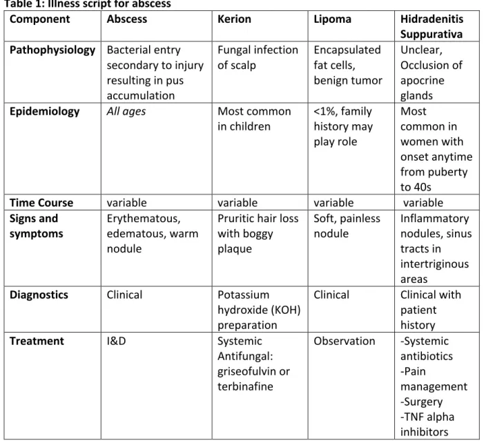
There are other diagnoses that may mimic an abscess. One must be able to differentiate
similar conditions that do not need incision and drainage, and therefore do not require wound
packing. Differential diagnoses to consider include kerion, lipoma and hidradenitis suppurativa.
An illness script is provided in Table 1 below. A kerion, also known as tinea capitis, is a fungal
infection that may present as a painful, boggy plaque that requires a systemic antifungal rather
nodule that usually does not require treatment.9 Hidradenitis suppurativa affects the apocrine glands resulting in chronic inflammatory abscesses of which incision may be beneficial, but also
requires dermatologic follow-up. 10 Table 1: Illness script for abscess
Component Abscess Kerion Lipoma Hidradenitis
Suppurativa Pathophysiology Bacterial entry
secondary to injury resulting in pus accumulation
Fungal infection
of scalp Encapsulated fat cells, benign tumor
Unclear, Occlusion of apocrine glands Epidemiology All ages Most common
in children <1%, family history may play role Most common in women with onset anytime from puberty to 40s
Time Course variable variable variable variable
Signs and
symptoms Erythematous, edematous, warm nodule
Pruritic hair loss with boggy plaque
Soft, painless
nodule Inflammatory nodules, sinus tracts in intertriginous areas
Diagnostics Clinical Potassium hydroxide (KOH) preparation
Clinical Clinical with patient history
Treatment I&D Systemic
Antifungal: griseofulvin or terbinafine
Observation -Systemic antibiotics -Pain management -Surgery -TNF alpha inhibitors Management
Incision and drainage is the appropriate management for abscesses, especially when
Ultrasonography may be used to guide. The incision should be large enough to allow for
continued drainage and should also be large enough to allow for destruction of loculations and
placement of packing, if desired.2
I&D leaves the patient with an open wound as the body responds with the healing
process. Wound healing consists of 4 stages; hemostasis, inflammation, proliferation and
maturation.12 The first step of wound healing is hemostasis which includes initiation of the clotting cascade creating fibrin. Inflammation then occurs with macrophage recruitment and
increased vascular permeability leading to edema. The proliferative phase includes proliferation
of fibroblast to allow for contraction of the wound. The final stage of healing is the maturation
phase which includes collagen to increase tensile strength.12
If any stage of wound healing is disrupted, complications can occur. The most common
cause of reoccurrence of an abscess is inadequate drainage.2 Packing a wound allows for absorption of drainage of the wound to allow for granulation tissue to form by preventing
wound margins from closing to a potential dead space.13 Packing of an abscess usually includes ¼-1/2 inch packing strips with or without iodoform. This material is placed inside the cavity,
without overpacking. This can prevent proper drainage and may result in ischemia.2 The packing is typically removed or changed in 2-3 days.
Antibiotics are generally not required for most abscesses following treatment with
incision and drainage. Antibiotics are not recommended if patients present with a mild skin
abscess without immunosuppression, age extremes, systemic infection, or more than one
METHODS
For the purpose of this paper, a search was conducted using the search databases of
PubMed, TRIP database and Cochrane Library. The following search terms were used: ‘skin AND
abscess AND packing,’ ‘abscess AND incision AND drainage’ and ‘incision AND drainage AND
packing.’ The initial search resulted in 1,236 studies. The search term “abscess AND incision
AND drainage AND packing’ was used to narrow results to 25 studies. Initial inclusion criteria
sought randomized control trials and systematic reviews, but few articles were found so criteria
was broadened to review the most reliable studies available based on design. Abstracts and
studies were excluded if wound packing versus not packing were not compared and if I&D did
not occur during the study. Quality evaluation was completed with either Cochrane tool for
assessing risk of bias and RoB 2.0 tool for randomized trials. The chosen articles with bias
evaluation are listed in the table in the results section.
RESULTS
After the database search was conducted, four studies were selected to be reviewed
with study details listed in Table 2. Results were divided based on outcomes of wound care at
48 hours, healing time and pain.
Wound care at 48 hours
O’Malley et al. recruited blinded emergency department physicians to evaluate wound
care 48 hours following I&D to further interventions.15 Data was recorded if extension of incision, packing, irrigation, change in antibiotics or surgical evaluation was required. No
significant difference was found for the need of intervention between the packed and
Kessler et al. found that overall failure rates were similar between the groups, with 19 of
27 subjects in the packed group needing an intervention at 48 hours compared with 13 of 22
subjects in the nonpacked group who required intervention (difference of means 11%; 95%
confidence interval [CI], -15% to 36%).16 Interventions post I&D required were similar between
groups, with 3 of 27 subjects in the packed group needing a major intervention compared with
5 of 22 subjects in the nonpacked group (difference, 12%; 95% CI, -12% to 36%).16
Healing time
Tonkin et al. reported median time to healing/complete epithelization similar between
24.5 days in packing group vs 21 days in nonpacking group (p=0.214).17 Upon further review of this analysis, the statistical analyses used are questionable according to Cochrane Systematic
Review.12 Smith et al. analyzed a study by Perera et al. that reported a mean time to wound healing of 26.8 days (95% confidence interval (CI) 22.7 to 30.7) in the packing group and 19.5
days (95% CI 13.6 to 25.4) in the non-packing group.18 It was concluded that the data was
difficult to determine if participants fully healed therefore compromising data. Data was
reanalyzed and no difference in healing time was found (7.30 days longer in the packing group,
95% CI -2.24 to 16.84; 14 participants).18 Both studies were reported with low quality evidence
due to bias risk and imprecision.18
Pain and analgesia requirements
O’Malley et al. evaluated pain and pain management at 48 hours via diaries and visual
analog scales. There was no significant difference found in pain in pre-I&D scores (difference of
hours post I&D was also significantly higher in packed group (difference of means=16.4mm,
95% CI=1.6 to 31.2 mm, p=0.03).15
Tonkin et al. pain scores were comparable between two group at initial dressing change
(P=0.296).17 At two weeks, the nonpacking group reported a pain score of 0 vs 2 in packing
group (p=0.004). 17 Analysis of the change in pain scores from initial assessment to the
two-week follow-up revealed no significant decrease in nonpacking group (p=0.916).17Smith et al.
also evaluated Perera 2015 study which reported pain scores as 3 in the packing group
compared to 2 in the non-packing group at the initial dressing change (P = 0.648).18
Table 2: Study details of reviewed articles
Study Set-Up Results Limitations Conclusions Bias
Routine packing of simple cutaneous abscesses is painful and probably unnecessary.
O’Malley et al. 200915 Randomized, single blinded, pilot study. Ages 18y.o.+ Sample size: 48, 23 to wound packing group and 25 to nonpacking group. No difference between groups. No significant difference in need for intervention between the packed (4 of 23 subjects) and
nonpacked (5 of 25 subjects) groups (p = 0.72, relative risk = 1.3, 95%
CI = 0.4–4.2).
Small pilot study leading to a small sample size, loss to f/u, poor validity This study concluded no difference in morbidity 48hr post I&D. Unpacked patients reported less pain and required less pain medications.
Low risk of bias judgements, some concern for bias from intended intervention due lack of information, low risk of bias due to missing outcome data, Low risk of bias for outcome measuremen t. Some concern for bias of reported results given small sample size. Randomized trial comparing wound packing Randomized, single-blinded, prospective Overall failure rates were similar Blinded assessors may not have been
Wound packing did not provide
Low risk of bias
to no wound packing following incision and drainage of superficial skin abscesses in the pediatric emergency department.
Kessler et al. 201216 Ages: pediatric patients 1-25 y.o. Sample size: 57, 27 to packing group, 22 in nonpacked group
between the groups, with 19 of 27 subjects in the packed group needing an intervention at 48 hours compared with 13 of 22 subjects in the nonpacked group who required intervention (difference of means 11%; 95% confidence interval [CI], -15% to 36%).
actually blinded, pain medication standardizations, wide CIs, small sample size benefit for need for intervention at 48hrs, shorter healing time, or rate of recurrence. some concern for bias from intended intervention due NI, low risk of bias due to missing outcome data. Low risk of bias for outcome measuremen t. Some concern for bias of reported results given small sample size. Perianal abscess: a pilot study comparing packing with nonpacking of the abscess cavity
Tonkin et al.l17
Sample size: 50 with 20 in the packing group and 23 in the nonpacking group, randomized, comparable groups Mean time to heal was similar between two groups: 24.5 (range, 10-150) days in the pack and 21 (range, 8-90) days in the nonpacking group (P=0.214.) Pain scores were comparable between two group at initial dressing change (P=0.296). Small sample
size, pilot study Safe management of perianal abscesses with I&D alone, no change in packing vs nonpacking for healing time
High risk of bias due to attrition, Some concerns for randomizatio n bias, Some concerns for intended interventions bias, some concern for missing outcome data, lwo risk of outcome measuremen t bias, some concerns for bias
Internal dressings for healing perianal abscess cavities
Smith et al. 201618
Cochrane Database of Systematic Review, 2 RCT studies reviewed (Tonkin and Perera)
Unable to provide clear results due to low quality of evidence between studies
Only 2 studies included
Unclear outcomes of packing vs nonpacking.
High risk of bias due to risk of attrition, performance and
detection bias
DISCUSSION
The need for wound packing in skin and soft tissue infections after incision and drainage
is unclear. The use of iodoform packing may improve proper wound healing while preventing
early closure of the wound and potential dead space allowing for recurrence of abscess. This
method requires increased resources and typically a return visit for packing removal. In
contrast, packing may not be necessary due to the natural wound healing process which may
result in reduced healthcare costs and provider efficiency. O’Malley et al. found no change in
wound healing, but increased pain levels in the packing group.15 Kessler et al. studied pediatrics and found no difference between groups in relation to pain.16 Tonkin et al. concluded packing not required post incision and drainage.17 Smith et al. found unclear evidence in regards to packing or not. 18
Current studies are limited revealing unclear information due to the lack of reliable
studies on wound packing for abscesses. This was primarily due to small sample size and low
validity. Blinding was a source of ascertainment bias for the reviewed studies. Blinding is
difficult for the question in review as patients and providers are aware if packing is
administered or not. Another source of bias includes the measurement of pain. Most studies
areas of the body were evaluated and may affect healing times as areas, such as perianal vs
axillary, which may require more healing time due to body mechanics. In addition, a variety of
age groups were evaluate including pediatrics and adults which may affect healing times, care
and pain, as well. Furthermore, imprecision was an issue in these studies due to small sample
sizes. Depending on study design and other variables, more precise statistical analyses would
result in more reliable data.
The strengths of this research revealed randomized and single-blinded studies.
Randomization of the reviewed studies were detailed and resulted to comparable groups for
each study. As mentioned above, blinding is difficult in the topics of abscesses, but single
blinding was attempted in the studies excluding Tonkin et al.17 The strengths were limited due to design and sample size as mentioned above.
Future studies need to be conducted to determine the use of packing or not in post
incision and drainage of abscesses. Larger sample sizes are a necessity to provide stronger
results and increased validity. A variety of ages of participants along with varying sizes and
locations of abscesses should be researched to determine if differences exist that may affect
outcomes. Another topic of interest to include would be the type of packing which may affect
healing, wound care and pain. Alimov et al. found that antimicrobial hydrofiber ribbon dressing
may result in faster wound healing and reduction in pain compared to iodoform dressing. 19 The results of these future studies will be essential in creating proper guidelines for the care of
CONCLUSION
This review resulted in unclear results that show packing may be unnecessary following
incision and drainage of abscesses in relation to the outcomes of wound care and healing time,
but may result in increased pain. More research is necessary in larger populations, with
improved adherence to further provide clearer answers as to what is the best care to provide
improved wound care for a problem common to those presenting with abscesses. Care
provided in the healthcare setting could be improved with less pain, less home care or specific
guidelines depending on future study outcomes. Cost effectiveness, poor outcomes such as
sepsis, home wound care adherence and other longer-term effects would be beneficial in
determining the most appropriate. The outcomes of this studied information can improve
overall a patient’s quality of life while possibly reducing healthcare costs. With the information
provided from this review it is unclear as to the true benefit of wound packing of an abscess
after and I&D. Given this, most medical experts recommend to continue to pack large wounds
after an I&D, but remains a clinical decision. Considering different types of dressings should also
REFERENCES
1. Wimberly, H. (n.d.). Incision and Drainage of Abscesses | Procedures | 5MinuteConsult.
Retrieved October 20, 2017, from
https://5minuteconsult.com/collectioncontent/30-156244/procedures/incision-and-drainage-of-abscesses
2. Fitch, M., Manthey, D., McGinnis, H., Nicks, B., & Pariyadath, M. (2007). Abscess Incision and
Drainage. New England Journal of Medicine,357, E20. doi: 10.1056/NEJMvcm071319
3. Kalyanakrishan, R et. al. (2015). Skin and Soft Tissue Infections. American Family Physician,
15(92), 6th ser., 474-483. Retrieved November 3, 2017.
4. Taira, B., Singer, A., Thode, H., & Lee, C. (2009). National epidemiology of cutaneous
abscesses: 1996 to 2005. American Journal of Emergency Medicine,27, 289-292. doi:
10.1016/j.ajem.2008.02.027
5. Kobayashi, S. D., Malachowa, N., & Deleo, F. R. (2015). Pathogenesis of Staphylococcus
aureus Abscesses. The American Journal of Pathology, 185(6), 1518-1527.
doi:10.1016/j.ajpath.2014.11.030
6. Magill S.S., Edwards J.R., Bamberg W., Beldavs Z.G., Dumyati G., Kainer M.A., Lynfield R.,
Maloney M., McAllister-Hollod L., Nadle J., Ray S.M., Thompson D.L., Wilson L.E., Fridkin
S.K., Emerging Infections Program Healthcare-Associated Infections and Antimicrobial
Use Prevalence Survey Team Multistate point-prevalence survey of health
care-associated infections. N Engl J Med. 2014;370:1198–1208. [PubMed]
7. Spelman, D. and Baddour, LM. Cellulitis and skin abscess: clinical manifestation and
diagnoses. Post TW, ed. UpToDate. Waltham, MA: UpToDate Inc.
8. Treat, J. (2017). Tinea Capitis. Post TW, ed. UpToDate. Waltham, MA: UpToDate Inc.
http://www.uptodate.com (Accessed on November 7, 2017.)
9. Goldstein, B. & Goldstein A. (2017). Overview of benign lesions of skin. Post TW, ed.
UpToDate. Waltham, MA: UpToDate Inc. http://www.uptodate.com (Accessed on
November 7, 2017.)
10. Ingram, J. Hidradenitis suppurativa: pathogenesis, clinical features, and diagnosis. Post TW,
ed. UpToDate. Waltham, MA: UpToDate Inc. http://www.uptodate.com (Accessed on
November 7, 2017.)
11. Downey, KA and Becker T. Techniques of incision and drainage for skin abscess. Post TW,
ed. UpToDate. Waltham, MA: UpToDate Inc. http://www.uptodate.com (Accessed on
August 24, 2017.)
12. Armstrong, DG and Meyer, AJ. Basic principles of wound healing. Post TW, ed. UpToDate.
Waltham, MA: UpToDate Inc. http://www.uptodate.com (Accessed on August 24, 2017.)
13. Guo, S., and Dipietro, L. (2010). Factors Affecting Wound Healing. Journal of Dental
Research,89(3), 219-229. doi:10.1177/0022034509359125
14. Spelman, D. and Baddour, LM. Cellulitis and skin abscess in adults: treatment. Post TW, ed.
UpToDate. Waltham, MA: UpToDate Inc. http://www.uptodate.com (Accessed on
August 24, 2017.)
15. O’Malley, G. F., Dominici, P., Giraldo, P., Aguilera, E., Verma, M., Lares, C., . . . Williams, E.
(2009). Routine Packing of Simple Cutaneous Abscesses Is Painful and Probably
Unnecessary. Academic Emergency Medicine,16(5), 470-473.
16. Kessler, D. O., Krantz, A., & Mojica, M. (2012). Randomized Trial Comparing Wound Packing
to No Wound Packing Following Incision and Drainage of Superficial Skin Abscesses in
the Pediatric Emergency Department. Pediatric Emergency Care,28(6), 514-517.
doi:10.1097/pec.0b013e3182587b20
17. Tonkin, D. M., Murphy, E., Brooke-Smith, M., Hollington, P., Rieger, N., Hockley, S., . . .
Wattchow, D. A. (2004). Perianal Abscess: A Pilot Study Comparing Packing With
Nonpacking of the Abscess Cavity. Diseases of the Colon & Rectum,47(9), 1510-1514.
doi:10.1007/s10350-004-0620-1
18. Smith, S. R., Pearce, L. E., Newton, K., Dumville, J. C., Smith, J. A., Barrow, P. J., . . . Hill, J.
(2014). Internal dressings for healing perianal abscess cavities. Cochrane Database of
Systematic Reviews. doi:10.1002/14651858.cd011193
19. Alimov, V., Lovecchio, F., Sinha, M., Foster, K. N., & Drachman, D. (2013). Use of a
Silver-Containing Hydrofiber Dressing for Filling Abscess Cavity Following Incision and Drainage
in the Emergency Department. Advances in Skin & Wound Care,26(1), 20-25.