International Journal of Research in Engineering & Applied Sciences
http://www.euroasiapub.org
47Synthesis and structural properties of Mg-Mn ferrites prepared by chemical
co-precipitation method.
SulinderKalia*a, Rohan Samkariab and Sunil Kumarc
aDepartment of Physics, S.M.D.R.S.D College, Pathankot, Punjab, India, 145001
bDepartment of Physics, National Institute of Technology, Hamirpur, Himachal Pradesh, India,
177005
cDepartment of Physics, Guru Nanak Dev University, Amritsar, Punjab India, 143005
Abstract: In the present study, MgxMn1-xFe2O4 (x = 0.00, 0.01, 0.03, 0.05) ferrites were prepared
by chemical co precipitation method using ammonia as a precipitating agent. XRD study of prepared samples reveals that all the samples have single phase cubic spinel structure. Broad peaks in XRD studies suggest that the prepared samples are in nano regime. Electron microscopic analysis confirm the nano-crystalline nature of prepared samples.
______________________________________________________________________________
1. Introduction
International Journal of Research in Engineering & Applied Sciences
http://www.euroasiapub.org
48 2. Experimental:All the compositions of nanocrystalline MgxMn1-xFe2O4 (x = 0.00, 0.01, 0.03, 0.05)system were
prepared from Merck Germany GR grade chemicals viz. Mg (NO3)2.6H2O, Fe (NO3)3.9H2O, Mn (NO3)2.6H2O and aqueous NH3 (Merck India, 30%). The samples were prepared using stoichiometric quantities of Mg(NO3)2·6H2O and Fe(NO3)3·9H2O then dissolving them separately in 100mL
deionized water and adding simultaneously into a flask containing 200mL deionized water. Ammonia solution was added drop wise till the pH value 10 was attained. The solution was continuously stirred by a magnetic stirrer for 1 h and aged at room temperature overnight. The precipitates were filtered and washed with deionized water and then dried at 120◦C for 16 h in a hot air oven. The dried samples were calcined at 950◦C in air in a tube furnace programmed at a fixed heating rate of 5◦C/min for 8 h. The Mn doped derivatives of Mg
xMn1-xFe2O4system were also
prepared by adding the appropriate stoichiometric quantities of dopant saltMn (NO3)2.6H2O and
following the same procedure as stated above.
3. Results & discussions
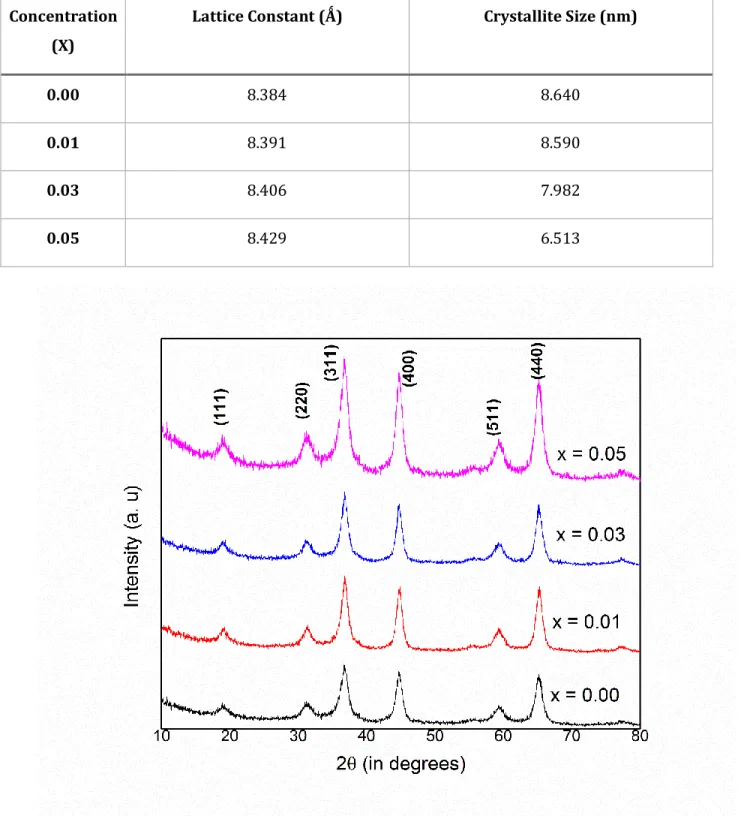
X- ray diffraction patterns were obtained for all the MgxMn1-xFe2O4 (x = 0.00, 0.01, 0.03, 0.05)
samples prepared by chemical co-precipitation method. The structural analysis and detection of phase present in these ferrites sampleswas done using Rigaku –Denki X-ray Diffractometer( Cu-Kα, λ = 1.154 Ǻ). The variation of x-ray intensities were studied between the ranges of 2θ from 10 to 80o. All the samples showed single phase spinel structure with several broad peaks. The most
intense peak were observed at 2θ = 36.75o.The lattice parameter of the MgxMn1-xFe2O4 ferrites was
found to reduce progressively from 8.482 Ǻ to 8.317 Ǻ with the increase in the concentration of the Mg2+ ions from x= 0.00 to x= 0.05. The decrease in the lattice parameter as tabulated in Table 1 is
attributed to the replacement of the Mn2+ ions of ionic radius 0.91 Ǻ, by the Mg2+ions of smaller
ionic radius 0.78Ǻ. The average grain size was found using Scherrer’s formula, D = Kλ/β cos θ,
whereβ is the full width at half maximum (FWHM) of diffraction peak,λ is the wavelength of X-ray, θ is the angle of diffraction, and K is the Scherrer’s constant of the order of unity for usual crystal. However, in case of nanoparticles, sizes calculated from Scherrer’s formula can vary fromthe real particle size because nanoparticles often produce associated structures [9]. The average grain thus found varies from 9 to 6 nm in the present studies.
Microstructural analysis of MgxMn1-xFe2O4 (x = 0.00, 0.01, 0.03, 0.05)ferrite samples prepared
International Journal of Research in Engineering & Applied Sciences
http://www.euroasiapub.org
49crystalline nature of the material. The average grain size has shown a significant decrease with increase in concentration as observed from SEM images shown in figures 2 and 3. This was in accordance with the XRD results where crystallite size was found to be decreasing. This can be attributed to the dissolution of grain boundaries due to the confinement of the heat energy released during sample preparation and thus forming small grains. A similar decrease in grain growth was evidenced from TEM analysis in the prepared samples. Here increasing dopant concentration was found to have significant decreasing effect on the grain size.
4. Conclusion
Chemical co-precipitation method is a very suitable technique for the formation of ferrites at relevant temperatures. Ferrites made by this technique have remarkably shown fine spinel crystalline structure in all the prepared samples.The same microstructural properties were observed in scanning electron microscopy (SEM) and Transmission electron microscopy (TEM)with decreasing grain sizewhen concentration increases which was also revealed from X-ray diffraction studies. Hence we can conclude that this technique is very useful for the fabrication of Mn-Mg based ferrites for application purpose where magnetic properties and electrical properties can be further explored.
5. References
[1] B.S. Chauhan, R. Kumar, K.M. Jadhav, M. Singh, J. Magn. Magn. Mater. 283 (2004) 71.
[2] M. Singh, S.P. Sud, R.K. Puri, J. Mat. Sci. 29 (1994) 2182.
[3] Y. Purushotam, M. Singh, S.P. Sud, P. Venugopal Reddy, Int. J. Mod. Phy. B 12 (1998) 2247.
[4] C. Heck, Magnetic Materials and their Applications, Butterworth and Co Ltd., London, 1974.
[5] N. Pezelson and D. Condurache, Phys. Stat. Solidi. (a) 26 (1974) K41
[6] G. F. Dionne, IEEE Yrans. Magn, Mag- 5 (1969) 596
[8] E. Moldovanu, Phys. Stat. Solidi. (a) 24 (1974) K55
[9] P.Scherrer, Bestimmung der Grösse und der InnerenStruktur von
International Journal of Research in Engineering & Applied Sciences
http://www.euroasiapub.org
50Table 1. Variation of lattice constant and crystallite size with Mn concentration in MnxMg
1-xFe2O4
Concentration (X)
Lattice Constant (Ǻ) Crystallite Size (nm)
0.00 8.384 8.640
0.01 8.391 8.590
0.03 8.406 7.982
0.05 8.429 6.513