MOLECULAR MECHANISMS OF FLEXIBILITY IN NONHOMOLOGOUS END JOINING
Michael Patrick Conlin
A dissertation submitted to the faculty at the University of North Carolina at Chapel Hill in partial fulfillment of the requirements for the degree of Doctor of Philosophy in the
Curriculum in Genetics and Molecular Biology in the College of Arts and Sciences.
Chapel Hill 2018
Approved by: Dale Ramsden
Dorothy Erie Jeff Sekelsy Shawn Ahmed
2018
ABSTRACT
Michael Patrick Conlin: Molecular Mechanisms of Flexibility in Nonhomologous End Joining
(Under the direction of Dale Ramsden)
DNA double strand breaks (DSBs) are highly toxic DNA lesions that play a critical role in human health and disease. The ability to repair these lesions is essential in all kingdoms of life, and in mammals is primarily attributed to the nonhomologous end joining (NHEJ) pathway. NHEJ faces a unique challenge: unlike other forms of DNA damage, DSBs are structurally heterogeneous, varying wildly in end chemistry. To address this problem, NHEJ has evolved uniquely flexible enzymes: DNA polymerases and a DNA ligase that can act on a remarkable variety of substrates, much more so than their counterparts in other pathways. The mechanistic basis of this flexibility, and its significance to biological repair, are unknown.
NHEJ employs two uniquely flexible polymerases to prepare ends for ligation: DNA polymerase μ (pol µ) and terminal deoxynucleotidyl transferase (TdT). These enzymes act on noncanonical substrates that other polymerases cannot engage. We show these polymerases primarily incorporate ribonucleotides (RNA), not
deoxynucleotides (DNA), during NHEJ, both during repair of chromosome breaks made by Cas9 and during V(D)J recombination. These ribonucleotides facilitate NHEJ by enabling ligation of ends with adjacent mispairs, and even single strand ligation.
Supplementing cells expressing TdT with deoxynucleotides thus blocks repair of Cas9-induced breaks, while ribonucleotide supplementation can improve Cas9-directed mutagenesis. Our results indicate cellular NHEJ often involves transiently embedded ribonucleotides, which promote flexibility in repair at the cost of more fragile
ACKNOWLEDGEMENTS
I thank my thesis advisor, Dale, for providing truly exemplary scientific
mentorship and training. Dale is the most engaged scientist I have ever met: his passion for the work is truly infectious and has been transformative in my own development both as a scientist and as a person.
I would like to thank each member of the Ramsden lab that I have had the pleasure of working with. I would especially like to thank Dr. David Wyatt, whose friendship and career guidance have been integral to my own success.
I am grateful to the members of my thesis committee, Dr. Dorothy Erie, Dr. R. Scott Williams, Dr. Shawn Ahmed and Dr. Jeff Sekelsky, who have been exceptionally engaged with my projects. Every one of our meetings has been productive, and I’ve never left one without ideas for new experiments.
I also thank my supportive group of friends, particularly Chris Rounds who has reviewed much of my scientific writing (including this dissertation) and Ryan Hurd who assisted me with bioinformatics analysis of high throughput sequencing data.
Additional acknowledgments specific to each chapter Chapter 2
This chapter was modified from its original version appearing in Cell Reports in 2017 entitled “DNA Ligase IV Guides End-Processing Choice during Nonhomologous End Joining,” in which Dylan Reid and I were co-first authors. I have divided the
publication here for the purpose of logical flow and have modified figure layouts for this dissertation. Supplemental figures and tables are included here. I, Dylan Reid, and George Small performed all experiments presented in this chapter and analyzed the data. I, Dale Ramsden, Dylan Reid, and Eli Rothenberg wrote the manuscript with input from Michael Lieber. I performed bioinformatics analysis of DNA sequences. Studies presented in this chapter were supported by CA203156 and GM007092, CA084442, and GM108118. I thank Dr. Eric Hendrickson for providing LIG4+/+ and LIG4-/- cell lines used in this work.
Chapter 3
This chapter was modified from its original version which is a manuscript currently under review, in which John Pryor is first author and I am second author. I have divided the publication here for the purpose of logical flow and have modified figure layouts for this dissertation. Supplemental figures and tables are included here. John Pryor, myself, George Small, and Juan Carvajal Garcia performed all experiments presented in this chapter and analyzed the data. In sum, 12 panels of the figures
presented here, will be presented in the final publication. Studies presented in this chapter were supported by CA203156, CA097096, and ACS PF-14-0438-01-DMC. I thank Dr. Luis Blanco for providing MEF cell lines used in this work, Dr. Eric
TABLE OF CONTENTS
LIST OF TABLES ... xi
LIST OF FIGURES ... xii
LIST OF ABBREVIATIONS AND SYMBOLS ... xiii
CHAPTER 1: INTRODUCTION ... 1
1.1 DNA Double Strand Break Repair Pathways ... 1
1.2 Sources of DNA Double Strand Breaks ... 2
1.3 Heterogeneity of Double Strand Breaks ... 3
1.4 Mechanism of NHEJ ... 5
1.5 Rejoining of break ends by DNA Ligase IV ... 6
1.6 End Processing During NHEJ ... 7
1.7 DNA Polymerase µ and Terminal deoxynucleotidyl Transferase ... 9
1.8 Summary ... 10
CHAPTER 2: MECHANISM OF LIGATION FLEXIBILITY IN NHEJ ... 11
2.1 Introduction ... 11
2.2 Methods ... 12
DSB Substrates ... 12
DNA Constructs and Protein Purification ... 12
Electrophoretic Mobility Shift Assay (EMSA) ... 14
smFRET Assays ... 14
Cell Lines ... 15
Cellular NHEJ Assays ... 16
Colony Formation and Cell Growth Assays ... 17
Statistical Analysis ... 18
2.3 Results... 18
LIG4 is specialized to directly ligate mismatched or damaged ends ... 18
Dynamic re-alignment of mismatched ends is required for their ligation .. 20
Cellular NHEJ of complex ends requires remodeling of the PEC ... 22
PEC remodeling guides end processing choice during cellular NHEJ ... 24
Cellular radioresistance requires tolerance of complex ends by LIG4 ... 26
2.4 Discussion ... 27
Mechanistic basis for repair of complex ends by NHEJ ... 27
Significance of LIG4 sensing complex ends ... 29
CHAPTER 3: RIBONUCLEOTIDES ENABLE FLEXIBILITY IN NHEJ ... 51
3.1 Introduction ... 51
3.2 Methods ... 52
Cell lines ... 52
Double strand break repair assays ... 53
Repair product analysis ... 54
Next-generation Sequencing ... 55
Cellular NHEJ products contain ribonucleotides ... 56
RNA is integrated into the genome during chromosome break repair ... 58
Ribonucleotides enable ligation of mismatched ends ... 59
Deoxynucleotides block mutagenic repair of Cas9 breaks ... 61
3.4 Discussion ... 61
CHAPTER 4: DISCUSSION ... 79
4.1 Tolerance of Mispairs and Damage at the Ligation Step ... 80
4.2 Structural Basis of Damage Tolerance ... 81
4.3 Mobilization of Ends within Repair Complexes ... 82
4.4 Biological Significance of Ligation Flexibility ... 83
4.5 Pol µ and TdT are RNA polymerases in cells ... 84
4.6 NHEJ and Ribonucleotide Excision Repair ... 85
4.7 Ribonucleotides Enable Ligation ... 86
4.8 Ribonucleotides in CRISPR-Cas9 Repair Products ... 87
4.9 Biotechnology Applications ... 88
4.10 Concluding Remarks ... 89
LIST OF TABLES
Table 2.1 – Sequences of DNA Reagents ... 48
Table 3.1 – Stimulation of NHEJ repair pathway by a terminal ribonucleotide ... 75
Table 3.2 – Frequencies of Cas9 repair products in TdT-expressing MEFs ... 76
LIST OF FIGURES
Figure 2.1 – Effect of LIG4 insert1 on NHEJ of complex ends in vitro ... 31
Figure 2.2 – Biochemical Characterization of LIG4 variants ... 32
Figure 2.3 – Effect of complex end structures on pairing dynamics of single molecule complexes with LIG4WT or LIG4Δi ... 34
Figure 2.4 – Effect of distorted ends on pairing dynamics of single molecule complexes with LIG4WT or LIG4Δi ... 35
Figure 2.5 – Effect of LIG4 insert1 on cellular joining of complex end structures ... 37
Figure 2.6 – Effect of LIG4 insert1 on cellular joining of complex ends ... 39
Figure 2.7 – Effect of PEC flexibility on nucleolytic end processing ... 40
Figure 2.8 – Effect of PEC flexibility on nucleolytic end processing ... 42
Figure 2.9 – Effect of LIG4 insert1 on cellular sensitivity to ionizing radiation ... 44
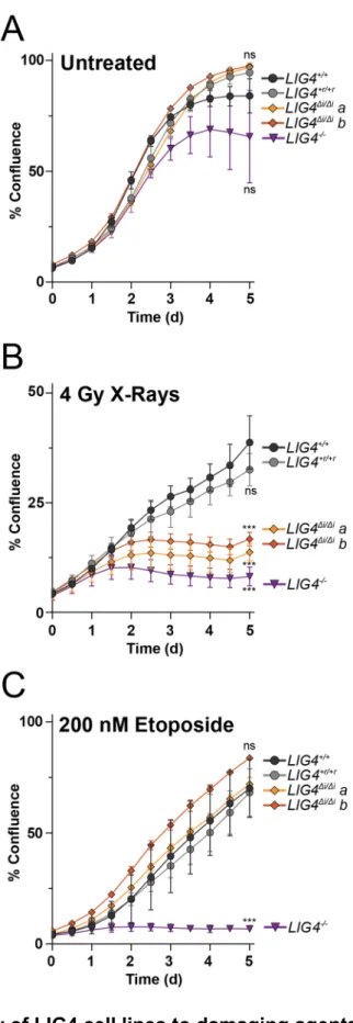
Figure 2.10 – Sensitivity of LIG4 cell lines to damaging agents ... 45
Figure 2.11 – Sensing of differences in end structure by LIG4 guides repair ... 47
Figure 3.1 – Ribonucleotide incorporation during cellular NHEJ ... 64
Figure 3.2 – Assays and cell lines to assess ribonucleotide content during NHEJ ... 65
Figure 3.3 – Detection of ribonucleotides in cellular NHEJ products ... 66
Figure 3.4 – Ribonucleotide incorporation during repair of chromosome breaks ... 67
Figure 3.5 – Detecting ribonucleotides in V(D)J recombination ... 68
Figure 3.6 – Ribo-NHEJ facilitates genome engineering ... 70
Figure 3.7 – Impact of ribonucleotide termini on the NHEJ ligation step ... 72
LIST OF ABBREVIATIONS AND SYMBOLS
Bp base pair
Cas9 CRISPR associated protein 9
CRISPR Clustered Randomly Interspaced Short Palindromic Repeats DNA deoxyribonucleic acid
DNA-PKcs DNA-dependent Protein Kinase catalytic subunit dNTP deoxynucleotide triphosphate
Ds double stranded DSB double strand break DTT dithiothreitol
EDTA ethylenediaminetetraacetic acid EFRET FRET energy
FRET Forster resonance energy transfer Go 8-oxo-7,8-dihydroguanine
HR homologous recombination Igk immunoglobulin kappa IR ionizing radiation Jk joining kappa LIG4 DNA Ligase IV
MEF mouse embryonic fibroblast NHEJ nonhomologous end joining
Nt nucleotide
PAGE polyacrylamide gel electrophoresis PAXX paralog of XLF and XRCC4
PBS phosphate buffered saline PCR polymerase chain reaction PEC paired end complex
Pol polymerase
qPCR quantitative polymerase chain reaction RAG1 recombinase activating gene 1
RAG2 recombinase activating gene 2 RER ribonucleotide excision repair RNA ribonucleic acid
RNP ribonucleoprotein
rNTP ribonucleotide triphosphate Sd standard deviation
SDS sodium dodecyl sulfate sgRNA CRISPR small guide RNA
sgRosa26 small guide RNA targeting Rosa26 smFRET single molecule FRET
Ss single stranded
TBE tris, boric acid, and ethylenediaminetetraacetic acid TdT terminal deoxynucleotidyl transferase
V(D)J variable, diversity, and joining VJk variable joining junction kappa Vk variable kappa
XLF XRCC4-like factor
XRCC4 X-ray cross complemented 4
µ mu
CHAPTER 1: INTRODUCTION
DNA double strand breaks (DSBs) are a ubiquitous and highly toxic form of DNA
damage. These lesions arise from a vast array of sources, including normal biological
processes and exogenous damaging agents. The frequency and myriad sources of
DSBs pose a serious threat to cells because unrepaired DSBs can lead to genome
instability and cell death1,2. Thus, the ability to repair DSBs is essential in all kingdoms
of life. In mammals, the dominant DSB repair pathway is nonhomologous end joining
(NHEJ)3. A central challenge for this pathway is that DSBs are highly heterogeneous
lesions and can have end chemistry that complicates repair4. To address this problem,
the NHEJ pathway has evolved highly flexible enzymes that work on noncanonical
substrates in unorthodox ways5. The mechanisms underlying this flexibility and their
significance for biological repair are poorly understood.
1.1 DNA Double Strand Break Repair Pathways
In addition to NHEJ, the other main DSB repair pathway is homologous
recombination (HR)6. HR is a template-based repair pathway: an intact second copy of
the broken DNA serves as a template for repair. Mechanistically, HR begins with
nucleolytic resection and proceeds through template-directed synthesis using a
A third repair pathway, DNA Polymerase Theta Mediated End Joining (TMEJ),
typically makes a relatively minor contribution to repair, although it becomes essential in
the absence of NHEJ or HR7,8. Like HR, TMEJ relies on resection and synthesis to
repair breaks7. These processing steps effectively convert a DSB into a substrate highly
amenable to ligation: a simple set of DNA nicks spread apart by fully complementary,
newly synthesized DNA. Because they involve such extensive processing, HR and
TMEJ face limitations on when they can be active within the cell9. Moreover, HR
requires a homologous template and TMEJ requires resected ends with embedded
homology. In contrast, NHEJ is free from these requirements; it is active throughout the
cell cycle and capable of robustly repairing DSBs irrespective of any homology9.
The NHEJ pathway has been highly conserved across evolution and is present in
bacteria and humans. Hypomorphic mutations in NHEJ genes cause human diseases
characterized by severe immunodeficiency, cancer predisposition, neurodevelopmental
disorders, and premature aging10. Repair by NHEJ is sometimes described as
error-prone because it can produce small insertions and deletions at the site of repair11. In the
event of multiple breaks, NHEJ can also improperly join different chromosomes
together, producing chromosomal translocations that can lead to cancer12. A key
challenge facing the NHEJ pathway is that DSBs are highly diverse lesions, both in
terms of their causes and their chemical structures.
1.2 Sources of DNA Double Strand Breaks
Chromosome DSBs arise from a vast array of sources, and are even induced as
introduce DSBs that are ultimately responsible for much of the genetic diversity of
gametes13. Similarly, rearrangement of the variable, joining, and diversity gene
segments (V(D)J recombination) underlies the vertebrate adaptive immune system and
proceeds through a DSB intermediate14. However, all DSBs outside of the germline and
lymphoid tissue are unintended, with dividing mammalian cells encountering around 10
breaks per cell cycle15. Genome replication is a major source of these breaks, which
occur when the replication fork encounters DNA damage that cannot be bypassed, or
when topoisomerase enzymes fail16,17.
Outside of those normally encountered throughout biology, the most important
sources of DSBs are exogenous damaging agents that include some of the most
commonly used cancer therapies. Ionizing radiation (IR), topoisomerase poisons, and
many other chemotherapeutics kill cancer cells primarily by inducing DSBs18,19. From a
biotechnology standpoint, DSBs are an essential tool used to specifically edit the
genome through guided nucleases, most notably in the CRISPR-Cas9 system20,21.
1.3 Heterogeneity of Double Strand Breaks
The many sources of DSBs result in breaks with even more diverse chemical
structures. Ionizing radiation (IR) alone produces breaks with a wide variety of
associated oxidative lesions including 5,6-thymine glycol,
5-hydroxymethyl-2’-deoxyuridine, 5-formyl-2’-5-hydroxymethyl-2’-deoxyuridine, 8-oxoguanine, and many chemically distinct
abasic sites22–25. Each of these forms of damage are common and in sum amount to
well over 1000 lesions per cell per Gray of radiation25. Moreover, IR produces these
no way to measure the exact fraction of IR-induced DSBs with associated oxidative
lesions, but estimates based on lesion frequency suggest the proportion to be around
one-third of all breaks23. Different sources of ionizing radiation are thought to vary in the
number and complexity of the oxidative lesions they produce, and thus create DSBs
with different end chemistry as well28.
In contrast with IR, the structures of breaks induced by V(D)J recombination are
well understood. V(D)J recombination occurs in developing lymphocytes and is
essential for the adaptive immune system in vertebrates14,29. In this pathway, DSBs are
intentionally induced by the recombination-activating genes RAG1 and RAG230–32. RAG
activity leaves one DSB end with a DNA hairpin that is cleaved imprecisely by the
Artemis nuclease resulting in heterogeneous end structures33. This heterogeneity is
further augmented by terminal deoxynucleotidyl transferase (TdT, see below)34. The
corresponding sequence heterogeneity in the repaired products of these breaks gives
rise to a diverse repertoire of antibody-encoding genes for an effective immune system.
Repair in V(D)J recombination is carried out entirely by NHEJ and its enzymes function
in this process largely as they do in nonlymphoid cells35,36. An exception is TdT, a
specialized DNA polymerase that is only expressed in lymphoid tissue and acts almost
exclusively during V(D)J recombination37,38.
Topoisomerase poisons such as etoposide are some of the most commonly used
cancer therapy agents. These drugs primarily kill cancer cells by generating DSBs with
topoisomerase protein adducts that block repair19. In sum, the striking heterogeneity of
1.4 Mechanism of NHEJ
The diversity of DSBs makes them unlike any other form of DNA damage, and
the way NHEJ addresses these breaks is fundamentally different from any other repair
pathway. While HR and TMEJ circumvent the heterogeneity of DSBs by extensively
resecting breaks to render them simple repair substrates6,7, NHEJ attacks complex
breaks directly, without resection11,39. To repair such a wide range of substrates, NHEJ
has evolved extraordinarily flexible enzymes that can act in unique ways and on
substrates that other enzymes in their respective classes cannot engage5.
The mechanism of NHEJ proceeds in 4 steps: 1) recognizing and binding the
DSB, 2) bridging of break ends by the NHEJ paired end complex (PEC), 3) optional
processing of the ends, and 4) ligation40. The first step, end recognition, is carried out by
the Ku70/80 heterodimer (Ku)41,42. Ku is often one of the first proteins that binds to the
ends of a double strand break; its binding then mediates the localization of other NHEJ
proteins to the break as well43,44. After the NHEJ machinery reaches the break, the ends
are juxtaposed and held in place to form a paired end complex (PEC)45. At a minimum,
formation of stable PECs requires the NHEJ ligase, DNA Ligase IV (LIG4), Ku, and the
scaffold proteins XRCC4 and XLF46–49. The DNA-dependent protein kinase catalytic
subunit (DNA-PKcs) may also participate in PEC formation50, but its specific role
remains controversial51.
Little is known about how efficiently PECs are formed on different kinds of breaks
(i.e. heterogeneous end structures), the dynamics of the DNA ends and the proteins
within the complex, and the relationship between PEC formation and ligation51. These
through direct ligation of the ends or optional processing of ends by polymerases or
nucleases4.
1.5 Rejoining of break ends by DNA Ligase IV
The ligation step of NHEJ is executed by LIG4, one of three DNA ligases in
mammals and the only one that participates in this pathway52. LIG4 is an
ATP-dependent enzyme consisting of a 3-domain N-terminal catalytic core and 2 C-terminal
domains that facilitate its interactions with other NHEJ factors53. The catalytic core of
LIG4, like those of most other ATP-dependent ligases, consists of a DNA binding
domain, an adenylation domain containing an active site lysine, and an OB-fold
domain54.
DNA ligases have been observed in two structural conformations: a “closed”
conformation in which the catalytic core domains wrap around a DNA substrate and an
“open” conformation in which the catalytic core is extended and largely unbound55–57.
Substrate binding is thought to dictate the transition between these states, and the
transition is believed to serve as a checkpoint for ligases to interrogate their substrates
for compatible termini to join58. This mechanism is highly conserved and is employed by
both bacterial and human DNA ligases; thus, LIG4 likely uses it as well, although this
remains to be demonstrated structurally. Since LIG4 is required for formation of the
NHEJ PEC, its open-closed transition is presumably key to stable PEC formation. Other
ligases probably do not form PECs; if and how LIG4 is specialized to act in this role is
After LIG4 engages the DSB ends, its active lysine covalently binds and then
transfers an adenylate group to the 5’ phosphate of the DSB, which is then resolved by
nucleophilic attack of the 3’ hydroxyl to form a phosphodiester bond54. LIG4 can carry
out this chemistry on a remarkable variety of substrates, including ends with mispairs,
oxidative damage, short patches of single strand DNA, and even 1-2 nucleotide gaps,
without any processing steps to remove these distortions11,59–61. In contrast, the other
mammalian ligases, and most ligases throughout biology, are high fidelity enzymes
unable to join these kinds of end structures60,62. Since the NHEJ ligation step shows low
fidelity, it is well adapted to the wide array of substrates it must join to allow for efficient
repair4. This ligase activity is extremely rare in biology; its only precedents are NHEJ
pathways in other organisms and some viral ligases63,64. While it is clear that NHEJ is
remarkably flexible in accommodating a wide range of substrates, it is unknown if this
activity is indeed important for cellular DSB repair in the context of the chromosome.
The mechanism underlying this flexibility is also unknown, and both of these issues are
addressed in Chapter 2. In addition to a flexible ligation step, NHEJ also addresses the
challenge of substrate diversity with a cast of end processing factors that add or
subtract nucleotides to generate more suitable ligation substrates4.
1.6 End Processing During NHEJ
The repair of any single NHEJ substrate can result in a large number of different
NHEJ products: heterogeneous substrates are repaired into even more heterogeneous
products11. This product heterogeneity is intrinsically linked to the complexity of the
products (e.g. fully complementary overhangs). As the severity of flanking mispairs and
damage increases, so do the number and frequency of different products recovered11.
To some extent, this phenomenon reveals the limitations of LIG4 flexibility: some ends
cannot be tolerated even by such a robust ligase and must instead be processed by
nucleases and polymerases prior to joining.
The Artemis endonuclease is the only nuclease known to act specifically within
NHEJ33. In fact, an Artemis fragment has been crystallized in complex with a fragment
of LIG4. Since Artemis cleaves V(D)J recombination intermediates, patients with defects
in this nuclease are severely immunodeficient65. Nonlymphoid cells derived from these
patients are sensitive to ionizing radiation, suggesting a role for Artemis in NHEJ
outside of V(D)J recombination66. Supporting this idea, a recent study showed in vitro
that Artemis cleaves flaps from 3’ overhangs during NHEJ67.
In addition to specialized nucleases, NHEJ has evolved the ability to utilize 3
X-family DNA polymerases to synthesize nucleotides onto 3’ termini of DSB ends in
preparation for ligation68. DNA polymerases λ (pol λ), µ (pol µ), and TdT all function in
this capacity and differ substantially from other polymerases. While replicative
polymerases are characterized by high fidelity and processivity, NHEJ polymerases lack
proofreading activity and rarely synthesize more than a few nucleotides69. Moreover, all
three NHEJ polymerases have N-terminal domains that facilitate their interactions with
the NHEJ core factors69. In addition to these differences, the NHEJ polymerases
possess the unique ability to incorporate nucleotides onto noncanonical polymerase
substrates69,70. Compared with pol λ, pol µ and TdT exhibit activity on an even wider
1.7 DNA Polymerase μ and Terminal deoxynucleotidyl Transferase
Pol µ and TdT possess noteworthy biochemical properties that render them
completely unique among nucleic acid polymerases. For instance, pol µ is the only
eukaryotic polymerase that enables rejoining of non-complementary, broken DNA ends
absent a paired primer terminus72. The unique ability of pol µ to function in this context
is evidently dependent upon loop 1, a conserved structural feature unique to the NHEJ
polymerases73. Additionally, pol µ carries out repair using a distinctive “skip ahead”
mechanism, wherein the polymerase fills gaps larger than one nucleotide by
synthesizing only one nucleotide immediately adjacent the 5’ DSB terminus74.
Consistent with this observation, the biological role of pol µ seems to be the
incorporation of single nucleotides almost exclusively72.
In contrast with pol µ and pol λ, TdT is only expressed in lymphoid tissue and
participates exclusively in V(D)J recombination75,76. Like pol µ, TdT’s possible primer
and template exhibit a remarkable level of flexibility71. Unlike all other DNA
polymerases, TdT synthesizes onto fully single stranded overhangs and is thus fully
template-independent71,77. In accordance with its role in introducing diversity during
V(D)J recombination, TdT will polymerize multiple nucleotides onto its primer78. Through
their unique biochemical activities, pol µ and TdT promote immune system diversity and
cellular radioresistance34,79–82. Investigations into the biochemical properties of these
polymerases have therefore focused on explaining these biological roles.
Perhaps the most intriguing and poorly understood biochemical property of pol µ
discriminate against incorporating ribonucleotides into the genome, with at least a
1,000-fold preference for deoxynucleotides83–85. This discrimination is vital because
genomic ribonucleotides are toxic and the pathway that removes them is essential in
mammals86. Surprisingly, TdT and pol µ demonstrate sugar selectivity 200-fold lower
than other polymerases, and only prefer deoxynucleotides about 5-fold more than
ribonucleotides37,87–90. It is unclear if these polymerases incorporate ribonucleotides into
the genome in cells or if their lack of sugar selectivity serves any biological role
whatsoever88. An investigation of this phenomenon is the subject of Chapter 3.
1.8 Summary
The NHEJ pathway must successfully repair a wide range of chromosome break
substrates to promote genome stability. The DNA ligase and DNA polymerases
employed by this pathway are the most flexible enzymes in their respective classes, in
terms of their ability to act on unorthodox substrates5. The mechanisms underlying this
flexibility are poorly understood and it is unclear if they are important for repair in cells.
My work has focused on answering these questions, initially focusing on the ligation
step (Chapter 2) and then on the interaction between polymerases and the ligation step
CHAPTER 2: MECHANISM OF LIGATION FLEXIBILITY IN NHEJ
2.1 Introduction
DNA double-strand breaks (DSBs) are genomic lesions that play an important
role in human health and disease. They are frequently generated by exogenous
damaging agents (e.g. ionizing radiation) or as programmed intermediates in meiosis
and V(D)J recombination15. The ends generated by these biological sources of
chromosome breaks are often “complex,” with DNA helix-distorting nucleotide damage,
mismatches, or chemical adducts that pose challenges to the ligases and polymerases
needed for DSB repair91. This problem is especially relevant to the nonhomologous end
joining (NHEJ) pathway since, unlike other DSB repair pathways, these complex ends
are not extensively resected prior to synthetic steps (polymerase and ligase activity).
Ligation is the only essential step in NHEJ, and is performed by one of the three
mammalian ligases, DNA ligase IV (LIG4)52. LIG4 is recruited to broken ends through
participation in a complex of core NHEJ factors including XRCC4, the Ku 70/80
heterodimer (Ku)44, and XLF. This NHEJ core complex is sufficient to physically link a
pair of broken ends together, and can thus be termed the paired end complex, or PEC.
The PEC is essential for repair of diverse end structures; for example, XLF is required
both for stable PEC formation51 and ligation of complex ends, but only modestly affects
can be modulated by ligation-compatible DNA end chemistry93. However, it is unclear
how differences in end structure trigger these changes in dynamics, and whether these
changes in dynamics impact cellular repair.
Here we address this problem by assessing the impact of diverse end structures
on in vitro functional assays, single-molecule analyses of end-pairing dynamics, and
cellular repair and survival. We show that mismatches near strand break termini trigger
extensive PEC remodeling. Moreover, a separation-of-function mutation in LIG4 links
this mispair-induced PEC remodeling to the sensing of these end structures by LIG4,
and argues PEC remodeling is essential to the proficiency of cellular NHEJ in repairing
these end structures.
2.2 Methods
DSB Substrates
DSB substrates were made by ligating the 15-30 bp double stranded
oligonucleotide “caps” described in Table 2.1 to a 285-bp PCR-generated common DNA
“core” segment that had been digested with BsaI to generate appropriate sticky ends.
Substrates were purified with the QiaQuick PCR purification kit (Qiagen), 5’
phosphorylated with T4 polynucleotide kinase (NEB), and substrate assembly validated
by gel electrophoresis.
DNA Constructs and Protein Purification
Constructs for expression after baculovirus delivery of human Ku, XLF, XRCC4,
and LIG4WT into Hi-5 insect cells have been previously described71,94. LIG4∆i was
The LIG3+4 chimera was generated by replacing amino acids 1-638 of LIG4WT with a
fusion of amino acids 170-862 of human LIG3 to the linker (GGGGS)x3 (Genewiz). Cell
pellets were extracted, lysed by sonification, and purified by sequential chromatography
on Histrap and MonoQ columns (GE Biosciences). Figure 2.1A structures were
prepared in Pymol and include hLIG1 (1X9N)56, hLIG3 (3L2P)55, and hLIG4 (3W1B)95,
with disordered insert1 modeled by the SWISSmodel server96.
in vitro Joining Assays
NHEJ reactions were initiated by incubating 2 nM DSB substrates, 25 nM Ku, 40
nM XLF, and 40 nM XRCC4-LIG4 in a buffer with 25 mM Tris pH 7.5, 100 uM EDTA, 1
mM DTT, 5 mM MgCl, 100 uM ATP, 150 mM KCl, 8.5% polyethelene glycol 3000, and
100 ng of supercoiled plasmid DNA. Reactions were carried out for 10 minutes at 37 C
and stopped with 0.1% SDS and 20 mM EDTA. Repair products were purified by
phenol-chlorform extraction and recovery was measured by real-time PCR (qPCR)
using a QuantStudio 6 system (Applied Biosciences), primers that amplify head-to-tail
junctions (Table 2.1), and VeriQuest SYBR Green master mix (Affymetrix). The relative
numbers of molecules recovered were quantified by a well characterized qPCR
assay11,72.
For nick sealing assays, a 5’Cy5 labeled, nicked 41 bp substrate was generated
by annealing three oligonucleotides (Table 2.1). 5 nM substrate was incubated with
XRCC4-LIG4 at 37 C and products were characterized by denaturing polyacrylamide
gel electrophoresis (PAGE). Wild-type XRCC4-LIG4 was titrated to determine that 0.5
nM ligase (1:10 enzyme:substrate) generates sub-saturating (19-21%) amounts of nick
triplicate to generate data presented in Figure 2.2B. Reaction velocity was determined
by quantifying band intensities using ImageJ software.
Electrophoretic Mobility Shift Assay (EMSA)
Substrates for EMSA were generated by annealing oligonucleotides (Table 2.1)
to produce a Cy5 labeled 15 bp substrate to assess DSB end binding (Figure 2.2C), as
well as a Cy5 labeled, 60 bp substrate to assess complex formation (Figure 2.2D). To
assess intrinsic end binding, the 15 bp substrate was incubated at 10 nM with 125, 250,
or 500 nM XRCC4-LIG4. For complex formation, the 60 bp substrate was incubated at
10 nM with 2 nM Ku, 40 nM XLF, and 40 nM XRCC4-LIG4. These samples were
incubated for 20 min on ice in EMSA buffer (50 mM NaCl, 75 mM KCl, 25 mM Tris pH 8,
13% Glycerol, and 0.015% X100). Samples were run on 4% (Figure 2.2C) or 6%
(Figure 2.2D) polyacrylamide gels in 0.5x TBE buffer and imaged using a Typhoon
(GE).
smFRET Assays
smFRET assays and analysis were performed as described previously51,93.
Briefly, NHEJ reactions composed of 50 nM Ku, LX, XLF gloxy (0.5 mg/mL glucose
oxidase and 0.4 μg/mL catylase), and 1 nM dsDNA were added stepwise to NEB4 (20
mM pH 7.5 TricAc, 50 mM KAc, 10 mM MgAc) supplemented with 0.8% glucose, ~5
mM Trolox, 1 mg/mL BSA, and 2 mM DTT. The reaction was immediately flowed into an
imaging chamber that had been prepared with surface dsDNA (~250 pM). Movies
consisting of 1000 frames (33Hz) were acquired for analysis of PECs. Trajectory
analysis, histogram assembly, and autocorrelation of PECs were performed in Matlab93.
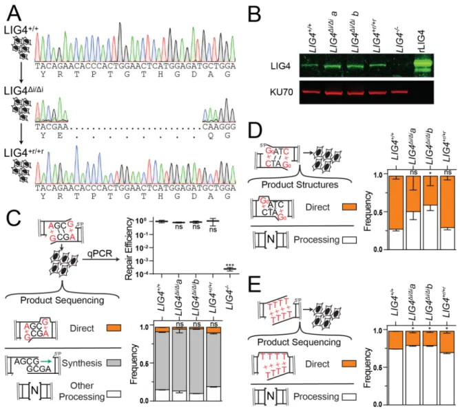
Cell Lines
LIG4-/- cells were generated from parental HCT116 human colorectal cancer cells
by conventional gene targeting and were the gift of Dr. Eric Hendrickson97. We
generated additional variants of the parental cells by CRISPR/Cas9 gene targeting. We
introduced by electroporation plasmids to express Cas998 (Addgene 44758; 5 µg) and
an sgRNA99 (Addgene 51133; 5 µg; guide sequence described in Table 2.1) that targets
insert1-encoding sequence from wild-type LIG4, as well as a gene-targeting donor
plasmid. The donor plasmid was engineered such that it contains 1.1kb of sequence
identical to the LIG4 gene except as modified such that gene targeting ablates the
sgRNA target site, generates the LIG4∆i mutation as described in Figure 2.1A, and
introduces synonymous mutations that result in a BsmFI site used for screening. The
native LIG4 sequence in this region and resulting LIG4∆i alleles are described in Figure
2.6 and Table 2.1. Targeted puromycin resistant clones were identified by amplification
of the insert1 region using primers specific to the native locus (i.e. originate outside of
donor sequence identity). Two independently generated clones, LIG4∆i/∆i a and b, were
produced that possessed only targeted alleles after sequencing (Figure 2.6A). To
generate LIG4+r/+r reverted cells we repeated gene targeting, but started with LIG4∆i/∆i a
cells and used an sgRNA specific for the LIG4∆i allele (Table 2.1) as well as a
gene-targeting donor with wild-type sequence in this region.
We verified LIG4 expression in all of these cell lines using standard western blot
techniques (Figure 2.6B) and antibodies against human LIG4 (Serotec cat no. AHP554)
and human Ku70 (Abcam cat no. ab62820). All 5 cell lines were cultured in McCoy’s 5A
mycoplasma contamination by PCR100; we additionally employed a third party to
validate the absence of mycoplasma by an alternate method for a randomly selected
cell line (Hoechst staining)101.
Cellular NHEJ Assays
Extrachromosomal DNA substrates described above (20 ng) were electroporated
into 2 x 10⁵ cells with pMAX-GFP plasmid (600 ng) at 1350 V in one 30 ms pulse in 10
µL (Neon, Invitrogen). Transfected cells were incubated for 30 minutes in antibiotic-free
McCoy’s 5A media with 10% fetal bovine serum. Cellular repair products were
harvested using a QIAamp DNA mini kit (Qiagen). Each electroporation was reproduced
in triplicate from 3 independent preparations of cells. Repair efficiency was quantified by
qPCR as described above for in vitro joining assays.
Repair product structures were determined by restriction digest for the
8-oxoguanine (2-Amino-7,9-dihydro-1H-purine-6,8-dione; Go) substrate, and by
high-throughput sequencing for all other substrates. For the Go substrate, harvested repair
products were amplified with Cy5-labeled primers (Table 2.1) and digested with BstZ17I
(New England Biolabs; recognizes transversion mutation after amplification of Go) and
BamHI (New England Biolabs; recognizes accurately amplified Go) to identify directly
ligated products. The intensities of digested and undigested bands were quantified
using ImageJ.
To determine repair product structures of all other substrates, sequencing
libraries were prepared by PCR amplification of repair products with primers containing
6-nucleotide indices on their 5’ ends (Table 2.1). Amplified DNA (40 ng per library) was
(NEB) to add dA to the 3’ termini. Ends were ligated to adapters for paired-end
sequencing (Illumina). Pooled libraries were purified from 3% agarose gels to remove
unligated adapters using the QIAquick gel extraction kit (Qiagen). Recovered samples
were amplified for an additional 9 cycles using enrichment primers (Illumina). Products
were again purified using Ampure XP beads (Beckman Coulter). 27.27 ng from each of
the 11 pools was combined (for a total of 300 ng of sample), supplemented with
PhiX174 (40% final concentration) and submitted to the UNC high throughput
sequencing facility for a 2 x 75-bp MiSeq run (Illumina). Genomics Workbench was
used to remove PhiX174 DNA, merge read pairs, de-index libraries, and remove low
quality sequences (CLC-Bio). Remaining sequences were analyzed using Microsoft
Excel.
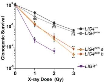
Colony Formation and Cell Growth Assays
For colony formation assays, seeding densities were determined independently
for each dose and cell line such that 50-150 colonies would be produced per 10 cm
dish. Cells were plated on 10 cm dishes in fresh McCoy’s 5A media with 10% fetal
bovine serum, incubated for 4 hours, and then irradiated with indicated doses of X-rays
using a RS 2000 irradiator (Rad Source Technologies). Colonies formed after 14 days
were stained with a solution of crystal violet (0.5%) and glutaraldehyde (6%). Colonies
were manually counted on three plates per dose and cell line. The surviving fraction of
LIG4-/-cells treated with 3 Gy of X-rays was much less than 10-3, and was excluded from
analysis because the resulting faint, small colonies could not be reliably discriminated
For live cell imaging, 2000 cells were plated into 96-well plates in triplicate for
each dose and cell line. After overnight incubation, cells were irradiated or treated with
etoposide and placed into the IncuCyte live cell imager (Essen biosciences). Four 215
mm2 images were taken per well at 10x objective every 4 hours for a total of 120 hours.
The confluence of each image was determined by generating a confluence mask with
IncuCyte software (Essen biosciences).
Statistical Analysis
For all experiments, means were tested for significance against a control (e.g.
LIG4WT, LIG4+/+ cells) using two-tailed t-tests for single comparisons, one-way ANOVA
for multiple comparisons, and two-way ANOVA for comparisons with multiple variables.
Dunnett’s correction for testing multiple hypotheses was applied as necessary. For each
experiment, the value and definition of n, the representation of error bars, the specific
tests used, the specific control tested, and the determination of statistical significance
are described in the figure legends.
2.3 Results
LIG4 is specialized to directly ligate mismatched or damaged ends
Activity of all three mammalian ligases requires the encircling of double stranded
DNA at a strand break54. Structural studies identified 6-10 amino acids inserted in LIG4
orthologs (residues 113-122 in human LIG4), relative to other eukaryotic ligases (Figure
2.1A; Figure 2.2A)95. This insert is located within the strand break-bound ligase on the
opposite side of the double helix from the strand break and site of catalysis, suggesting
damage. Consistent with this idea, we purified LIG4 with this element specifically
deleted (LIG4∆i) in a complex with XRCC4. We determined that insert1 had no
significant impact on LIG4-XRCC4 intrinsic nick sealing activity (Figure 2.2B), DNA
binding (Figure 2.2C), or ability to form a higher-order complex with NHEJ core factors
Ku and XLF on DNA (Figure 2.2D). In vitro NHEJ activity was also similar comparing
LIG4∆i to LIG4WT when ends had complementary overhangs (Figure 2.1B; 5’ G:C, 3’
G:C). In contrast, when ends had mispairs or damage at strand-break termini, in vitro
NHEJ activity using LIG4∆i was reduced 21-66-fold relative to LIG4WT (Figure 2.1B; 5’
GoxC, 3’ GxT, 3’ GxA). LIG4∆i is thus specifically defective in supporting in vitro NHEJ
when substrates have helix-distorting 8-oxoguanine (Go) damage or mispairs near
strand termini.
Ends with mispaired nucleotides are critical NHEJ substrates that arise during
V(D)J recombination and after nucleolytic processing of radiation-induced breaks. They
also presumably act as a model for ends with other sources of helical distortion,
including nucleotide damage. To validate this inference, we measured in vitro NHEJ of
ends with 8-oxoguanine (Figure 2.1B, 5’ GoxC), the most common form of oxidative
base damage. NHEJ activity on this substrate was reduced over 50-fold with LIG4∆i,
which was comparable to the effect of a terminal G:A mispair. Therefore, insert1 is
required for direct ligation of end structures with flanking helical distortions, whether the
distortions are due to mispairs or nucleotide damage. To further explore the extent to
which ligation of ends with terminal mispairs or damage is specific to wild-type LIG4, we
generated a chimera (LIG3+4) with all three LIG4 catalytic sub-domains replaced with the
LIG3+4 physically associates with XRCC4 and was fully competent in Ku- and
XLF-dependent ligation of ends with complementary overhangs. However, end joining with
this chimera was even more sensitive than LIG4∆i to terminal nucleotide damage
(activity reduced more than 100-fold, relative to LIG4WT; Figure 2.2E). This result
consistent with the argument that LIG4 is unique amongst mammalian ligases in its
ability to repair damaged termini. Additionally, the impact of LIG4∆i on repair of damaged
ends is less severe than that of the LIG3+4 chimera, suggesting that insert1 is not
entirely responsible for the unique ability of LIG4 to tolerate mispairs and damage. We
therefore sought to use the LIG4∆i separation-of-function mutation to investigate both
the mechanistic basis for the unique ability of NHEJ to tolerate helix-distorting mispairs
or damage at the ligation step, as well as its significance to cellular double strand break
repair.
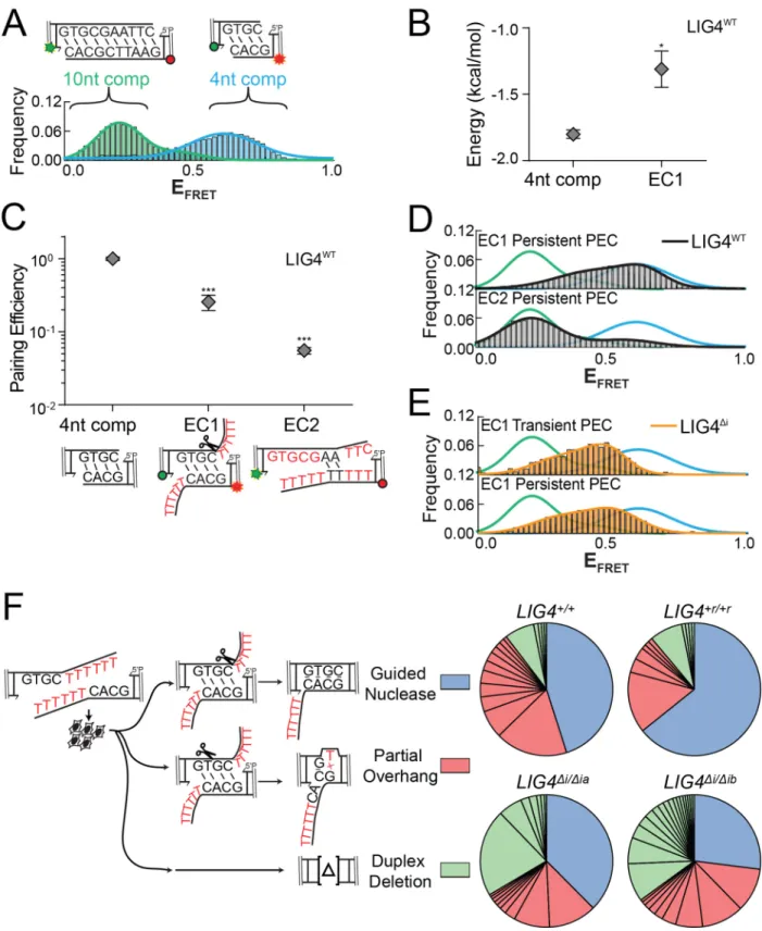
Dynamic re-alignment of mismatched ends is required for their ligation
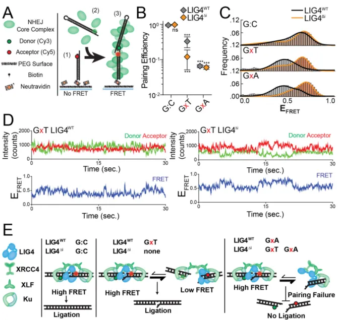
We previously described a single-molecule fluorescent resonance energy
transfer (smFRET) assay that reports on pairing of DNA ends as mediated by a
complex of Ku, XRCC4, LIG4, and XLF51,93. These PECs are apparent as FRET pairs
generated when a Cy3 labeled donor dsDNA fragment in solution stably associates with
a Cy5 labeled acceptor dsDNA fragment immobilized on a surface (Figure 2.3A). In
accord with the in vitro ligation assay, LIG4∆i and LIG4WT similarly promote stable PEC
formation when ends have complementary 4-nucleotide overhangs (G:C, Figure 2.3B).
In contrast, pairing of overhangs with 3’ terminal G:T mismatches is significantly
reduced when comparing LIG4Δi to LIG4WT; this reduced pairing efficiency represents a
even less efficient when termini have a bulkier purine:purine G:A mismatch, but is
similarly inefficient for both LIG4WT and LIG4Δi (Figure 2.3B). PECs thus form less
efficiently with increasing terminal helical distortion, and PECs formed with LIG4∆i are
more sensitive to this challenge.
Changes in FRET efficiency (EFRET) reflect dynamic repositioning of DNA ends
relative to each other within individual PECs51,93. When using complementary ends (3’
G:C; Figure 2.3C), EFRET distributions were not significantly different when comparing
PECs formed with LIG4WT (black line) vs. LIG4∆i (orange line). LIG4∆i PECs also had
similar FRET distributions when ends had terminal mispairs (Figure 2.3C); importantly,
LIG4WT PECs formed on ends with mispaired termini more often had lower EFRET (DNA
labels located further apart; black lines for G:A and G:T mispairs, Figure 2.3C), and
consequently overall wider distributions of EFRET (Figure 2.3C; Figure 2.4A) when
compared to paired temini (G:C). Ends with terminal distortions thus trigger PECs to
sample a wider variety of end-alignment configurations to remain efficiently paired, but
only when using LIG4WT.
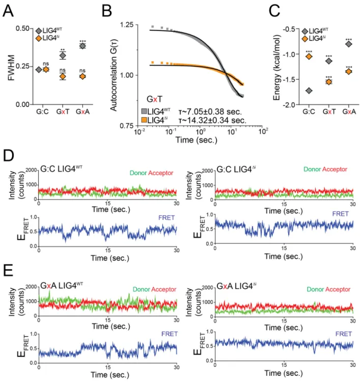
Examination of smFRET trajectories from individual PECs also shows the
transition frequency between FRET states increases when comparing LIG4WT and
LIG4∆i (Figure 2.3D, Figure 2.4C). We quantified this difference by using autocorrelation
of individual FRET trajectories to calculate the average transition times - “lag times” (τ) -
between FRET states93. For ends with G:T mismatches, these were approximately
two-fold lower for PECs formed with LIG4WT, compared to PECs formed with LIG4∆i (Figure
2.4B). These values are then used to calculate the relative stability of the DNA ends in
PECs have lower energetic barriers in assuming new conformations, compared to
LIG4∆i PECs, but again only when ends have terminal mismatches.
PECs containing LIG4∆i and mispaired ends are thus formed less efficiently
(Figure 2.3B), and even when formed do not acquire the high degree of conformational
plasticity observed when PECs are formed with LIG4WT (Figure 2.3C-D, Figure 2.4D-E).
We argue the inability of LIG4∆i to allow for mispair-induced PEC remodeling accounts
for its specific defect in direct ligation of such end structures (Figure 2.1B). There are
also limits to the extent to which remodeling enables ligation, as even LIG4WT is
inefficient in joining ends with bulky G:A mismatches (Figure 2.1B). PECs formed with
paired termini favor a narrow distribution of high FRET end alignments that more closely
resemble FRET distributions observed with products of ligation93; these alignments thus
likely directly juxtapose strand-break termini in anticipation of catalytic steps
(“pre-catalytic”, Figure 2.3E). We attribute the LIG4WT-specific, insert1-dependent flexibility in
accommodation of mispaired termini to a favoring of end alignments that both have
lower FRET (more distally-located labels) and are more dynamic. These more dynamic
and lower FRET PECs – “remodeling PECS” - may be catalytically incompetent, but
allow for iterative attempts at the now transient (but occasionally catalytically
competent) high-FRET intermediate (Figure 2.3E).
Cellular NHEJ of complex ends requires remodeling of the PEC
We next addressed whether the differences in PEC flexibility described above
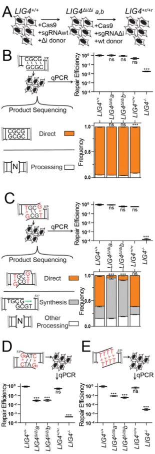
significantly impact cellular NHEJ. We employed scar-free gene targeting to exchange
LIG4WT for LIG4∆i alleles within the native LIG4 locus of a human cell line (Figure 2.5A).
confirmed they express only LIG4∆i from endogenous loci (Methods; Figure 2.6A-B). We
then generated a cell line by another round of gene targeting where the LIG4 locus of
LIG4∆i/∆ia was reverted back to wild type sequence (LIG4+r/+r), as a means of assessing
the effects of possible off-target mutations incurred in the original round of gene
targeting (Figure 2.5A, Figure 2.6A-B). Both LIG4∆i/∆i subclones acted equivalently in
functional assays below. Similarly, results using parental wild type cells (LIG4+/+)
matched those from the LIG4+r/+r reversion, confirming the differences observed in the
LIG4∆i/∆i cells could be attributed to the 8 amino acids deletion.
DSB substrates with varied end structures were introduced into these cells, after
which efficiency of repair was determined by qPCR and product structures were
characterized by high-throughput sequencing. In accord with in vitro results, ends with
complementary overhangs were efficiently joined almost entirely by direct ligation in
both wild-type and LIG4∆i/∆i cells (Figure 2.5B). Also in accord with in vitro data, ends
with terminal G:T mispairs were efficiently repaired by direct ligation (accounts for 60%
of all repair) in both LIG4+/+ and LIG4+r/+r cells, while this class of product is rarely
(<10%) seen in LIG4∆i/∆i clones (Figure 2.5C). Instead, repair in LIG4∆i/∆i cells typically
requires re-alignment of overhangs and gap-repair synthesis prior to ligation of the now
“sticky” end. This alternate pathway is sufficient to fully compensate for the inability of
LIG4∆i to directly ligate terminal mispairs, since overall joining efficiency was
comparable for LIG4+/+ vs. LIG4∆i/∆i cells. Considering repair of ends with bulkier G:A
mispairs, both wild-type and LIG4∆i/∆i cells rely on this compensating pathway (Figure
2.6C), consistent with in vitro observations that neither LIG4WT nor LIG4∆i can ligate this
reduced in LIG4-/-cells (Figure 2.5C; approximately 0.0005 products per cell), to the
extent that we could not recover sufficient repair products to accurately assess product
spectra.
Additional substrates were introduced into cells to assess whether barriers to
mispair tolerance are routinely bypassed by cellular end processing. Similar to 3’ G:T
mispairs, ends with 5’ Go terminal damage are primarily repaired by direct ligation in
LIG4+/+ cells. Importantly, joining of 5’ GoxC in LIG4∆i/∆i cells is over 10-fold less efficient
(Figure 2.5D), even though what little repair does occur is processing-dependent
(Figure 2.6D). We also investigated cellular NHEJ of end structures with entirely
non-complementary overhangs (TTTT). Joining efficiency was again severely reduced in
LIG4∆i/∆i cells, relative to wild-type cells (Figure 2.5D). For this substrate, the rare
products recovered from LIG4∆i/∆i cells only subtly differed from wild-type controls in
terms of junction structure (Figure 2.6E). Thus, in contrast to previously tested
substrates (Figure 2.5C), end processing was not sufficient to rescue repair of TTTT
and 8-oxoguanine substrates in LIG4∆i/∆i cells. We initially linked LIG4WT PEC flexibility
only to the ability of cellular NHEJ to directly ligate ends with terminal mispairs (Figures
2.1, 2.3, 2.5C); these latter results identify additional important contributions to cellular
NHEJ associated with end processing.
PEC remodeling guides end processing choice during cellular NHEJ
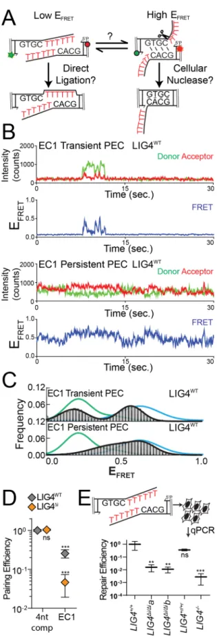
We generated the substrate “EC1” (embedded complementarity 1) to further
explore the relationship between PEC flexibility, cellular end processing, and ligation.
EC1 has long (10-nt) non-complementary overhangs that can plausibly be aligned to
Alternatively, EC1 can be re-aligned to pair complementary sequence embedded within
the overhang, where unpaired tails are a presumptive substrate for nucleolytic end
processing. These two alignments are readily distinguished by smFRET (Figure 2.8A);
PECs formed with a control substrate (with fully complementary 10nt overhangs) had
low EFRET ranges, expected for EC1 alignments that juxtapose 3’OH:5’P termini (green
lines), while PECs formed with 4-nt complementary overhangs had a clearly distinct
population of high EFRET (blue lines) expected for EC1 alignments that pair embedded
complementary sequence. Analysis of individual smFRET trajectories of PECs formed
with EC1 and NHEJ core factors identified a much larger than typical fraction of
transient complexes (lifetimes <5 seconds; Figure 2.7B). Transient PECs had two
distinct populations of EFRET distributions, each roughly corresponding to the two
alignment classes predicted above (Figure 2.7C). Long-lived PECs (persistent) favor
only the high EFRET state, but sample both a wider range of alignment configurations
(Figure 2.7C) and are more dynamic (have lower energetic barriers to transition; Figure
2.8B) than PECs formed with complementary overhangs. To further address if the
persistent PECs frequently involve pairing at embedded complementary sequence (as
suggested by comparison to substrate standards; Figure 2.7C) we used a substrate
where the complementary sequence was both reduced and re-located (“EC2”). As
expected, PECs formed less efficiently with EC2 (Figure 2.8C), and when formed had
mostly lower EFRET (Figure 2.8D). Importantly, LIG4∆i was largely unable to form PECs
with the EC1 substrate (Figure 2.7D), and the rare PECs that do form primarily have
intermediate EFRET states that are inconsistent with either alignment (Figure 2.8E).
PECs formed with LIG4WT that juxtapose strand termini were “filtered out"; only the most
plausibly productive alignments, i.e. those that could lead to ligation after nucleolytic
end processing, were stable (Figure 2.7C).
We next assessed how the EC1 substrate was resolved by cellular NHEJ. Nearly
all products (>99%) were indeed consistent with ligation after nuclease activity, with the
dominant product guided by the alignment at embedded complementary sequence also
favored in smFRET analysis (Figure 2.8F). By comparison, direct ligation of EC1
accounted for less than 0.1% of all cellular repairs. Importantly, joining efficiency of this
substrate was reduced over 60-fold in LIG4∆i/∆i cells, relative to LIG4+/+ cells (Figure
2.7E), even though LIG4∆i/∆i cells are fully proficient at ligating the inferred product of
alignment-guided nuclease activity (a 4bp complementary overhang; Figure 2.5B). This
result suggests that for this substrate, LIG4∆i fails to efficiently mediate repair because it
is defective at an earlier step than ligation – specifically, stable accommodation of
end-alignments required for nucleolytic end processing (Figure 2.7D).
Cellular radioresistance requires tolerance of complex ends by LIG4
LIG4WT thus uniquely accommodates diverse end structures during end pairing.
However, there is wide variation in how this flexibility impacts cellular NHEJ. Depending
on the starting end structure it can be dispensable (Figure 2.5B, Figure 2.6C), alter
product spectra (Figure 2.5C), or can be critical for efficient repair (Figure 2.5D-E,
Figure 2.7E). We therefore addressed the extent to which the inability of LIG4 to tolerate
structural diversity impacts cell growth and survival after ionizing radiation. Using both
standard colony forming assays and real-time imaging of cell growth, LIG4+/+ and
comparison, LIG4∆i/∆i cells were radiosensitive to a degree intermediate between LIG4+/+
and LIG4-/- cells (Figure 2.9, Figure 2.10), a result strikingly similar to joining efficiencies
described for the majority of substrates with complex ends (Figure 2.5D-E, Figure 2.7E).
In contrast with ionizing radiation, LIG4∆i/∆i and LIG4+/+ cells are equally resistant to
etoposide (Figure 2.10). This is consistent with specific requirement for insert1 in repair
of ends with mispairs or damage, since etoposide induced breaks can be processed by
tyrosine phosphodiesterase 2 such that overhangs are undamaged and fully
complementary102. These results show that the ability of LIG4 to sense distortions
facilitates cell survival following treatment with ionizing radiation.
2.4 Discussion
Repair by NHEJ implicitly requires the pairing together of broken chromosome
ends. A complex of Ku, XRCC4, DNA Ligase IV, and XLF (paired end complex, or PEC)
is necessary and sufficient for this purpose51; we describe here dynamic changes in this
complex that are triggered by differences in end structure, and show that this response
is essential for efficient cellular repair.
Mechanistic basis for repair of complex ends by NHEJ
Ends with complementary (“sticky”) overhangs are aligned efficiently and with
little mobility, to the extent that pairing EFRET more closely resemble the narrow
distributions observed in products of ligation, relative to other end structures tested
here. We suggest these PECs describe “pre-catalytic” end alignments, where strand
break termini are directly juxtaposed in anticipation of ligase-mediated catalytic steps
termini – complex ends - induce the sampling of a much wider variety of alignment
configurations, most or all of which no longer juxtapose strand termini.
We use a LIG4 separation of function mutation (LIG4∆i) to identify an essential
role for this second, more dynamic “remodeling” class of PECs in cellular NHEJ for the
repair of complex ends. We show LIG4∆i is specifically unable to accommodate PEC
remodeling in response to complex ends. As a consequence, PECs formed with LIG4∆i
are unable to directly ligate such substrates, but are also – with rare exception (Figure
2.5C) – unable to couple ligation to end processing when end complexity is sufficient to
block direct ligation.
Notably, the exceptions are restricted to contexts where alignment-directed
synthesis generates a fully complementary 6-nt overhang, a substrate expected to be
especially permissive for the ligation step.
By comparison, insert1 is dispensable for the XLF-, XRCC4-, and Ku-dependent
alignment of ends with complementary overhangs, as well as catalytic activity on this
conventional ligase substrate. Moreover, a chimeric ligase with all three LIG4 catalytic
subdomains replaced with LIG3 counterparts is equally effective in ligation of “sticky”
ends (and is similarly stimulated by Ku and XLF), but is even less able to repair complex
ends. Prior work emphasized the importance of a variety of NHEJ proteins, including
PAXX103,104, end processing factors33,67,72, and especially the end-bridging filament of
XRCC4-XLF59,105,106 in repairing complex breaks. Indeed, we previously reported that
these filaments form on bleomycin-induced DSBs and orchestrate their repair51. Here
we identify a critical role for specialization of LIG4 catalytic subdomains in repair of
How does insert1 contribute to PEC remodeling? The three subdomains of
eukaryotic ligases are extended in the absence of DNA (“open” conformation), and
engage substrates by forming a ring around double stranded DNA (“closed”
conformation)55–57. In the closed conformation, the central catalytic subdomain is bound
to strand break termini while insert1 is located in the N-terminal subdomain on the
opposite side of the double helix95. Though not resolved in current apo-enzyme crystal
structures, its location suggests that insert1 helps LIG4 maintain a closed conformation,
either by stabilizing the ring-closing interactions between N and C terminal catalytic
subdomains or by interacting with DNA107. We suggest stable end-pairing is dependent
on LIG4 maintaining a closed conformation, even if LIG4 can directly interact with only
the 5’ phosphate side of a strand break (“half-site” binding). LIG4∆i instead transitions to
an open conformation in this context (like conventional ligases), which leads to failure of
end pairing.
Significance of LIG4 sensing complex ends
Prior work indicates that LIG4 has functions in NHEJ distinct from the ligation
step, most clearly in promoting end pairing51,93,108–110. Data presented here identify a
much more sophisticated function. Differences in how LIG4 catalytic domains interact
with different end structures trigger dramatic changes in the dynamics of the entire
paired-end complex – i.e. including Ku, XRCC4, and XLF paired ends – and these
altered dynamics determine the steps taken to complete repair. This role is distinct from
critical LIG4 roles in catalysis and end pairing, since both of the latter functions are fully
intact in PECs formed with LIG4∆i. LIG4 can thus be identified as the PEC “sensor,”
extent that how LIG4 interacts with aligned ends may dictate the identity of the end
processing factor that next engages the end.
Inhibitors of LIG4 are being explored for their potential to sensitize tumors to
radiation therapy111. Here we identify a role of LIG4 that is specific to the ability of cells
to repair complex damage, identify a structural element required for this role, then show
deletion of this element leads to cellular sensitivity to ionizing radiation. Since this
structural element is unique to LIG4 and required for radioresistance, it presents a
promising therapeutic target, as it is less likely to engender the off-target effects
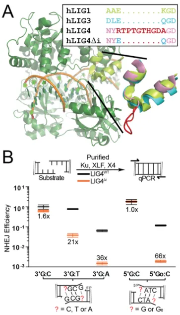
Figure 2.1: Effect of LIG4 insert1 on NHEJ of complex ends in vitro
(A) Structure of human LIG1 bound to DNA (1X9N; green), with inset emphasizing sequence and structural alignments of human LIG1 α helices 5-6 to human LIG3 (3L2P; blue) and human LIG4 (based on 3W1B; pink), with sequence and a modeled location of LIG4 insert1 (disordered in the 3W1B apoenzyme) in red. (B) Ku, XLF, and either XRCC4-LIG4WT (gray) or XRCC4-LIG4Δi (orange) were incubated with substrates
containing different complementary (5’ G:C, 3’ G:C) or non-complementary (5’ GoxC, 3’ GxT, 3’ GxA) overhangs as noted. Joining efficiency is expressed as a fraction of the total junctions recovered using the 5’ G:C substrate with NHEJ reactions containing LIG4WT. Ligation reactions were performed in triplicate and the mean joining efficiencies
are shown, along with the fold difference between LIG4WT and LIG4Δi for each substrate.
Figure 2.2: Biochemical Characterization of LIG4 variants
(A) Amino acid sequence alignment of LIG4 orthologs, with insert1 in orange. (B) A nicked, Cy5-labeled 41 bp substrate was incubated with XRCC4-LIG4WT or
XRCC4-LIG4Δi in triplicate and joining was assessed by stand-denaturing gel electrophoresis.
Error bars represent standard error of the mean for 3 experiments. The mean for LIG4Δi
was assessed by t-test as not statistically significantly different (ns) from control (LIG4WT). (C) A 15 bp Cy5-labeled substrate was incubated with XRCC4-LIG4WT or
XRCC4-LIG4Δi and substrate binding was assessed by native gel electrophoresis. (D) A
60 bp Cy5 labeled substrate was incubated with indicated NHEJ factors and NHEJ complex formation was assessed by native gel electrophoresis. (E) LIG3+4 chimera was
generated by fusing catalytic domains of LIG3 with C-terminal domains of LIG4 and purified after co-expression with XRCC4. NHEJ reactions were performed in vitro as in Figure 2.1B using undamaged (5’ G:C) and damaged (5’ GoxC) substrates. Joining efficiency is expressed as a fraction of the total junctions recovered using the 5’ G:C substrate with LIG4WT. Ligation reactions were performed in triplicate and error bars
Figure 2.3: Effect of complex end structures on pairing dynamics of single molecule complexes with LIG4WT or LIG4Δi
(A) smFRET NHEJ assay: (1) dsDNA with a Cy5 acceptor is tethered to a bitonylated PEG surface via a biotin-neutravadin linkage, (2) dsDNA with a Cy3 donor and NHEJ proteins (green) are added to the chamber, and (3) ends are paired and FRET is observed. (B) Quantitation of pairing efficiency of ends with complementary (G:C) or mismatched (GxT, GxA) overhangs by Ku, XLF, XRCC4 and either LIG4WT (gray) or
LIG4Δi (orange). Error bars represent standard error of the mean for 3 experiments.
Means were assessed by two-way ANOVA as significantly different from control (LIG4WT on G:C substrate) with confidence p<0.001 (***). (C) Histograms of observed
EFRET for PECs formed as in (B). (D) Representative smFRET trajectory for LIG4WT and
LIG4Δi PECs formed with GxT ends demonstrating altered transition frequency and
Figure 2.4: Effect of distorted ends on pairing dynamics of single molecule complexes with LIG4WT or LIG4Δi
(A) Full width at half maximum (FWHM) of peaks was calculated from EFRET histograms
for G:C, GxT, and GxA substrates. For (A) and (C), error bars represent standard error of the mean for 3 experiments, and means were assessed for significance as in Figure 2.3B with confidence p<0.01 (**), p<0.001 (***) or not significantly different (ns). (B)
Autocorrelation of individual FRET trajectories was used to calculate average transition times () between FRET states of PECs formed on the GxT substrate with LIG4WT or
LIG4Δi. (C) Transition energy between FRET states calculated from autocorrelation. (D)
Representative smFRET trajectory for LIG4WT and LIG4Δi PECs formed with G:C
complementary ends (E) Representative smFRET trajectories of LIG4WT and LIG4Δi