Viral Modulators of KSHV Lytic Replication
Carlos M. González
A dissertation submitted to the faculty of the University of North Carolina at Chapel Hill in partial fulfillment of the requirements for the degree of Doctor of Philosophy in the Department of Microbiology and Immunology (Virology).
Chapel Hill 2009
Approved by:
Blossom Damania, PhD
Dirk Dittmer, PhD
Eng-Shang Huang, PhD
Steven Bachenheimer, PhD
ii ABSTRACT
CARLOS M. GONZÁLEZ: Viral Modulators of KSHV Lytic Replication
(Under the direction of Blossom Damania)
Kaposi’s Sarcoma and the B-cell derived primary effusion lymphoma and multicentric Castleman’s Disease are all outcomes of infection with Kaposi’s Sarcoma– associated Herpesvirus (KSHV). A predominantly latent virus, KSHV also requires a subpopulation of cells to undergo lytic reactivation in order to generate an environment conducive to neoplastic growth. The replication and transcription activator (RTA, Orf50) is necessary and sufficient to initiate the lytic cascade. We have studied two modulators of lytic replication encoded by KSHV Orf49 and Orf64 that support RTA during lytic reactivation.
iii
KSHV Orf64 encodes a large, 2636 amino acid tegument protein. We have found that Orf64 encodes deubiquitinase (DUB) activity in its amino-terminal 200 amino acids in vitro and in vivo. Whereas all other herpesviral DUBs cleave Lysine 48 (K48)-linked ubiquitin, KSHV Orf64 was capable of cleaving both K48 and K63-linked ubiquitin chains. Cleavage of K63-linked ubiquitin is associated with functional regulation whereas K48-ubiquitination leads to proteosomal degradation. Mutation of a cysteine residue at amino acid position 29 in the catalytic core of the DUB ablated DUB activity. Orf64 also synergized with RTA to augment transcription from RTA-responsive promoters. These data suggest that the deubiquitination function of Orf64 may play an important role in viral replication.
iv
DEDICATION
v
ACKNOWLEDGEMENTS
First and foremost I would like to thank my advisor, Dr. Blossom Damania, for the guidance, encouragement and generous support throughout this endeavor.
My committee members: Dr. Steve Bachenheimer, Dr. Lishan Su, Dr. Eng-Shang ‘Clark’ Huang, and Dr. Dirk Dittmer. Thank you. You have all been a great source of stimulating discussions, direction and ideas that greatly enriched the work as well as my experience here.
The members of the Damania lab have consistently made the lab a place I want to go in the morning. Thanks to Christine Tomlinson, Brian Bowser and Scott DeWire, all since graduated, for blazing the path in establishing our lab. Special thanks to Prasanna Bhende, Ling Wang and Kwun Wah Wen for all the engaging discussions, laughter and support, technical and otherwise.
vi
TABLE OF CONTENTS
LIST OF FIGURES………...x
LIST OF ABREVIATIONS………...xiii
CHAPTER I. INTRODUCTION……….1
Kaposi’s Sarcoma………...2
Kaposi’s Sarcoma Associated Herpesvirus………....4
KSHV-related diseases………..4
Primary Effusion Lymphoma….………...4
Multicentric Castleman’s Disease………...5
Epidemiology ………...5
Disease Manifestations of Human Herpesviruses……….6
vii
Lytic Replication………9
Latency…………..………...10
Reactivation from Latency………...11
The viral Genome………...11
Model Systems………...12
RRV……….………..16
KSHV viral proteins involved in the modulation of the lytic cycle……….………..17
KSHV Orf50………18
KSHV Orf49………20
KSHV Orf64………...…...20
The NFκB pathway……..………23
OBJECTIVES………..26
viii
II. IDENTIFICATION AND CHARACTERIZATION
OF THE KSHV ORF49 PROTEIN……….43
ABSTRACT………...44
INTRODUCTION……….45
MATERIALS AND METHODS………...47
RESULTS………..51
DISCUSSION………...74
ACKNOWLEDGEMENTS………...78
REFERENCES………..79
III.KSHV ENCODES A VIRAL DEUBIQUITINASE………...84
ABSTRACT………...85
INTRODUCTION……….86
MATERIALS AND METHODS………...89
RESULTS………...94
DISCUSSION………..116
ACKNOWLEDGEMENTS……….120
ix
IV. MULTIPLE FUNCTIONS OF ORF64……….126
ABSTRACT……….127
INTRODUCTION………...128
MATERIALS AND METHODS………135
RESULTS………136
DISCUSSION……….142
ACKNOWLEDGEMENTS……….147
REFERENCES………148
V. GENERAL CONCLUSIONS………...150
x
LIST OF FIGURES
CHAPTER 1
Figure 1: Classification of herpesviruses. ………...7
Figure 2: Schematic Representation of the KSHV Genome………..13
Figure 3: Alignment of KSHV and RRV26-95 Genomes.………...18
Figure 4: Comparison of the BRLF-1 and RTA loci of EBV and KSHV. ………...21
CHAPTER 2 Figure 1: Delineation of the full-length ORF49 transcript………53
Figure 2: Expression Kinetics of ORF49………...56
Figure 3: Cell fractionation of ORF49-transfected cells…………...59
Figure 4: Cellular localization of ORF49………..60
Figure 5: ORF49 enhances ORF50-mediated induction of K8, Orf57 and vGPCR lytic promoters…..……….63
xi
Figure 7: ORF49 induces phosphorylation of c-Jun
by activation of JNK and p38……….69
Figure 8: JNK and p38 activation is necessary
for lytic gene expression……….71
CHAPTER 3
Figure 1: Sequence analysis predicts a
deubiquitinase in KSHV ORF64………95
Figure 2: Expression and intracellular localization of ORF64………..97
Figure 3: The N-terminus of ORF64 encodes deubiquitinating (DUB) activity for both K48- and K63-linked
ubiquitin chains in vitro………100
Figure 4: ORF64 exerts DUB activity in vivo……….103
Figure 5: ORF64 can effectively cleave both K48 and K63
ubiquitin linkages………...106-107
Figure 6: KSHV ORF64 can enhance RTA-mediated
transcriptional activation of lytic promoters………..111 Figure 7: Knock-down of ORF64 affects transcription
and protein expression of lytic genes………114 CHAPTER 4
Figure 1: Diagram of the canonical and non-canonicalNFκB signaling
xii
Figure 2: KSHV Orf64 down-regulates IκBα……….138
Figure 3. Expression of KSHV Orf64 targets various proteins
in the NFκB pathway towards degradation.………141
Figure 4: Reactivation from latency triggers the degradation of
NFκB-1 p105 and IKKγ and stabilization of NFκB-2………..145
Figure 5: Model of Orf64 mediated activation of the
canonical NF-κB pathway………146
CHAPTER 5
Figure 1: Model of Orf49- and Orf64-mediated support
xiii
LIST OF ABREVIATIONS
AIDS Acquired immunodeficiency syndrome
AMC 7-amido-4-methylcoumarin
bp Base pairs of DNA
BC-1 EBV+, KSHV+ BCBL-derived B-cell line
BCBL Body cavity based lymphoma
BJAB Burkitt lymphoma-derived B-cell line
BL Burkitt Lymphoma
CAT Chloramphenicol acetyl-transferase
cDNA Complementary DNA
CIP Calf intestinal phosphatase
CV-1 African green monkey kidney-derived cell line
DAPI 4'-6-Diamidino-2-phenylindole
DBP DNA-binding protein
DMEM Dulbecco’s modified eagle media
DMSO Dimethyl-sulfoxide
xiv DUB Deubiquitinase
DTT Dithiothreitol
E Early class of lytic transcription
EDTA Ethyl-diamine tetra-acetic acid
EBV Epstein-Barr virus
EDTA Ethyl-diamine tetra-acetic acid
EGFP Enhanced green fluorescent protein
ELISA Enzyme-Linked Immunosorbant Assay
ER Endoplasmic reticulum
FLICE Fas-associated death domain-like interleukin-1beta-converting enzyme
FLIP Fas-associated death domain-like interleukin-1beta-converting enzyme-inhibitory protein
GAPDH Glyceraldehyde 3-phosphate dehydrogenase
gB glycoprotein B
GST Glutathione S-transferase
GRP-78 glucose regulated protein 78
hCMV Human cytomegalovirus
xv HEK-293 Human endothelial kidney- 293 cells
HeLa Human liver-derived cell line
HEPES 4-(2-hydroxyethyl)-1-piperazineethanesulfonic acid
HIV Human immunodeficiency virus
HSV Herpes simplex virus
HHV8 Human herpesvirus 8 (KSHV)
IE Immediate early class of lytic transcription
IL Interleukin
IP Immunoprecipitation
IPTG Isopropyl β-D-1-thiogalactopyranoside
IsoT Isopeptidase T
JNK c-Jun N-terminal kinase
kDa Kilodalton
KS Kaposi’s Sarcoma
KSHV Kaposi’s sarcoma-associated herpesvirus
L Late class of lytic transcription
xvi MCD Multicentric Castleman’s disease
mCMV Murine cytomegalovirus
mCP minor Capsid Protein
MHV-68 Murine herpesvirus-68
mRNA Messenger RNA
MTA mRNA transport and accumulation protein
NERPRC New England Regional Primate Research Center
nt Nucleotide of DNA or RNA
ORF Open reading frame
ORPRC Oregon Regional Primate Research Center
PAGE Polyacrylamide gel electrophoresis
PBS Phosphate-buffered saline
PCR Polymerase chain reaction
PEL Primary effusion lymphoma
PRV Pseudorabies Virus
RACE Rapid amplification of cDNA ends
xvii RhF Rhesus fibroblast cell line
RNA Ribonucleic acid
RRE RTA-responsive element
RRV Rhesus monkey rhadinovirus
RTA Replication and transcription activator
RT-PCR Reverse-trancriptase PCR
siRNA Short interfering RNA
SDS Sodium dodecyl sulfate
TBS Tris Buffered Saline
TBSt Tween-20, Tris-Buffered Saline
TPA 12-O-tetradecanoylphorbol-13-acetate
TR Terminal repeat
Ub-Al Ubiquitin aldehyde
Ub Ubiquitin
USP Ubiquitin-specific protease
UTR Untranslated region
xviii
Vero African green monkey kidney-derived cell line
vGPCR viral G protein-coupled receptor
vIL-6 Viral interleukin-6
CHAPTER 1
2 Kaposi’s Sarcoma
Austro-Hungarian physician Moritz Kaposi worked for two years in the University of Vienna’s syphilis clinic before deciding to change to dermatology. In the General Hospital’s School of Dermatology and during the years 1868 and 1871 he treated 5 patients, all male and over 40 years of age, for what he called “idiopathisches multiples pigmentsarcoma der haut” or ‘idiopathic multiple pigmented sarcoma of the skin (26). In his seminal 1872 paper he described the lesions as “Nodules the size of peas or hazelnuts, brown-red to blue-red in colour. They are either isolated or form groups; in the latter case, the central nodules of a plaque retrogress and generate a pitted, dark-pigmented depression.” He notes that the lesions typically appear first on the feet and hands and later appear on the upper extremities, trunk, and face. “…Finally, identical nodules appear on the mucous linings of the larynx, trachea, stomach, intestines and liver. The disease is rapidly lethal, within two or three years.” Dr. Kaposi determined it to be a systemic disease based on the simultaneous appearance of nodules on both feet and hands. The disease is now known as Kaposi’s sarcoma or KS.
The KS lesion is composed mostly of proliferating spindle cells and is highly vascularized. The cellular lineage of these spindle cells is now thought to be endothelial or vascular endothelial in origin (55, 60, 127, 131). However, the lesion itself is complex and includes other cell types such as macrophages and inflammatory cells.
3
type observed by Dr. Kaposi. The disease is mostly indolent, slow and characterized by lymphedema, and venous stasis followed by painful swelling of the hands and feet in the latter stages. Secondary malignancies develop in nearly thirty percent of Classic KS patients (56).
The second clinical type known as ‘Endemic KS’ mostly involves younger men and boys from tropical and southern Africa. Endemic KS presents with a far more aggressive disease and is associated with a high fatality rate (146). It also results in a widely varied neoplastic disease across central Africa and is characterized by involvement of multifocal mucocutaneous, lymphatic and visceral tissues (142, 147). Aggressive endemic forms of KS have been documented in children from central Africa (9, 166). A known risk factor for the Endemic subtype of KS is exposure to high levels of iron (167). Iron-rich soils in central Africa tightly correlate with a much higher incidence of endemic KS and the appearance of KS in the feet and lower legs, the areas in regular direct constant contact with the soil (167).
Iatrogenic or post-transplant KS is characterized by the onset of KS after solid organ transplants. Up to forty-five percent of patients will develop KS in the viscera in an average of 30 months post-transplantation (122). The development of post-transplant KS carries a poor prognosis (63, 122). Treatment includes altering or discontinuing the immuno-suppresive therapy and chemotherapeutics like paclitaxel (Taxol) (59, 120).
The advent of the AIDS epidemic also brought a sharp increase of KS cases
4
will develop AIDS-related KS (16). Epidemic KS is also the most aggressive form of KS, achieving a high degree of occurrence in heterosexual adults in developing nations like Africa and lower occurrence in children (16).
Kaposi’s Sarcoma-Associated Herpesvirus
The exponential increase in KS cases during the 1980s suggested that KS could be transmitted by an infectious agent. Bacterial (74), chemical (115) as well as viral (71-73, 119, 152) culprits were considered including human cytomegalovirus (hCMV) and HIV.
Identification of the actual infectious agent was first accurately reported by Chang, et al in 1994 (41). Using representational difference analysis they found herpes-like sequences in KS biopsies that were specific to KS tumors when compared to normal tissues. The sequences were most closely related to Herpesvirus Saimiri (HVS) a gammaherpesvirus that induces lymphoma in New World monkeys and less so to Epstein-Barr Virus (EBV). Based on the limited homology to herpesviruses the authors proposed that the causative agent of KS represented a herpesvirus that was related, but distinct, from previously known herpesviruses. This etiologic agent of KS was named Kaposi’s Sarcoma Associated Herpesvirus, or human herpesvirus eight (HHV8).
KSHV-related diseases
KSHV has since been shown to be present in all forms of KS lesions (21). More recently KSHV has also been implicated as the etiologic agent of two lymphoproliferative diseases; Primary Effusion Lymphoma (PEL) and Multicentric Castleman’s Disease (MCD).
5
clear (34). PEL is a non-Hodgkin’s, diffuse-large B-cell lymphoma of clonal origin. It is characterized by liquid growth in the visceral cavities of the pleura, peritoneum and pericardium. An HIV-positive status and homosexual behaviors are two prominent risk factors. Upon development of PEL most patients face a negative prognosis, with a mean survival time between three to six months post-diagnosis (42).
Multicentric Castleman’s Disease (MCD) is a multifocal hyperplasia of polyclonal origin that is clinically characterized by systemic manifestations. These include fever, weakness, generalized lymphadenopathy, and hyper-gamma-globulinemia which are due in part to raised serum concentrations of interleukin 6 (75, 117). Exacerbation of the disease correlates with an increase in KSHV viral loads in peripheral blood mononuclear cells (PBMCs). Patients also suffer from cytopenias, rashes, and recurrent infections and some have been reported to develop non-Hodgkin lymphoma. KSHV-associated MCD is in most cases refractory to corticosteroids and chemotherapy. The resistance of MCD to systemic treatments makes MCD potentially lethal.
Epidemiology
KSHV seroprevalence varies widely and is not as widespread as that of other herpesviruses.
In African regions where KSHV is endemic, fifty to seventy percent of individuals may be positive
(76, 135). The incidence of KS roughly correlates with seroprevalence of the disease. However, gross
disparities between seroprevalence and incidence in certain populations point to the importance of
other potential aggravating factors in the progression from KSHV infection to KS (148).
6
increases with the number of male partners (35, 38). Pre-existing sexually transmitted diseases (STDs) and intravenous drug use increase the likelyhood of infection. The risk of developing KS is also significantly increased with HIV co-infection (109). These data suggest that sexual transmission may be a viable route of infection. However, heterosexual couples have a low degree of prevalence (143) that is thought to be due to KSHV being poorly infectious (25, 78). Interestingly, saliva seems to be a more infectious body fluid than semen. KSHV viral load in semen from HIV-positive or HIV-negative patients is low (84) while viral loads in saliva can be quite high (121). Moreover, transmission via salivary secretions has been documented (154).
Areas with endemic KSHV produce cases where virus shedding occurs from the oropharynx of immunocompetent and immunocompromised men and women alike (36, 37, 151). In these areas transfusion recipients show an increased risk of KSHV transmission (33, 83). In addition, transmission of KSHV through solid organ transplants has also been
documented (12, 106). Transmission through exposure to saliva has been shown between mother and child, and between siblings (108, 125).
Disease Manifestations of Human Herpesviruses
7
8
pox as well as shingles, which is associated with reactivation of VZV. The prototype for alpha-herpesviruses, HSV-1, infects epithelial cells on the mouth and lips and travels to the dorsal root ganglion where it can establish a latent infection. From there it periodically reactivates and infects innervated epithelial cells.
Human Cytomegalovirus or HCMV causes disease in small children, unborn babies and transplant recipients. HCMV belongs to the beta family of herpesviruses as does HHV-6 and HHV-7. HCMV infection is a leading cause of birth defects and a major cause of
morbidity and mortality in immune-compromised individuals (48, 66). HCMV prevalence can be found world-wide. Disease manifestations in immune-compromised patients include infectious mononucleosis, pneumonia, hepatitis, CNS involvement (Guillain-Barré
syndrome) aseptic meningitis, and immunologic abnormalities. In contrast, the immune-competent host has a mostly asymptomatic infection.
Gamma-herpesviruses are further classified into two subclasses, the genus lymphocryptovirus (Gamma-1) and the genus rhadinovirus (Gamma-2). While KSHV belongs to the rhadinovirus or gamma-2 grouping of herpesviruses, Epstein-Barr Virus (EBV) is the archetypal lymphocryptovirus and can be found world-wide. EBV can cause a wide range ofB-cell lymphoproliferative disorders, including Burkitt lymphoma (BL), classicHodgkin lymphoma (HL), and lymphomas arising in immune-compromised
9
Lifecycle of Herpesviruses: Latency and Lytic replication
Herpesviruses are distinguished from other viruses by their inherent ability to exist in bimodal replicative states. The herpesviral characteristic state is that of latency which
alternates with the state of productive lytic replication. Herpesviral infection of the host cell is initiated by specific interactions between viral envelope proteins and cell surface receptors as well as non-specific binding to heparin sulfate moieties. In the case of KSHV, integrin alpha3beta5 has been identified as a cellular receptor (3). The initial binding of virus to receptor triggers fusion of the viral and plasma membranes which liberates the virion into the cytoplasm. Once inside the cell, the virion proteins are shed allowing the linear viral genome to circularize and travel to the nucleus. After nuclear entry the viral genome can go into either lytic replication or latency.
Lytic Replication
Productive lytic replication is characterized by replication of viral genomes, followed by assembly and release of viral progeny resulting in cell lysis. However, these processes occur in a succession of steps dictated by a highly-coordinated set of transcriptional waves known as immediate early (IE), early (E) and late (L). The temporal regulation of
transcription ensures that the IE, E, and L proteins required at each step are available when needed. Moreover, uncontrolled expression of all viral ORFs would be massively inefficient and counterproductive to viral replication and survival.
10
primase-associated factor, and a ssDNA binding protein. These in addition to the IE protein KSHV RTA and K8 are required for the replication of the viral genome from two distinct origins of lytic replication (ori-lyt) (AuCoin-04Virol). The protein complexes formed on these ori-lyts mediate replication via the rolling circle mode characteristic of herpesviruses.
After the onset of viral DNA replication, the L genes are transcribed which code for the structural components of progeny virions. Once L proteins have been expressed the nascent virions are assembled in the nucleus, and start to egress. During egress the capsids will acquire an array of viral and host proteins known as the tegument, which decorate the outer capsid. The capsid and tegument then acquire an outer membrane embedded with viral glycoproteins. Mature virions reach the plasma membrane and are released to infect
neighboring cells.
Latency
11
cells after mitosis. During latency the viral genome is replicated by the cellular machinery and LANA is essential in this respect as well (141).
Reactivation
Certain stimuli can trigger latent cells to undergo reactivation. Reactivation generally results in the viral genome entering the lytic phase of replication, although at time this may be abortive rather than complete replication. Latency allows for viral persistence and evasion of immune surveillance, but reactivation is viral replication and dissemination to new cells and tissues. Although latent virus can be reactivated in vitro by phorbol esters (TPA) (105) or histone de-acetylase (HDAC) inhibitors (N-butyrate), the in vivo factors that act as triggers of reactivation remain elusive. Reactivation is enhanced by immune-deficiencies and hypoxia may be another such factor. Expression of the lytic switch protein, RTA, is induced under hypoxic conditions (30, 47, 81). This finding correlates very well with the fact that classic KS develops first in the feet and hands, sites where older patients frequently show poor circulation. Solid tumors also face the prospect of hypoxia as the tumor grows and requires more nutrients and oxygen. It is possible that in this scenario hypoxia induces reactivation of some KSHV-infected cells and may contribute to the neoplastic process.
The Viral Genome
12
genes tend to be located towards the ends where rates of recombination are higher (Fig. 2). There are close to eighty open reading frames encoded by KSHV. The functions for most ORFs have been predicted from sequence homology to proteins from other herpesviruses and most ORFs have had some empirically-confirmed function. Still, some ORFs remain to be functionally described. The KSHV genome includes core herpesviral genes, KSHV-specific genes and genes that are homologous to cellular gene. These viral homologs have most likely been “stolen” from the host in what is referred to as “molecular piracy” (111). The virus has altered these genes in order to subvert host control mechanisms and thus modify the cellular environment to suit its particular needs. Cellular homologs in KSHV include a viral cyclin (vCyclin), viral FLICE inhibitor (vFLIP), viral bcl-2 (vbcl-2), viral IL-6 (vIL-6), four viral IRFs (vIRFs), two viral MIPs (vMIP1 and 2), and a constitutively active G protein-coupled receptor (vGPCR). They mediate viral-induced disruption of cell cycle progression and cellular signaling pathways, and can also down-modulate the immune response.
Model Systems
14
spontaneously enter productive lytic replication. Lytic reactivation can be induced in these cell lines by ectopic expression of RTA or chemical treatments such as phobol ester or HDAC inhibitors (104, 128). The proportion of cells that are induced is nonetheless quite low, peaking at twenty percent. Thus, the study of KSHV reactivation and pathogenesis in this system has been performed in the presence of a high degree of background latency. Likewise, the study of latency is hampered by the small degree of spontaneous lytic reactivation that occurs with these cell lines. Moreover, because they are already transformed, this system cannot help to delineate the mechanism of KSHV-induced transformation.
Endothelial cells (EC), believed to be the primary target of de novo infection and the precursors of spindle cells, have been shown to be infected and transformed by KSHV in vitro (7, 65). These cells also displayed the classic markers of KS and the development of the characteristic spindle cell morphology. However, immortalized ECs as well as primary ECs infected in vitro quickly switch to a latent state and shed the viral episome making this system inadequate for the study of lytic replication and transformation. B-cell lines either established or primary can be infected in vitro but cannot maintain the episome after extended passages (13, 18). Intriguingly, cell lines established from KS lesions are also unable to maintain the viral episome (5, 6, 14, 98, 144).
Limits in existing cell culture systems have hampered the study of viral replication and transformation. The development of a bacterial artificial chromosome (BAC) system for KSHV (163) has proved to be useful in allowing the study of individual ORFs in the context of the whole viral genome (94, 159, 160). In addition, this system makes possible the
15
viruses we are now able to explore the contributions of individual genes to the biology of the entire virus.
Previously, studies of individual ORFs were limited to cloning and over-expressing ORFs in the absence of the viral genome. This approach can delineate the effects of
individual proteins only if they are capable of acting in the absence of other viral proteins. Cooperative actions of several ORFs or all ORFs encoded by the viral genome are
cumbersome and inefficient to study this way. Moreover, since these experiments are also confined to tissue culture systems, these types of experiments cannot predict how the gene or genes under study will affect host pathogenesis.
Model animal systems for KSHV are lacking. The extent of animal use for KSHV research has been limited to recombinant inbred mice. Xenograft injection of KSHV infected B-cells (BCBL-1 or BCP-1) into NOD/SCID and PEL cells into C.B.17 SCID mice (21, 45, 123, 145) have been documented. In addition, human cells or tissues have been
transplantated into mice and have been shown to develop human, KSHV-specific antibodies and identified CD34+ hematopoietic progenitor cells (HPC) as a possible reservoir for KSHV infection (44, 58, 67, 118, 158). Daily systemic treatment with Azidothymidine (AZT) and IFN-alpha curtailed viral replication and increased mean survival times in these animals.
16
Finally, although progress has been made in the in vitro and in vivo fronts, we are still lacking a robust animal model system for the study of KSHV-induced tumorgenesis. Mice, in addition to being inbred and thus not representative of a heterogeneous human population, are only distantly related to humans. A better, more closely related animal system like non-human primate would be best, although significantly more costly.
RRV
Another member of the gamma-2 or rhadinovirus sub-family is Rhesus monkey rhadinovirus (RRV). Immunocompromised rhesus macaques infected with RRV develop diseases that resemble KSHV-associated multicentric Castleman's disease and non-Hodgkin's lymphoma. Rhesus macaques are naturally infected with RRV.
In 1997, the New England Primate Research Center (NEPRC) and the Oregon Regional Primate Research Center (ORPRC) practically simultaneously reported the discovery of two distinct strains of a monkey rhadinovirus termed rhesus monkey
rhadinovirus or RRV (strains H26-95 and 17577, respectively) (15, 53). The H26-95 strain was isolated from primary rhesus fibroblasts exhibiting cytopathic effects (CPE) after co-culture with PBMCs from healthy rhesus macaques. Strain 17577 was isolated from an SIV-infected macaque presenting with a lymphoproliferative disease. Both strains were fully sequenced less than three years after their discovery (4, 136).
17
manipulation of the viral genome and establishment of different assays to measure viral replication (54). The advent of BAC-assisted RRV genome (61) now allows more convenient manipulation of the viral genome for further study.
As discussed previously, efforts to investigate KSHV pathogenesis have been
hampered by the virus’ slow growth kinetics, limited infectivity in cell culture and lack of an animal model. RRV, in contrast can grow to high titers in cell culture of RhF and naturally infects rhesus macaques. Moreover, The RRV genome shows a high degree of homology and colinearity with the KSHV genome (Fig. 3.). Every gene in RRV is represented in KSHV. Finally, SIV plus RRV co-infection recapitulates the lymphoproliferative diseases seen in AIDS patients. Taken together these finding indicate that RRV provides a suitable animal system for the study of KSHV pathogenesis.
KSHV viral proteins involved in the modulation of the lytic cycle
KSHV RTA/Orf50
The KSHV RTA/ORF50 encodes a replication and transcription activator. The RTA protein is one of several core herpesviral proteins that is conserved across the
19
The RTA protein is multifunctional and is termed the lytic switch protein due to its ability to single-handedly induce the lytic replication cycle (105, 149). Indeed, RTA has been shown to be necessary and sufficient for lytic induction. Recently, a KSHV-BAC
recombinant virus lacking ORF50 (BAC36Δ50) was reported to be tightly latent (159). The ability of RTA to single-handedly activate the entire lytic cascade depends on two separate mechanisms. RTA can directly bind promoters through RTA-responsive
elements (RREs) to induce transcription from them (101, 103). This is true for Orf57 (MTA), K8 (K-bZIP, ZTA) (103), K12 (kaposin), and polyandelytated nuclear RNA (Pan) (40). In all cases electrophoretic mobility shift assays (EMSA) have shown that transcription from these promoters requires direct binding by RTA. A second mechanism of transcriptional activation involves binding to cellular components. RTA can bind to recombination signal-binding protein Jkappa (RBP-Jk). RBP-Jk signal-binding sites in the targeted promoters recruit an RTA-RBP-Jk complex to the promoter where RTA can induce transcription (100). Another cellular RTA binding partner is the transcription factor CCAAT/enhancer-binding protein-alpha (CEPB-α). Similar to RBP-Jk, CEPB-α binds both K8 and RTA and upregulates transcription from the RTA, Orf57 and K8 promoters. Again, EMSA studies showed that CEPB-α recruits RTA and K8 to CEPB-α-binding sites in these promoters and that binding is required for optimal induction (156). Additional binding partners of RTA during
20 KSHV ORF49
ORF49 is an open reading frame in the RTA locus that is encoded on the
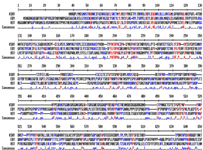
complementary strand. It is flanked by a short exon and the second long exon encoding the RTA gene. The ORF49 gene shows limited sequence homology to the EBV BRRF1 gene. The EBV BRRF1 is also contained within the EBV RTA homolog, BRLF-1 locus. These loci are collinear and are subject to a splicing event resulting in the removal of the ORF49 and BRRF-1 sequences from the ORF50 and BRLF-1 transcripts, respectively (104, 105, 137) (Fig. 4.). The BRRF-1transcript was detected in Raji and Akata cells treated with TPA/Na-butyrate or anti-IgG respectively. The BRRF-1 protein was found predominantly in the nucleus of transfected cells.
KSHV ORF64
The gene encoded by KSHV ORF64 is predicted to be a large tegument protein due to homology to other herpesviruses. It is conserved across the alpha, beta and
22
treatment of the virion. This finding suggests that ORF64 is in contact with the capsid and the envelope of KSHV virions where it is exposed to protease and bind glycoproteins. The significance of these findings is currently under investigation.
ORF64 and its homologues in all the herpesviruses have predicted deubiquitinase (DUB) domains encoded in their N-termini. DUB activity has now been identified in several herpesviruses (134). In Herpes Simplex virus-1 (HSV-1), and Pseudorabies virus (PRV) (α-subfamily) the UL36 gene encodes a multifunctional, large tegument protein that are DUBs. In human Cytomegalovirus (β-subfamily), the homologous UL48 protein also encodes DUB activity (155).
Although the targets of the herpesviral deubiquitinases remain to be discovered, studies of HSV-1 mutants have implicated the HSV DUB in virion transport through microtubules (139), release of the viral genome into the nucleus (1, 89), tegumentation and envelopment (52) as well as virion egress (112). A DUB-null mutant of HSV UL36
23 The NFκB pathway
Nuclear factor (NF)-κB is an evolutionarily conserved signaling molecule that plays a central role in the regulation of a variety of cellular processes including the immune system (70) (99) (19). The NF-κB family is made up of p50/p105 κB1), p52/p100 (NF-κB2), p65 (RelA), RelB, and c-Rel. In their steady state they form homo- or heterodimers in
the cytoplasm. The inhibitor of κB (IκB) proteins binds to and prevents NF-κB translocation to the nucleus. Signaling through NF-κB follows two parallel pathways, the canonical or non-canonical (19). Pro-inflammatory cytokines, pathogens or antigenic peptides can activate this pathway. Once induced, the classical pathway activates the β subunit of the IκB kinase (IKK) complex, which can phosphorylate IκB proteins and trigger their ubiquitination and degradation.
The non-canonical or alternative pathway, is induced by activation of the α subunit (IKKα), which in turn phosphorylates p100. IκB proteins are phosphorylated and
subsequently ubiquitinated through lysine 48 (K48) by the Skp1–Cullin1– F-box (SCF)/-transducin repeat-containing protein (TrCP) E3-ligase complex.
Poly-ubiquitination targets IκB molecules for processing by the preteosome into p50, p65 and c-Rel. The resulting subunits form NF-κB heterodimers that translocate to the nucleus. Once in the nucleus, the heterodimers bind to NF-κB-responsive promoters of target genes.
24
and IKKγ, or NEMO (NF-κB essential modifier), a regulatory subunit (19, 69). IKKγ, considered the ‘molecular switch of NF-κB activation’, has been shown to be
non-destructively regulated through ubiquitination through different lysine residues (2, 85, 164). The TNF receptor (TNFR)-associated factor 6 (TRAF-6) is a lysine-63 (K63)-specific E3 ligase which is required for IKK complex function in in vitro assays (43). TRAF-6
mediated ubiquitination of IKKγ is essential for IL-1 receptor (IL-1R) and Toll-like receptor (TLR) signaling (51, 90). TRAF-2 is another K63-specific E3 ligase that mediates
polyubiquitination of RIP (receptor-interacting protein) (157) which plays a pivotal role in receptor complex formation after TNF-α-stimulation. TRAF2/TRAF5 doubly deficient mice (150) display marked inhibition of NF-κB activation during TNF receptor (TNFR) induction. Thus, TRAFs 2 and 5 could function in TNF signaling as analogs to TRAF6 during IL-1 activation. IL-1 induction causes TRAF6 binding to TGF-β-activated kinase 1 (TAK1) which activates the NFκ-B pathway (116). TAK1 kinase activity is regulated by TAK1-binding (TAB) proteins TAB2 and TAB3. TABs recognize proteins ubiquitinated by TRAF2 and TRAF6 (90).
CYLD is a cellular, K63-specific DUB capable of inhibiting NF-κB signaling (28, 93, 153). Mutations in the CYLD gene result in an autosomal-dominant disease familial
cylindromatosis. The disease is characterized by benign tumors arising from hair follicles or eccrine gland cell types. NF-κB activation induces CYLD expression which inhibits IKK activity (88). The mechanism of NF-κB signaling inhibition by CYLD involves the cleavage of K63-linked Ub chains from TRAF2, TRAF6 and IKKγ.
25
TRAF6. The deubiquitination of RIP and TRAF6 block signaling to NF-κB (20, 157). A specific Ub E3 ligase is also encoded by A20. The E3 module can catalyze the K48-linked poly-ubiquitination of RIP (157). Cezanne is another DUB that may regulate TRAF6 (62). Cezanne blocks NF-κB signaling possibly through IKK deubiquitination.
26 OBJECTIVES
1) Characterization of KSHV ORF49
There have been no previous studies on KSHV ORF49 or its role in the viral life cycle. The initial study of KSHV ORF49 aimed to characterize both transcript and protein. The kinetics of KSHV Orf49 transcription and the 5’ and 3’ ends of the transcript (RACE) were identified. The properties and function of the encoded protein were investigated as well as the contribution to lytic replication.
2) Characterization of KSHV ORF64
KSHV ORF64 has been shown to be present in the tegument of virions and to associate with several tegument and glycoproteins but has not been ascribed a function. We aimed to identify potential roles of KSHV ORF64 in the viral life cycle. DUB enzymatic activity was identified in the N-terminus of KSHV ORF64 by a number of different assays. The role of virus-encoded DUB activity in the context of lytic replication was assessed.
3) KSHV ORF64 DUB activity and signaling
The robust DUB activity encoded by KSHV Orf64 suggests a large pool of potential protein targets of Orf64-mediated deubiquitination. Since several signaling pathways pertinent to viral infection are regulated through
27
REFERENCES
1. Abaitua, F., and P. O'Hare. 2008. Identification of a highly conserved, functional nuclear localization signal within the N-terminal region of herpes simplex virus type 1 VP1-2 tegument protein. J Virol 82:5234-44.
2. Abbott, D. W., A. Wilkins, J. M. Asara, and L. C. Cantley. 2004. The Crohn's disease protein, NOD2, requires RIP2 in order to induce
ubiquitinylation of a novel site on NEMO. Curr Biol 14:2217-27.
3. Akula, S. M., N. P. Pramod, F. Z. Wang, and B. Chandran. 2002. Integrin alpha3beta1 (CD 49c/29) is a cellular receptor for Kaposi's
sarcoma-associated herpesvirus (KSHV/HHV-8) entry into the target cells. Cell 108:407-19.
4. Alexander, L., L. Denekamp, A. Knapp, M. R. Auerbach, B. Damania, and R. C. Desrosiers. 2000. The primary sequence of rhesus monkey rhadinovirus isolate 26-95: sequence similarities to Kaposi's
sarcoma-associated herpesvirus and rhesus monkey rhadinovirus isolate 17577. J Virol 74:3388-98.
5. Aluigi, M. G., A. Albini, S. Carlone, L. Repetto, R. De Marchi, A. Icardi, M. Moro, D. Noonan, and R. Benelli. 1996. KSHV sequences in biopsies and cultured spindle cells of epidemic, iatrogenic and Mediterranean forms of Kaposi's sarcoma. Res Virol 147:267-75.
6. Ambroziak, J. A., D. J. Blackbourn, B. G. Herndier, R. G. Glogau, J. H. Gullett, A. R. McDonald, E. T. Lennette, and J. A. Levy. 1995. Herpes-like sequences in HIV-infected and uninfected Kaposi's sarcoma patients. Science 268:582-3.
7. An, F. Q., H. M. Folarin, N. Compitello, J. Roth, S. L. Gerson, K. R. McCrae, F. D. Fakhari, D. P. Dittmer, and R. Renne. 2006. Long-term-infected telomerase-immortalized endothelial cells: a model for Kaposi's sarcoma-associated herpesvirus latency in vitro and in vivo. J Virol 80: 4833-46.
8. Arvanitakis, L., E. A. Mesri, R. G. Nador, J. W. Said, A. S. Asch, D. M. Knowles, and E. Cesarman. 1996. Establishment and characterization of a primary effusion (body cavity-based) lymphoma cell line (BC-3) harboring kaposi's sarcoma-associated herpesvirus (KSHV/HHV-8) in the absence of Epstein-Barr virus. Blood 88:2648-54.
28
10. Ballestas, M. E., and K. M. Kaye. 2001. Kaposi's sarcoma-associated
herpesvirus latency-associated nuclear antigen 1 mediates episome persistence through cis-acting terminal repeat (TR) sequence and specifically binds TR DNA. J Virol 75:3250-8.
11. Barbera, A. J., J. V. Chodaparambil, B. Kelley-Clarke, V. Joukov, J. C. Walter, K. Luger, and K. M. Kaye. 2006. The nucleosomal surface as a docking station for Kaposi's sarcoma herpesvirus LANA. Science 311:856-61. 12. Barozzi, P., M. Luppi, F. Facchetti, C. Mecucci, M. Alu, R. Sarid, V.
Rasini, L. Ravazzini, E. Rossi, S. Festa, B. Crescenzi, D. G. Wolf, T. F. Schulz, and G. Torelli. 2003. Post-transplant Kaposi sarcoma originates from the seeding of donor-derived progenitors. Nat Med 9:554-61.
13. Bechtel, J. T., Y. Liang, J. Hvidding, and D. Ganem. 2003. Host range of Kaposi's sarcoma-associated herpesvirus in cultured cells. J Virol 77:6474-81. 14. Benelli, R., A. Albini, C. Parravicini, S. Carlone, L. Repetto, G.
Tambussi, and A. Lazzarin. 1996. Isolation of spindle-shaped cell populations from primary cultures of Kaposi's sarcoma of different stage. Cancer Lett 100:125-32.
15. Bergquam, E. P., N. Avery, S. M. Shiigi, M. K. Axthelm, and S. W. Wong. 1999. Rhesus rhadinovirus establishes a latent infection in B lymphocytes in vivo. J Virol 73:7874-6.
16. Biggar, R. J., and C. S. Rabkin. 1996. The epidemiology of AIDS--related neoplasms. Hematol Oncol Clin North Am 10:997-1010.
17. Birch, N. H. 1967. A case of Kaposi's Sarcoma of the small intestine. East Afr Med J 44:343-5.
18. Blackbourn, D. J., E. Lennette, B. Klencke, A. Moses, B. Chandran, M. Weinstein, R. G. Glogau, M. H. Witte, D. L. Way, T. Kutzkey, B.
Herndier, and J. A. Levy. 2000. The restricted cellular host range of human herpesvirus 8. Aids 14:1123-33.
19. Bonizzi, G., and M. Karin. 2004. The two NF-kappaB activation pathways and their role in innate and adaptive immunity. Trends Immunol 25:280-8. 20. Boone, D. L., E. E. Turer, E. G. Lee, R. C. Ahmad, M. T. Wheeler, C.
Tsui, P. Hurley, M. Chien, S. Chai, O. Hitotsumatsu, E. McNally, C. Pickart, and A. Ma. 2004. The ubiquitin-modifying enzyme A20 is required for termination of Toll-like receptor responses. Nat Immunol 5:1052-60. 21. Boshoff, C., S. J. Gao, L. E. Healy, S. Matthews, A. J. Thomas, L.
29
(BCP-1) from peripheral blood and characterizing its growth in Nod/SCID mice. Blood 91:1671-9.
22. Bottcher, S., H. Granzow, C. Maresch, B. Mohl, B. G. Klupp, and T. C. Mettenleiter. 2007. Identification of functional domains within the essential large tegument protein pUL36 of pseudorabies virus. J Virol 81:13403-11. 23. Bottcher, S., C. Maresch, H. Granzow, B. G. Klupp, J. P. Teifke, and T.
C. Mettenleiter. 2008. Mutagenesis of the active-site cysteine in the ubiquitin-specific protease contained in large tegument protein pUL36 of pseudorabies virus impairs viral replication in vitro and neuroinvasion in vivo. J Virol 82:6009-16.
24. Bowser, B. S., S. M. DeWire, and B. Damania. 2002. Transcriptional
regulation of the K1 gene product of Kaposi's sarcoma-associated herpesvirus. J Virol 76:12574-83.
25. Brambilla, L., V. Boneschi, S. Ferrucci, M. Taglioni, and E. Berti. 2000. Human herpesvirus-8 infection among heterosexual partners of patients with classical Kaposi's sarcoma. Br J Dermatol 143:1021-5.
26. Braun, M. 1982. Classics in Oncology. Idiopathic multiple pigmented sarcoma of the skin by Kaposi. CA Cancer J Clin 32:340-7.
27. Brown, H. J., M. J. Song, H. Deng, T. T. Wu, G. Cheng, and R. Sun. 2003. NF-kappaB inhibits gammaherpesvirus lytic replication. J Virol 77:8532-40. 28. Brummelkamp, T. R., S. M. Nijman, A. M. Dirac, and R. Bernards. 2003.
Loss of the cylindromatosis tumour suppressor inhibits apoptosis by activating NF-kappaB. Nature 424:797-801.
29. Bryk, D., J. Farman, S. Dallemand, M. A. Meyers, and A. Wecksell. 1978. Kaposi's sarcoma of the intestinal tract: roentgen manifestations. Gastrointest Radiol 3:425-30.
30. Cai, Q., K. Lan, S. C. Verma, H. Si, D. Lin, and E. S. Robertson. 2006. Kaposi's sarcoma-associated herpesvirus latent protein LANA interacts with HIF-1 alpha to upregulate RTA expression during hypoxia: Latency control under low oxygen conditions. J Virol 80:7965-75.
31. Cai, X., S. Lu, Z. Zhang, C. M. Gonzalez, B. Damania, and B. R. Cullen. 2005. Kaposi's sarcoma-associated herpesvirus expresses an array of viral microRNAs in latently infected cells. Proc Natl Acad Sci U S A 102:5570-5. 32. Cannon, J. S., D. Ciufo, A. L. Hawkins, C. A. Griffin, M. J. Borowitz, G.
30
33. Cannon, M. J., S. C. Dollard, D. K. Smith, R. S. Klein, P. Schuman, J. D. Rich, D. Vlahov, and P. E. Pellett. 2001. Blood-borne and sexual
transmission of human herpesvirus 8 in women with or at risk for human immunodeficiency virus infection. N Engl J Med 344:637-43.
34. Carbone, A., G. Gaidano, A. Gloghini, L. M. Larocca, D. Capello, V. Canzonieri, A. Antinori, U. Tirelli, B. Falini, and R. Dalla-Favera. 1998. Differential expression of BCL-6, CD138/syndecan-1, and Epstein-Barr virus-encoded latent membrane protein-1 identifies distinct histogenetic subsets of acquired immunodeficiency syndrome-related non-Hodgkin's lymphomas. Blood 91:747-55.
35. Casper, C., D. Carrell, K. G. Miller, F. D. Judson, A. S. Meier, J. S. Pauk, R. A. Morrow, L. Corey, A. Wald, and C. Celum. 2006. HIV
serodiscordant sex partners and the prevalence of human herpesvirus 8 infection among HIV negative men who have sex with men: baseline data from the EXPLORE Study. Sex Transm Infect 82:229-35.
36. Casper, C., E. Krantz, S. Selke, S. R. Kuntz, J. Wang, M. L. Huang, J. S. Pauk, L. Corey, and A. Wald. 2007. Frequent and asymptomatic
oropharyngeal shedding of human herpesvirus 8 among immunocompetent men. J Infect Dis 195:30-6.
37. Casper, C., M. Redman, M. L. Huang, J. Pauk, T. M. Lampinen, S. E. Hawes, C. W. Critchlow, R. A. Morrow, L. Corey, N. Kiviat, and A. Wald. 2004. HIV infection and human herpesvirus-8 oral shedding among men who have sex with men. J Acquir Immune Defic Syndr 35:233-8. 38. Casper, C., A. Wald, J. Pauk, S. R. Tabet, L. Corey, and C. L. Celum.
2002. Correlates of prevalent and incident Kaposi's sarcoma-associated herpesvirus infection in men who have sex with men. J Infect Dis 185:990-3. 39. Cesarman, E., Y. Chang, P. S. Moore, J. W. Said, and D. M. Knowles.
1995. Kaposi's sarcoma-associated herpesvirus-like DNA sequences in AIDS-related body-cavity-based lymphomas. N Engl J Med 332:1186-91.
40. Chang, P. J., D. Shedd, L. Gradoville, M. S. Cho, L. W. Chen, J. Chang, and G. Miller. 2002. Open reading frame 50 protein of Kaposi's sarcoma-associated herpesvirus directly activates the viral PAN and K12 genes by binding to related response elements. J Virol 76:3168-78.
41. Chang, Y., E. Cesarman, M. S. Pessin, F. Lee, J. Culpepper, D. M. Knowles, and P. S. Moore. 1994. Identification of herpesvirus-like DNA sequences in AIDS-associated Kaposi's sarcoma. Science 266:1865-9. 42. Chen, Y. B., A. Rahemtullah, and E. Hochberg. 2007. Primary effusion
31
43. Chen, Z. J., L. Parent, and T. Maniatis. 1996. Site-specific phosphorylation of IkappaBalpha by a novel ubiquitination-dependent protein kinase activity. Cell 84:853-62.
44. Chronowski, G. M., C. S. Ha, R. B. Wilder, F. Cabanillas, J. Manning, and J. D. Cox. 2001. Treatment of unicentric and multicentric Castleman disease and the role of radiotherapy. Cancer 92:670-6.
45. D'Agostino, G., E. Arico, L. Santodonato, M. Venditti, P. Sestili, L. Masuelli, A. Coletti, A. Modesti, G. Picchio, D. E. Mosier, M. Ferrantini, and F. Belardelli. 1999. Type I consensus IFN (IFN-con1) gene transfer into KSHV/HHV-8-infected BCBL-1 cells causes inhibition of viral lytic cycle activation via induction of apoptosis and abrogates tumorigenicity in sCID mice. J Interferon Cytokine Res 19:1305-16.
46. Damania, B., and J. U. Jung. 2001. Comparative analysis of the
transforming mechanisms of Epstein-Barr virus, Kaposi's sarcoma-associated herpesvirus, and Herpesvirus saimiri. Adv Cancer Res 80:51-82.
47. Davis, D. A., A. S. Rinderknecht, J. P. Zoeteweij, Y. Aoki, E. L. Read-Connole, G. Tosato, A. Blauvelt, and R. Yarchoan. 2001. Hypoxia induces lytic replication of Kaposi sarcoma-associated herpesvirus. Blood 97:3244-50. 48. de Jong, M. D., G. J. Galasso, B. Gazzard, P. D. Griffiths, D. A. Jabs, E.
R. Kern, and S. A. Spector. 1998. Summary of the II International Symposium on Cytomegalovirus. Antiviral Res 39:141-62.
49. Delecluse, H. J., R. Feederle, B. O'Sullivan, and P. Taniere. 2007. Epstein Barr virus-associated tumours: an update for the attention of the working pathologist. J Clin Pathol 60:1358-64.
50. Deng, H., J. T. Chu, M. B. Rettig, O. Martinez-Maza, and R. Sun. 2002. Rta of the human herpesvirus 8/Kaposi sarcoma-associated herpesvirus up-regulates human interleukin-6 gene expression. Blood 100:1919-21.
51. Deng, L., C. Wang, E. Spencer, L. Yang, A. Braun, J. You, C. Slaughter, C. Pickart, and Z. J. Chen. 2000. Activation of the IkappaB kinase complex by TRAF6 requires a dimeric ubiquitin-conjugating enzyme complex and a unique polyubiquitin chain. Cell 103:351-61.
52. Desai, P. J. 2000. A null mutation in the UL36 gene of herpes simplex virus type 1 results in accumulation of unenveloped DNA-filled capsids in the cytoplasm of infected cells. J Virol 74:11608-18.
53. Desrosiers, R. C., V. G. Sasseville, S. C. Czajak, X. Zhang, K. G.
32
54. DeWire, S. M., E. S. Money, S. P. Krall, and B. Damania. 2003. Rhesus monkey rhadinovirus (RRV): construction of a RRV-GFP recombinant virus and development of assays to assess viral replication. Virology 312:122-34. 55. Dictor, M. 1997. Human herpesvirus 8 and Kaposi's sarcoma. Semin Cutan
Med Surg 16:181-7.
56. DiGiovanna, J. J., and B. Safai. 1981. Kaposi's sarcoma. Retrospective study of 90 cases with particular emphasis on the familial occurrence, ethnic background and prevalence of other diseases. Am J Med 71:779-83.
57. Dittmer, D., M. Lagunoff, R. Renne, K. Staskus, A. Haase, and D. Ganem. 1998. A cluster of latently expressed genes in Kaposi's sarcoma-associated herpesvirus. J Virol 72:8309-15.
58. Dittmer, D., C. Stoddart, R. Renne, V. Linquist-Stepps, M. E. Moreno, C. Bare, J. M. McCune, and D. Ganem. 1999. Experimental transmission of Kaposi's sarcoma-associated herpesvirus (KSHV/HHV-8) to SCID-hu Thy/Liv mice. J Exp Med 190:1857-68.
59. Duman, S., H. Toz, G. Asci, S. Alper, M. Ozkahya, I. Unal, A. Celik, E. Ok, and A. Basci. 2002. Successful treatment of post-transplant Kaposi's sarcoma by reduction of immunosuppression. Nephrol Dial Transplant 17:892-6.
60. Dupin, N., C. Fisher, P. Kellam, S. Ariad, M. Tulliez, N. Franck, E. van Marck, D. Salmon, I. Gorin, J. P. Escande, R. A. Weiss, K. Alitalo, and C. Boshoff. 1999. Distribution of human herpesvirus-8 latently infected cells in Kaposi's sarcoma, multicentric Castleman's disease, and primary effusion lymphoma. Proc Natl Acad Sci U S A 96:4546-51.
61. Estep, R. D., M. F. Powers, B. K. Yen, H. Li, and S. W. Wong. 2007. Construction of an infectious rhesus rhadinovirus bacterial artificial chromosome for the analysis of Kaposi's sarcoma-associated herpesvirus-related disease development. J Virol 81:2957-69.
62. Evans, P. C., E. R. Taylor, J. Coadwell, K. Heyninck, R. Beyaert, and P. J. Kilshaw. 2001. Isolation and characterization of two novel A20-like proteins. Biochem J 357:617-23.
63. Farge, D., C. Lebbe, Z. Marjanovic, P. Tuppin, C. Mouquet, M. N.
33
64. Field, N., W. Low, M. Daniels, S. Howell, L. Daviet, C. Boshoff, and M. Collins. 2003. KSHV vFLIP binds to IKK-gamma to activate IKK. J Cell Sci 116:3721-8.
65. Flore, O., S. Rafii, S. Ely, J. J. O'Leary, E. M. Hyjek, and E. Cesarman. 1998. Transformation of primary human endothelial cells by Kaposi's sarcoma-associated herpesvirus. Nature 394:588-92.
66. Forbes, B. A. 1989. Acquisition of cytomegalovirus infection: an update. Clin Microbiol Rev 2:204-16.
67. Foreman, K. E., J. Friborg, B. Chandran, H. Katano, T. Sata, M. Mercader, G. J. Nabel, and B. J. Nickoloff. 2001. Injection of human herpesvirus-8 in human skin engrafted on SCID mice induces Kaposi's sarcoma-like lesions. J Dermatol Sci 26:182-93.
68. Gaidano, G., K. Cechova, Y. Chang, P. S. Moore, D. M. Knowles, and R. Dalla-Favera. 1996. Establishment of AIDS-related lymphoma cell lines from lymphomatous effusions. Leukemia 10:1237-40.
69. Ghosh, S., and M. Karin. 2002. Missing pieces in the NF-kappaB puzzle. Cell 109 Suppl:S81-96.
70. Ghosh, S., M. J. May, and E. B. Kopp. 1998. NF-kappa B and Rel proteins: evolutionarily conserved mediators of immune responses. Annu Rev Immunol 16:225-60.
71. Giraldo, G., and E. Beth. 1972. [Kaposi's sarcoma: presence of herpes type virus in tissue culture in 5 different cases. A possible new model in the study of viruses associated with human tumors]. C R Acad Sci Hebd Seances Acad Sci D 275:289-92.
72. Giraldo, G., E. Beth, W. Henle, G. Henle, V. Mike, B. Safai, J. M. Huraux, J. McHardy, and G. deThe. 1978. Antibody patterns to
herpesviruses in Kaposi's sarcoma. II. Serological association of American Kaposi's sarcoma with cytomegalovirus. Int J Cancer 22:126-31.
73. Glaser, R., L. Geder, S. St Jeor, S. Michelson-Fiske, and F. Haguenau. 1977. Partial characterization of a herpes-type virus (K9V) derived from Kaposi's sarcoma. J Natl Cancer Inst 59:55-60.
74. Gougerot, H., and J. Hewit. 1950. [Unna-Kaposi's acrosarcoma ameliorated by massive doses of penicillin.]. Bull Soc Fr Dermatol Syphiligr 57:502-3. 75. Grandadam, M., N. Dupin, V. Calvez, I. Gorin, L. Blum, S. Kernbaum,
34
high increase in Kaposi's sarcoma herpesvirus DNA load in peripheral blood mononuclear cells. J Infect Dis 175:1198-201.
76. Grossman, Z., J. Iscovich, F. Schwartz, E. Azizi, A. Klepfish, A.
Schattner, and R. Sarid. 2002. Absence of Kaposi sarcoma among Ethiopian immigrants to Israel despite high seroprevalence of human herpesvirus 8. Mayo Clin Proc 77:905-9.
77. Grossmann, C., and D. Ganem. 2008. Effects of NFkappaB activation on KSHV latency and lytic reactivation are complex and context-dependent. Virology 375:94-102.
78. Guttman-Yassky, E., Z. Kra-Oz, J. Dubnov, R. Friedman-Birnbaum, I. Segal, N. Zaltzman, T. Roth, F. Schwartz, S. Linn, D. Rozenman, M. David, M. Silbermann, M. Barchana, R. Bergman, and R. Sarid. 2005. Infection with Kaposi's sarcoma-associated herpesvirus among families of patients with classic Kaposi's sarcoma. Arch Dermatol 141:1429-34.
79. Gwack, Y., H. Byun, S. Hwang, C. Lim, and J. Choe. 2001. CREB-binding protein and histone deacetylase regulate the transcriptional activity of Kaposi's sarcoma-associated herpesvirus open reading frame 50. J Virol 75:1909-17. 80. Haque, M., J. Chen, K. Ueda, Y. Mori, K. Nakano, Y. Hirata, S.
Kanamori, Y. Uchiyama, R. Inagi, T. Okuno, and K. Yamanishi. 2000. Identification and analysis of the K5 gene of Kaposi's sarcoma-associated herpesvirus. J Virol 74:2867-75.
81. Haque, M., D. A. Davis, V. Wang, I. Widmer, and R. Yarchoan. 2003. Kaposi's sarcoma-associated herpesvirus (human herpesvirus 8) contains hypoxia response elements: relevance to lytic induction by hypoxia. J Virol 77:6761-8.
82. Herndier, B. G., L. D. Kaplan, and M. S. McGrath. 1994. Pathogenesis of AIDS lymphomas. Aids 8:1025-49.
83. Hladik, W., S. C. Dollard, J. Mermin, A. L. Fowlkes, R. Downing, M. M. Amin, F. Banage, E. Nzaro, P. Kataaha, T. J. Dondero, P. E. Pellett, and E. M. Lackritz. 2006. Transmission of human herpesvirus 8 by blood transfusion. N Engl J Med 355:1331-8.
84. Howard, M. R., D. Whitby, G. Bahadur, F. Suggett, C. Boshoff, M. Tenant-Flowers, T. F. Schulz, S. Kirk, S. Matthews, I. V. Weller, R. S. Tedder, and R. A. Weiss. 1997. Detection of human herpesvirus 8 DNA in semen from HIV-infected individuals but not healthy semen donors. Aids 11:F15-9.
35
ubiquitin mediates NF-kappaB activation by genotoxic stress. Cell 115: 565-76.
86. Jarosinski, K., L. Kattenhorn, B. Kaufer, H. Ploegh, and N. Osterrieder. 2007. A herpesvirus ubiquitin-specific protease is critical for efficient T cell lymphoma formation. Proc Natl Acad Sci U S A 104:20025-30.
87. Jeong, J., J. Papin, and D. Dittmer. 2001. Differential regulation of the overlapping Kaposi's sarcoma-associated herpesvirus vGCR (orf74) and LANA (orf73) promoters. J Virol 75:1798-807.
88. Jono, H., J. H. Lim, L. F. Chen, H. Xu, E. Trompouki, Z. K. Pan, G. Mosialos, and J. D. Li. 2004. NF-kappaB is essential for induction of CYLD, the negative regulator of NF-kappaB: evidence for a novel inducible
autoregulatory feedback pathway. J Biol Chem 279:36171-4.
89. Jovasevic, V., L. Liang, and B. Roizman. 2008. Proteolytic cleavage of VP1-2 is required for release of herpes simplex virus 1 DNA into the nucleus. J Virol 82:3311-9.
90. Kanayama, A., R. B. Seth, L. Sun, C. K. Ea, M. Hong, A. Shaito, Y. H. Chiu, L. Deng, and Z. J. Chen. 2004. TAB2 and TAB3 activate the NF-kappaB pathway through binding to polyubiquitin chains. Mol Cell 15: 535-48.
91. Katano, H., Y. Hoshino, Y. Morishita, T. Nakamura, H. Satoh, A.
Iwamoto, B. Herndier, and S. Mori. 1999. Establishing and characterizing a CD30-positive cell line harboring HHV-8 from a primary effusion lymphoma. J Med Virol 58:394-401.
92. Keller, S. A., E. J. Schattner, and E. Cesarman. 2000. Inhibition of NF-kappaB induces apoptosis of KSHV-infected primary effusion lymphoma cells. Blood 96:2537-42.
93. Kovalenko, A., C. Chable-Bessia, G. Cantarella, A. Israel, D. Wallach, and G. Courtois. 2003. The tumour suppressor CYLD negatively regulates NF-kappaB signalling by deubiquitination. Nature 424:801-5.
94. Krishnan, H. H., N. Sharma-Walia, L. Zeng, S. J. Gao, and B. Chandran. 2005. Envelope glycoprotein gB of Kaposi's sarcoma-associated herpesvirus is essential for egress from infected cells. J Virol 79:10952-67.
95. Kurgan, J. 1972. [Disseminated pulmonary lesions in the course of Kaposi's sarcoma]. Gruzlica 40:751-4.
36
97. Lee, J. I., G. W. Luxton, and G. A. Smith. 2006. Identification of an essential domain in the herpesvirus VP1/2 tegument protein: the carboxy terminus directs incorporation into capsid assemblons. J Virol 80:12086-94. 98. Li, J. J., Y. Q. Huang, C. J. Cockerell, and A. E. Friedman-Kien. 1996.
Localization of human herpes-like virus type 8 in vascular endothelial cells and perivascular spindle-shaped cells of Kaposi's sarcoma lesions by in situ hybridization. Am J Pathol 148:1741-8.
99. Li, Q., and I. M. Verma. 2002. NF-kappaB regulation in the immune system. Nat Rev Immunol 2:725-34.
100. Liang, Y., and D. Ganem. 2004. RBP-J (CSL) is essential for activation of the K14/vGPCR promoter of Kaposi's sarcoma-associated herpesvirus by the lytic switch protein RTA. J Virol 78:6818-26.
101. Liao, W., Y. Tang, Y. L. Kuo, B. Y. Liu, C. J. Xu, and C. Z. Giam. 2003. Kaposi's sarcoma-associated herpesvirus/human herpesvirus 8 transcriptional activator Rta is an oligomeric DNA-binding protein that interacts with tandem arrays of phased A/T-trinucleotide motifs. J Virol 77:9399-411.
102. Liu, L., M. T. Eby, N. Rathore, S. K. Sinha, A. Kumar, and P. M.
Chaudhary. 2002. The human herpes virus 8-encoded viral FLICE inhibitory protein physically associates with and persistently activates the Ikappa B kinase complex. J Biol Chem 277:13745-51.
103. Lukac, D. M., L. Garibyan, J. R. Kirshner, D. Palmeri, and D. Ganem. 2001. DNA binding by Kaposi's sarcoma-associated herpesvirus lytic switch protein is necessary for transcriptional activation of two viral delayed early promoters. J Virol 75:6786-99.
104. Lukac, D. M., J. R. Kirshner, and D. Ganem. 1999. Transcriptional activation by the product of open reading frame 50 of Kaposi's sarcoma-associated herpesvirus is required for lytic viral reactivation in B cells. J Virol 73:9348-61.
105. Lukac, D. M., R. Renne, J. R. Kirshner, and D. Ganem. 1998. Reactivation of Kaposi's sarcoma-associated herpesvirus infection from latency by
expression of the ORF 50 transactivator, a homolog of the EBV R protein. Virology 252:304-12.
106. Luppi, M., P. Barozzi, G. Santagostino, R. Trovato, T. F. Schulz, R. Marasca, D. Bottalico, L. Bignardi, and G. Torelli. 2000. Molecular evidence of organ-related transmission of Kaposi sarcoma-associated
herpesvirus or human herpesvirus-8 in transplant patients. Blood 96:3279-81. 107. Luxton, G. W., J. I. Lee, S. Haverlock-Moyns, J. M. Schober, and G. A.
37
108. Lyall, E. G., G. S. Patton, J. Sheldon, C. Stainsby, J. Mullen, S. O'Shea, N. A. Smith, A. De Ruiter, M. O. McClure, and T. F. Schulz. 1999. Evidence for horizontal and not vertical transmission of human herpesvirus 8 in children born to human immunodeficiency virus-infected mothers. Pediatr Infect Dis J 18:795-9.
109. Martin, J. N., D. E. Ganem, D. H. Osmond, K. A. Page-Shafer, D.
Macrae, and D. H. Kedes. 1998. Sexual transmission and the natural history of human herpesvirus 8 infection. N Engl J Med 338:948-54.
110. Matsumura, S., Y. Fujita, E. Gomez, N. Tanese, and A. C. Wilson. 2005. Activation of the Kaposi's sarcoma-associated herpesvirus major latency locus by the lytic switch protein RTA (ORF50). J Virol 79:8493-505.
111. Moore, P. S., and Y. Chang. 1998. Antiviral activity of tumor-suppressor pathways: clues from molecular piracy by KSHV. Trends Genet 14:144-50. 112. Morrison, E. E., A. J. Stevenson, Y. F. Wang, and D. M. Meredith. 1998.
Differences in the intracellular localization and fate of herpes simplex virus tegument proteins early in the infection of Vero cells. J Gen Virol 79 ( Pt 10):2517-28.
113. Mutlu, A. D., L. E. Cavallin, L. Vincent, C. Chiozzini, P. Eroles, E. M. Duran, Z. Asgari, A. T. Hooper, K. M. La Perle, C. Hilsher, S. J. Gao, D. P. Dittmer, S. Rafii, and E. A. Mesri. 2007. In vivo-restricted and reversible malignancy induced by human herpesvirus-8 KSHV: a cell and animal model of virally induced Kaposi's sarcoma. Cancer Cell 11:245-58.
114. Nash, G., and S. Fligiel. 1984. Kaposi's sarcoma presenting as pulmonary disease in the acquired immunodeficiency syndrome: diagnosis by lung biopsy. Hum Pathol 15:999-1001.
115. Newell, G. R., S. C. Adams, P. W. Mansell, and E. M. Hersh. 1984. Toxicity, immunosuppressive effects and carcinogenic potential of volatile nitrites: possible relationship to Kaposi's sarcoma. Pharmacotherapy 4:284-91. 116. Ninomiya-Tsuji, J., K. Kishimoto, A. Hiyama, J. Inoue, Z. Cao, and K.
Matsumoto. 1999. The kinase TAK1 can activate the NIK-I kappaB as well as the MAP kinase cascade in the IL-1 signalling pathway. Nature 398:252-6. 117. Oksenhendler, E., M. Duarte, J. Soulier, P. Cacoub, Y. Welker, J.
Cadranel, D. Cazals-Hatem, B. Autran, J. P. Clauvel, and M. Raphael. 1996. Multicentric Castleman's disease in HIV infection: a clinical and pathological study of 20 patients. Aids 10:61-7.
38
119. Pastinszky, I. 1950. [Herpetic virus in the pathogenesis of Kaposi's disease and differential diagnosis.]. Borgyogy Venerol Sz 4:146-55.
120. Patel, N., M. Salifu, N. Sumrani, D. Distant, J. Hong, M. Markell, and A. S. Braverman. 2002. Successful treatment of post-renal transplant Kaposi's sarcoma with paclitaxel. Am J Transplant 2:877-9.
121. Pauk, J., M. L. Huang, S. J. Brodie, A. Wald, D. M. Koelle, T. Schacker, C. Celum, S. Selke, and L. Corey. 2000. Mucosal shedding of human herpesvirus 8 in men. N Engl J Med 343:1369-77.
122. Penn, I. 1997. Kaposi's sarcoma in transplant recipients. Transplantation 64:669-73.
123. Picchio, G. R., R. E. Sabbe, R. J. Gulizia, M. McGrath, B. G. Herndier, and D. E. Mosier. 1997. The KSHV/HHV8-infected BCBL-1 lymphoma line causes tumors in SCID mice but fails to transmit virus to a human peripheral blood mononuclear cell graft. Virology 238:22-9.
124. Piolot, T., M. Tramier, M. Coppey, J. C. Nicolas, and V. Marechal. 2001. Close but distinct regions of human herpesvirus 8 latency-associated nuclear antigen 1 are responsible for nuclear targeting and binding to human mitotic chromosomes. J Virol 75:3948-59.
125. Plancoulaine, S., L. Abel, M. van Beveren, D. A. Tregouet, M. Joubert, P. Tortevoye, G. de The, and A. Gessain. 2000. Human herpesvirus 8
transmission from mother to child and between siblings in an endemic population. Lancet 356:1062-5.
126. Rainbow, L., G. M. Platt, G. R. Simpson, R. Sarid, S. J. Gao, H. Stoiber, C. S. Herrington, P. S. Moore, and T. F. Schulz. 1997. The 222- to 234-kilodalton latent nuclear protein (LNA) of Kaposi's sarcoma-associated herpesvirus (human herpesvirus 8) is encoded by orf73 and is a component of the latency-associated nuclear antigen. J Virol 71:5915-21.
127. Regezi, J. A., L. A. MacPhail, T. E. Daniels, Y. G. DeSouza, J. S. Greenspan, and D. Greenspan. 1993. Human immunodeficiency virus-associated oral Kaposi's sarcoma. A heterogeneous cell population dominated by spindle-shaped endothelial cells. Am J Pathol 143:240-9.
128. Renne, R., W. Zhong, B. Herndier, M. McGrath, N. Abbey, D. Kedes, and D. Ganem. 1996. Lytic growth of Kaposi's sarcoma-associated herpesvirus (human herpesvirus 8) in culture. Nat Med 2:342-6.
129. Rozen, R., N. Sathish, Y. Li, and Y. Yuan. 2008. Virion-wide protein interactions of Kaposi's sarcoma-associated herpesvirus. J Virol 82:4742-50. 130. Russo, J. J., R. A. Bohenzky, M. C. Chien, J. Chen, M. Yan, D.
39
Moore. 1996. Nucleotide sequence of the Kaposi sarcoma-associated herpesvirus (HHV8). Proc Natl Acad Sci U S A 93:14862-7.
131. Rutgers, J. L., R. Wieczorek, F. Bonetti, K. L. Kaplan, D. N. Posnett, A. E. Friedman-Kien, and D. M. Knowles, 2nd. 1986. The expression of endothelial cell surface antigens by AIDS-associated Kaposi's sarcoma. Evidence for a vascular endothelial cell origin. Am J Pathol 122:493-9. 132. Sadler, R., L. Wu, B. Forghani, R. Renne, W. Zhong, B. Herndier, and D.
Ganem. 1999. A complex translational program generates multiple novel proteins from the latently expressed kaposin (K12) locus of Kaposi's sarcoma-associated herpesvirus. J Virol 73:5722-30.
133. Sakakibara, S., K. Ueda, J. Chen, T. Okuno, and K. Yamanishi. 2001. Octamer-binding sequence is a key element for the autoregulation of Kaposi's sarcoma-associated herpesvirus ORF50/Lyta gene expression. J Virol
75:6894-900.
134. Schlieker, C., G. A. Korbel, L. M. Kattenhorn, and H. L. Ploegh. 2005. A deubiquitinating activity is conserved in the large tegument protein of the herpesviridae. J Virol 79:15582-5.
135. Schulz, T. F. 1999. Epidemiology of Kaposi's sarcoma-associated herpesvirus/human herpesvirus 8. Adv Cancer Res 76:121-60.
136. Searles, R. P., E. P. Bergquam, M. K. Axthelm, and S. W. Wong. 1999. Sequence and genomic analysis of a Rhesus macaque rhadinovirus with similarity to Kaposi's sarcoma-associated herpesvirus/human herpesvirus 8. J Virol 73:3040-53.
137. Segouffin-Cariou, C., G. Farjot, A. Sergeant, and H. Gruffat. 2000. Characterization of the epstein-barr virus BRRF1 gene, located between early genes BZLF1 and BRLF1. J Gen Virol 81:1791-9.
138. Sengupta, P., and M. M. Das. 1978. Kaposi's sarcoma of small intestine. J Indian Med Assoc 71:120-2.
139. Shanda, S. K., and D. W. Wilson. 2008. UL36p is required for efficient transport of membrane-associated herpes simplex virus type 1 along microtubules. J Virol 82:7388-94.
140. Sharp, T. V., H. W. Wang, A. Koumi, D. Hollyman, Y. Endo, H. Ye, M. Q. Du, and C. Boshoff. 2002. K15 protein of Kaposi's sarcoma-associated herpesvirus is latently expressed and binds to HAX-1, a protein with antiapoptotic function. J Virol 76:802-16.