THE DOCK FAMILY OF ATYPICAL GUANINE NUCLEOTIDE EXCHANGE FACTORS: REGULATION BY ELMO1 AND RHOG
Cynthia P. Holley
A dissertation submitted to the faculty of the University of North Carolina at Chapel Hill in partial fulfillment of the requirements for the degree of Doctor of Philosophy in the Department of Biochemistry and Biophysics.
Chapel Hill 2008
Approved by:
ABSTRACT
CYNTHIA P. HOLLEY: The Dock Family of Atypical Guanine Nucleotide Exchange Factors: Regulation by ELMO1 and RhoG
(Under the direction of John Sondek)
The Dock family of proteins regulates diverse biological processes including cell migration, phagocytosis and neuronal polarization. These proteins contain a unique type of guanine nucleotide exchange factor (GEF) domain, and function as GEFs for Rho-family GTPases. Several Dock-Rho-family proteins form complexes with ELMO proteins and the Dock/ELMO complex acts as a bi-partite GEF for Rac. Molecular details of how the Dock/ELMO complexes bind and exchange nucleotide on Rac are critical for our understanding of their biological effects, yet remain poorly defined.
As described here, purified Dock2/ELMO1 complex is a stable heterotetramer composed of two molecules each of Dock2 and ELMO1. This heterotetramer
coordinates a single molecule of nucleotide-free Rac. We identify an inhibitory
conformation within ELMO1 mediated through contacts between the N- and C-terminal regions of ELMO1 and describe a mechanism for relief of this inhibition through the binding of RhoG, another Rho-family GTPase. The interaction between RhoG and ELMO1 is both nucleotide-dependent, and dependent upon the C-terminal polybasic region of RhoG. These data provide fundamentally important molecular insights into the composition of the Dock/ELMO complex and regulation of nucleotide exchange via the Dock/ELMO proteins.
For Grant, without you, this would never have been possible.
ACKNOWLEGEMENTS
I would like to thank the many people whose efforts have contributed to this work, both directly and indirectly. First, I would like to thank the members of the Sondek lab for their support, both scientific and emotional, during my tenure in the lab. Their willingness to help and offer advice at any time and their excitement about science is what makes science such a special and rewarding pursuit. Marielle Yohe and Rafael Rojas were both instrumental in helping me learn the intricacies of exchange assays, and discussions with Matt Cheever and Kent Rossman about phosophoinositide binding were immensely helpful. Brant Hamel provided excellent discussions on the Dock family of proteins, and was a source of ideas and inspiration for this project. The emotional support of Marielle Yohe, Stephanie Hicks and Mariya Chhatriwala during this process was invaluable.
Our collaborators at the University of Virginia, the Ravichandran Lab, have been a source of ideas, excitement and data. Without their efforts, this project would not have existed, and I am indebted to them. In particular, Colin deBakker and Lisa Haney provided information and data which were included in this dissertation. All
immunoprecipitated proteins were provided by Lisa, and in-cell assays, yeast-two hybrid assays and radioactive GEF assays were performed by Lisa and Colin. Kodi
Ravichandran was an excellent source of ideas and inspiration, and it was a pleasure to collaborate with him and his lab.
v
Others at the University of North Carolina have been of great help as well. Ashutosh Tripathy, from the Macromolecular Interactions Facility helped with light scattering analyses and discussions on light scattering, fluorescence polarization and analytical ultracentrifugation. Brian Kuhlman’s lab, in the Department of Biochemistry and Biophysics generously allowed me to use their fluorimeter for the fluorescence polarization assays, and Deanne Sammond, Carrie Purbeck and Ramesh Jha in particular took time out of their days to help me with those experiments.
I would like to thank John Sondek for the opportunity to work in his lab, and for the excitement for good science that he fosters in the laboratory. I appreciate his
enthusiasm for this project, and his willingness to commit time out of a busy schedule whenever I had questions or needed help. My appreciation also goes to my committee members for their help and guidance along the way, and to the Program in Molecular and Cellular Biophysics for their support and assistance.
TABLE OF CONTENTS
LIST OF FIGURES ... viii
LIST OF ABBREVIATIONS ... x
CHAPTER 1: INTRODUCTION ... 1
Small GTPases ... 1
Guanine Nucleotide Exchange Factors ... 3
Dock-family GEFs ... 6
Dock A subfamily: Dock1, Dock2 and Dock5 ... 6
Dock B subfamily: Dock3 and Dock4 ... 13
Dock C subfamily: Dock6, Dock7 and Dock8 ... 15
Dock D subfamily: Dock9, Dock10 and Dock11 ... 18
Dock-family GTPase recognition ... 21
Concluding remarks ... 22
CHAPTER 2 – DOCK2/ELMO1: MOLECULAR INTERACTIONS OF A HETEROTETRAMERIC GEF ... 26
Background ... 26
Experimental procedures ... 29
Results ... 36
Discussion ... 46
CHAPTER 3 – RHOG DIRECTLY REGULATES THE ACTIVITY OF THE
DOCK/ELMO COMPLEX VIA INTERACTION WITH ELMO PROTEINS ... 60
Background ... 60
Experimental Procedures ... 62
Results ... 68
Discussion ... 74
CHAPTER 4 – CONCLUSIONS AND FUTURE DIRECTIONS ... 88
Conclusions ... 88
Future Directions ... 90
REFERENCES ... 95
LIST OF FIGURES
Figure 1: Overview of the Rho-family GTPase cycle. ... 24
Figure 2: Overview of the Dock family of proteins (mammalian). ... 25
Figure 3: Schematic representation of Dock1, Dock2 and ELMO1 constructs and domains. ... 50
Figure 4: Purified ELMO1 is a monomer in solution. ... 51
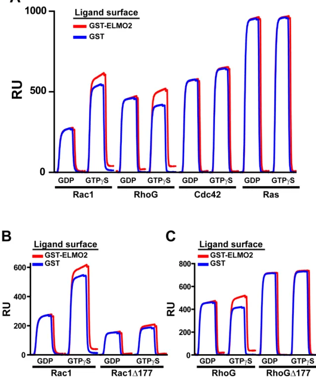
Figure 5: Binding of Rac1 and RhoG to ELMO1 is both nucleotide-dependent and PBR-dependent. ... 52
Figure 6: ELMO1 binding to RhoG and Rac1 is nucleotide and PBR dependent. ... 53
Figure 7: ELMO2 binding to RhoG and Rac1 is nucleotide and PBR dependent. ... 54
Figure 8: Dock2/ELMO1 is an effective GEF for Rac1, Rac1Δ177, Rac2 and Rac3. ... 55
Figure 9: Dock2/ELMO1 does not catalyze exchange on other Rho-family GTPase members. ... 56
Figure 10: Dock2/ELMO1 does not catalyze exchange on other Ras superfamily GTPase members. ... 57
Figure 11: Dock2/ELMO1 is a heterotetramer. ... 58
Figure 12: The Dock2/ELMO1 tetramer binds a single Rac1 molecule. ... 59
Figure 13: ELMO1 can interact with itself in cells... 79
Figure 14: The N-and C-terminal regions of ELMO1 interact ... 80
Figure 15: Deletion of the N-terminus of ELMO1 hyper-activates the Dock1/ELMO1 complex. ... 81
Figure 16: Deletion of the N-terminus of ELMO1 hyper-activates the Dock2/ELMO1 complex. ... 82
Figure 17: Active RhoG enhances the exchange activity of the Dock1/ELMO1 complex. ... 83
Figure 18: Active RhoG requires the N-terminus of ELMO1 to enhance the exchange activity of the Dock1/ELMO1 complex. ... 84
Figure 19: B-D. Active RhoG requires the N-terminus of ELMO1 to enhance the exchange activity of the Dock2/ELMO1 complex. ... 85
Figure 20: Dock2/ELMO1ΔN specifically binds more PI(3,4,5)P3 than Dock2/ELMO1. ... 86 Figure 21: Model of direct activation of the Dock/ELMO tetramer by RhoG. ... 87
LIST OF ABBREVIATIONS
3-AT 3-amino-1,2,4-triazole
A600 Absorbance at 600 nm
ACG Activated Cdc42-associated GEF
AD Alzheimer’s disease
ADHD Attention deficit hyperactivity disorder
ADP Adenoside 5’-diphosphate
APP Amyloid precursor protein
ARF6 ADP-ribosylation factor 6
ArhGEF8 (Net1) (Neuroepithelial transforming protein-1) ARM Armadillo ARNO ARF nucleotide binding site opener BAI1 Brain-specific angiogenesis inhibitor 1 BLAST Basic local alignment search tool
BODIPY Dipyrromethene boron difluoride
BODIPY-FL BODIPY fluorescein
BSA Bovine serum albumin
C Cysteine
C2 Ca2+binding domain
CD4+ Cluster of differentiation 4 positive
CDM Ced-5/Dock180/Myoblast city
Ced-5 Cell death abnormality 5
COS-7 African green monkey SV40 kidney fibroblast cell line C-terminus Carboxy-terminus
CZH-1 CDM-zizimin homology 1
CZH-2 CDM-zizimin homology 2
DAD Diaphanous autoregulatory domain
Dbl Diffuse B-cell lymphoma
DH Dbl-homology
DHR-1 Dock homology region 1
DHR-2 Dock homology region 2
DNA Deoxyribonucleic acid
Dock180 180 kDa protein downstream of Crk DTT Dithiothreitol
E Glutamic acid
ECL Enhanced chemiluminescence
EDTA Ethylenediamine tetra-acetic acid
ELMO Engulfment and cell motility
F Phenylalanine
FBS Fetal bovine serum
FCS Fetal calf serum
G Glycine
GAP GTPase accelerating protein
GBD GTPase binding domain
GDI Guanine dissociation inhibitor
GDP Guanosine 5’-diphosphate
GEF Guanine nucleotide exchange factor
GFP Green fluorescent protein
GST Glutathione S-transferase
GTP Guanosine 5’-triphosphate
GTPase Guanosine triphosphatase
GTPγS Guanosine 5'-(γ-thio)triphosphate HA Hemagglutinin HEK293T Human embryonic kidney 293T cell line HeLa Henrietta Lacks cervical cancer cell line
HEPES 4-(2-hydroxyethyl)-1-piperazineethanesulfonic acid
HIV-1 Human immunodeficiency virus 1
HRP Horseradish peroxidase
IBP IRF-4 binding protein
IL-4Rα Interleukin 4 receptor alpha
IPTG Isopropyl-β-D-thiogalactopyranoside ITSN Intersectin
JNK C-Jun N-terminal kinase
kDa Kilodalton
kobs Observed exchange rate
L Leucine
LB Luria-Bertani media
LIC Ligation-independent cloning
MBC Myoblast city
mDia1 Mammalian ortholog of Diaphanous 1
MES 2-(N-morpholino)ethanesulfonic acid MFG-E8 Milk fat globule EGF factor 8
MOCA Modifier of cell adehesion
MOPS 3-(N-morpholino)propanesulfonic acid
mRNA Messenger ribonucleic acid
N Asparagine NIH3T3 National Institutes of Health 3T3 cell line
NP-40 Nonidet P-40 detergent
N-terminus Amino-terminus
Op18 Oncoprotein 18
p130Cas P130 Crk-associated substrate
PBP presinilin binding protein
PBR Polybasic region
PBS Phosphate-buffered saline
PCR Polymerase chain reaction
PH Pleckstrin homology
Pi Inorganic phosphate
PI(3,4,5)P3 Phosphatidylinositol (3,4,5)-trisphosphate PMSF Phenylmethanesulphonylfluoride PRONE Plant-specific Rop nucleotide exchanger
PS Phosphatidylserine
xiv
PS1 Presinilin 1
Q Glutamine
RANKL Receptor Activator for Nuclear Factor κ B Ligand
Rop RHO-related proteins from plants
RU Response units
SDS-PAGE Sodium dodecylsulfate-polyacrylamide gel electrophoresis
SEC Size exclusion chromatography
SH2 Src-homology 2
SH3 Src-homology 3
SmgGDS Guanine nucleotide dissociation stimulator for smg p21
SopE Salmonella outer protein-E
Sos Son of sevenless
SPR Surface plasmon resonance
T Threonine
TB Terrific broth
TEV Tobacco etch virus
Tiam1 T-cell invasion and metastasis factor
Tris Tris-hydroxymethylaminomethane
TSC Tuberous sclerosis
CHAPTER 1: INTRODUCTION
Small GTPases
Proteins from the Ras superfamily of small guanosine triphosphatases (GTPases) function as binary molecular switches within cellular signaling pathways. These proteins cycle from an inactive GDP-bound state to an active GTP-bound state. There are over 150 human proteins within this family, with orthologs in organisms ranging from roundworms and fruit flies to slime mold and plants [1].
Within this larger superfamily, the Rho family of small GTPases comprises one of five major branches grouped by sequence similarity [2]. Rho-family GTPases regulate cellular signaling pathways that coordinate actin reorganization and gene expression. Diverse cellular processes including phagocytosis, adhesion, migration, neurite extension and retraction, growth, survival and cell polarization are controlled through intricate regulation of these 22 Rho-family GTPases and the numerous effector proteins to which they bind [2].
involved in membrane association as well, through electrostatic interactions with the generally negatively-charged phospholipid head groups of the membrane surface [4].
As the name implies, small GTPases have intrinsic GTP hydrolysis activity, and are capable of self-inactivation through hydrolysis of the gamma phosphate in GTP. The mechanism of activation involves dissociation of the bound GDP molecule, and then binding of the more prevalent GTP located within the cytosol [1]. Most small GTPases have a higher affinity for GTP compared to GDP. Two regions within the GTPases, called switch 1 and switch 2, change conformations depending upon the bound nucleotide [1]. The movement of the switch regions is what allows for binding to effector proteins in the GTP-bound state. The loading of GTP and hydrolysis of GTP occur relatively slowly, and thus, a variety of proteins act as catalysts and regulators of the GTPase cycle [1] (Fig. 1).
GTPase activating proteins, or GAPs, increase the intrinsic hydrolysis activity of the GTPase by stabilizing the GTPase active site in a conformation needed for hydrolysis [5]. The GAPs provide a system of control whereby the GTPases are rapidly inactivated, preventing overstimulation of signaling pathways. Highly transforming mutations in GTPases often disable the ability of the GTPase to hydrolyze GTP, or interfere with GAP binding [6,7].
Guanine dissociation inhibitors, or GDIs, prevent activation of GTPases by sequestering the inactive, GDP-bound form through specific binding and removal from the membrane [8]. Without proper localization to the membrane, the GTPases are unable to function in the proper signaling pathways, and therefore, signaling through those pathways is suppressed.
Guanine nucleotide exchange factors, or GEFs, catalyze the exchange of GDP for GTP, thereby activating the GTPase, enabling signaling to downstream effectors. The exchange reaction is catalyzed by the destabilization of nucleotide binding caused by the GEF, and the exchange of GDP for GTP is driven by the substantially higher ratio of GTP:GDP within the cell [9,10]. Once activated, the GTPases are able to bind to
downstream effector proteins, setting off a chain-reaction-like string of events within the cell.
Guanine Nucleotide Exchange Factors
Although there are only 22 known human Rho-family members, there are
approximately four times as many known human GEFs for these proteins [9]. Obviously, Rho-family GTPases can function in more than one cellular signaling pathway, and it has been suggested that exchange factors can determine in which pathway a specific GTPase will act by localization and even acting as a scaffold to bring requisite components of a specific pathway together [9].
The Dbl-family of GEFs is well-studied and the largest of the known Rho-family GEFs. Composed of 69 human members, Dbl-family GEFs can be identified by the presence of a Dbl homology (DH) domain followed by a pleckstrin homology (PH) domain [9]. Nucleotide exchange is catalyzed by the DH domain, while the purpose of the PH domain is less certain. In some cases, the PH domain participates in the GTPase binding surface, and in others it is far removed from the GTPase. Membrane
localization, allosteric modulation through phospholipid binding, and protein interactions are all suggested roles for the PH domain [9-11].
The mechanism for exchange by Dbl-family GEFs involves significant change to the nucleotide-binding pocket of the GTPase caused by the binding of the exchange factor to the switch regions [9]. The remodeling of the switch regions destabilizes the required Mg2+ ion cofactor (which helps coordinate the nucleotide in the binding pocket) which, in turn, destabilizes the nucleotide, allowing for dissociation of GDP and
subsequent association of Mg2+ and GTP. This general mechanism is common to the Dbl-family GEFs as well as the non-homologous bacterial toxin SopE [9,10]. This protein does not have the DH and PH domains of the Dbl-family GEFs, yet the similarity of the mechanisms shown by crystal structures would suggest a common mechanism may be seen even for other GEFs without a DH domain.
No Dbl-family GEFs have been identified for plant Rho-family GTPases, or Rops. Instead, a new family of RopGEFs, containing a domain named PRONE (for plant-specific Rop nucleotide exchanger) has been identified [12]. These GEFs do not possess any sequence homology with the Dbl-family GEFs. A structure of the PRONE domain from RopGEF8 has recently been solved both in isolation and in a complex with Rop4 [13]. Although the structure of the PRONE domain is unlike any GEF structure solved to date, the mechanism of activation as determined by these structures is similar to that seen with Dbl-family GEFs and SopE. The primary method of catalyzing nucleotide exchange involves manipulation of the switch residues of the GTPase to destabilize the nucleotide binding pocket [12-14].
Other mammalian Rho-family-specific GEFs have been identified that do not contain the DH/PH domains of the Dbl-family GEFs. SWAP-70 and related IBP are proteins that contain a PH domain, but lack a DH domain [15-17]. SWAP-70 has
specific GEF activity and lies downstream of phosphatidylinositol 3-kinase (PI3K). A region with low homology to DH domains has been predicted to exist N-terminal to the PH domain, but that prediction is not statistically significant. Alternatively, these
proteins are predicted to contain a coiled-coil region C-terminal to the PH domain. These atypical GEFs are poorly characterized [9,15-17].
SmgGDS is an atypical GEF for Rho-family GTPases. The literature on this particular GEF identifies a number of different Rho and Ras-family GTPase substrates for this exchange factor, but recent studies in our lab have determined that SmgGDS is specific for RhoA and RhoB [18]. SmgGDS is an armadillo (ARM)-repeat containing protein, and has no detectable DH or PH domains. The polybasic regions of the GTPases play an active role in the binding and exchange activity of this particular GEF [18-24].
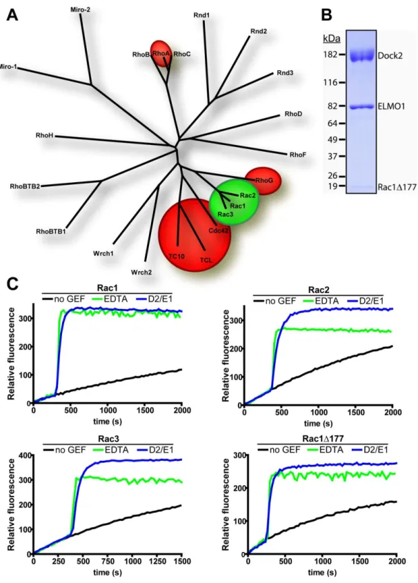
The Dock1-related family of proteins contains 11 mammalian members, with orthologs in humans, roundworms, slime mold, fruit flies, yeast and plants, among others (referred to as the Dock family of proteins in the rest of this document) [25-29]. This family also contains no obvious homology to the Dbl-family GEFS, but are, instead, defined by two regions of homology within the family, DHR-1 and DHR-2 for Dock homology region 1 and 2. The DHR-2 region is the center of exchange activity for these atypical exchange factors for Rho-family GTPases. While these proteins have been shown to be bona fide GEFs, information on how these proteins are regulated is relatively sparse and the mechanism by which they catalyze exchange is still unknown [9,27,28]. The Dock family has been grouped into four different subfamilies (Fig. 2), based on sequence identity over the entire sequence of the proteins, with 50-65% sequence identity between subfamily members[26,27,29]. The following sections review each subfamily.
Dock-family GEFs
Dock A subfamily: Dock1, Dock2 and Dock5
The Dock A subfamily of proteins includes two of the most well-studied of the Dock-family proteins, Dock1 and Dock2. This subfamily has several features in
common. The domain structure consists of the typical Dock-family DHR-1 and DHR-2 regions, along with an N-terminally located SH3 domain and a C-terminally located, variable proline and serine-rich region in most members [26-29]. Rac is the known Rho-family GTPase substrate for two of the three proteins within the Dock A subRho-family [26-29].
Dock1, originally identified as Dock180 (for 180-kDa protein downstream of Crk), is a prototype member of the Dock family of proteins [30,31]. Identified as a binding partner for the SH2 and SH3-domain containing adaptor protein, Crk, Dock1 mRNA was expressed strongly in the placenta, lungs, kidneys, pancreas and ovaries than in the thymus testes and colon. In contrast, expression was not detected in blood cells, suggesting that Dock1 was expressed only in adherent cells [30]. Subsequent analysis found that Dock1 association with CrkII was stimulated by integrin in NIH3T3 cells and that this binding correlated with CrkII binding to p130Cas, a protein that localizes to focal adhesions [32]. Consequently, coexpression of p130cas and CrkII with Dock1 induced local membrane spreading and accumulation of complexes containing
Dock1/CrkII/p130cas at focal adhesions. The C-terminal proline and serine-rich region of Dock1 was determined to be necessary for the Dock1-CrkII interaction [30,32].
Dock1 was found to increase active Rac1 in cells and was capable of direct interaction with Rac1, but not Cdc42 or RhoA, and Dock1-induced membrane spreading
was inhibited by dominant-negative Rac1 [33]. Two homologs of Dock1 in Drosophila
(myoblast city, MBC) and C. elegans (ced-5) were identified, and were grouped into a family of proteins, CDM, for ced-5, MBC, and Dock180 [31]. This family of proteins was suggested to be an evolutionarily conserved family involved in the extension of cell surfaces. MBC was necessary for dorsal closure and myoblast fusion in Drosophila
embryos [34], and ced-5 was necessary for cell corpse engulfment and distal tip cell migration [31]. The αvβ5 integrin was shown to mediate phagocytosis of apoptotic cells through recruitment of the Dock1/CrkII/p130cas complex, which in turn triggered Rac1 activation and phagosome formation [35]. A later study demonstrated that a secreted glycoprotein, MFG-E8 which can bind phosphatidylserine (PS) on apoptotic cells, is a ligand for the αvβ5 integrin, and can trigger activation of Rac1 through Dock1 for phagocytosis of apoptotic cells [36].
The identification of Dock1, and determination that it and its homologs participated in the activation of Rac1 and extension of cell surfaces paved the way for identification of Dock1 and homologs as Rho-family specific GEFs. The ability to directly activate Rac1 by a portion of Dock1 called the Docker domain led to the
proposal that Dock1 was an atypical, non-Dbl-family Rac-specific GEF [25]. About the same time, the other mammalian members of the Dock-family of proteins were identified through homology searches, and several were shown to have direct GEF activity on Rho-family GTPases [26,28]. The Docker domain was found to be within a region of
homology between the family members and was named DHR-2 [26] or CZH-2[28]. Another region of homology between the Dock-family proteins was N-terminal to the DHR-2 region, and was named DHR-1 [26], or CZH-1 [28]. Dock1 and its homolog,
MBC, can bind phosphatidylinositol 3,4,5-trisphosphate (PI(3,4,5)P3) through the DHR-1 region. This binding mediates localization of Dock1 to the membrane [37-39].
The ELMO (ced-12) family of proteins were identified as Dock1/ced-5 binding partners [25,40]. These proteins were shown to functionally cooperate with Dock1, although they had no exchange activity alone, leading to the suggestion that the
Dock1/ELMO complex functioned as a bipartite GEF for Rac1 [25]. This finding was not universally accepted, as Dock1 was capable of acting as an exchange factor in vitro
without ELMO1 [27,38]. Also, it was found by some groups that efficient activation of Rac1 by Dock1 required ELMO1 in cells [25,41], and others found the ELMO1
interaction dispensable [26].
Interactions between Dock1 and ELMO proteins include at least two direct interactions between the N-terminal 350 residues of Dock1 and the C-terminal 100 residues of ELMO1, with one of those interactions involving the Dock1 SH3 domain and the ELMO1 PxxP motif [25,41,42]. Separate from these interactions, the isolated PH domain of ELMO1, although missing the regions of ELMO1 necessary for direct
interaction with Dock1, is capable of binding, in trans, to a complex of Dock1 and Rac1 [41]. This interaction between the PH domain and the Dock1/Rac1 complex is capable of enhancing the exchange activity of Dock1. It was suggested that the PH domain
stabilizes the nucleotide-free transition state of Rac1 bound to Dock1, allowing for enhanced exchange activity[41]. The SH3 domain of Dock1 is also capable of binding to the DHR-2 region of Dock1 and competing for binding with Rac1 [42]. This SH3/DHR-2 interaction is relieved upon binding of ELMO1. The authors suggested this was an inhibitory mechanism for regulation of the Dock1 molecule, although it is unclear if the
two proteins exist separately within cells [42]. The assembly of the Dock1/ELMO complex may be regulated by the C-terminal SH3 domain of CrkII [43]. Endogenous Dock1/ELMO complex has been immunoprecipitated from HeLa and CHO-K1 nuclear extracts using an anti-Dock1 antibody [44]. The resulting Dock1/ELMO complexes contained more than one Dock1 molecule and more than one ELMO molecule per complex. The isoform of ELMO (1, 2 or 3) immunoprecipitated with Dock1 depended on the cell line. Multiple isoforms of ELMO1 were precipitated with Dock1 in each cell line [44].
Other functions for the interaction between Dock1 and ELMO proteins are evident. ELMO1 is capable of inhibiting ubiquitylation of Dock1, thereby protecting Dock1 from degradation [45]. Brain-specific angiogenesis inhibitor 1 (BAI1), a PS-sensitive receptor, can interact with the Dock1/ELMO1 complex through ELMO1 [46]. Recruitment of Dock1/ELMO1 to the BAI1 receptor mediates phagocytosis of apoptotic cells. RhoG, another Rho-family small GTPase has been shown to bind specifically to the N-terminus of ELMO1 in a GTP-dependent manner [47]. This interaction can localize Dock1/ELMO1 to the plasma membrane. Activation of Rac1-mediated cell migration is mediated by RhoG through the Dock1/ELMO1 complex. Interestingly, the interaction of Dock1 with CrkII is dispensable for this function [48]. Phagocytosis of apoptotic cells is also mediated by this interaction, and this mechanism is conserved in C. elegans as well as humans [49]. The interaction between RhoG and ELMO1 is exploited by the bacterial pathogen, Shigella. The bacterial-produced IpgB1 protein binds to ELMO1 in a manner mimicking RhoG, and activates production of membrane ruffles through the Dock1/ELMO complex, enabling bacterial entry into epithelial cells [50].
The Dock1/ELMO complex has also been implicated in other pathways promoting cell motility. A recent study implicates Dock1 in mediating attractive responses by neurons to netrin-1, an axon guidance cue [51]. It has also been reported that the Dock1/ELMO complex mediates the activation of Rac1 by ARNO, a GEF for the ADP-ribosylation factor 6 (ARF6) at the leading edge of migrating cells [52].
Despite the multitude of interacting proteins and pathways in which Dock1 is involved, it is clear that Dock1 is active in pathways that promote cell migration and phagocytosis within adherent cells. The evidence that ELMO proteins are a key
component of Dock1 activity whether by direct activation or indirect activation through localization and protein binding is also clear.
Dock2 was identified through homology to Dock1, and the expression pattern of Dock2 in tissues is nearly opposite that of Dock1 [53]. While Dock1 is present in mostly adherent cells, Dock2 is expressed mainly in non-adherent, hematopoietic cells. Dock2 mRNA expression was detected in peripheral blood cells, with slight expression in spleen and thymus. Dock2 was expressed only in lymphocytes and macrophages of various organs as detected by immunostaining of human cadaver tissue [53]. Dock2 is a specific exchange factor for Rac1, and does not activate Cdc42 or RhoA and is at least capable of binding to Rac2 [26,53].
Unlike Dock1, Dock2 does not bind to CrkII. It does, however, associate with a hematopoietic-specific Crk-like protein, CrkL, which can induce activation of Rac1 [53]. Interruption of this interaction inhibited CrkL activation of Rac1. Dock2 is essential for lymphocyte chemotaxis [54]. In Dock2-deficient cells, Rac activation and actin
polymerization induced by chemokines was nearly abolished.
ELMO proteins are capable of interacting with Dock2, and this interaction is critical for Dock2/ELMO1-mediated Rac1 activation[55]. For instance, expression of Dock2 in T-hybridoma cells lacking endogenous Dock2 induced Rac activation and actin polymerization. In addition, expression of a Dock2 mutant incapable of binding ELMO1 failed to effect these changes [55]. In plasmacytoma cells expressing Dock2 but not ELMO1, expression of ELMO1 induces Rac activation [55].
Dock2 has been implicated in a variety of processes critical for the survival of hematopoietic cells. For instance, Dock2 is essential for the activation of Rac1 required for the formation of immunological synapses mediated by T-cell receptors [56]. In this case, Dock2 regulates antigen-induced translocation of T-cell receptors and lipid rafts during synapse formation. Similarly, deletion of Dock2 has been shown to suppress cardiac allograft rejection by eliminating lymphocyte homing and immunological synapse formation [57]. Furthermore, chemokine-stimulated adhesion of lymphocytes under shear stress requires Dock2 for efficient attachment to VCAM-1 [58]. Vav1, a Dbl-family GEF that specifically activates Rac GTPases, is also required for this process, but the interplay between Vav1 and Dock2 is not well understood. Dock2 expression is also required for development of mouse Vα14 natural killer T cells [59], which play important roles in host defense against pathogens, immune regulation, and tumor surveillance. Dock2 is necessary for another type of T-cell differentiation in CD4+ T cells. Dock2 links T-cell receptor signals to IL-4Rα downregulation to control lineage commitment of these cells [60]. Finally, spingosine-1-phosphate-mediated egress of lymphocytes from peripheral lymph nodes and interstitial mobility of lymphocytes also depend on Dock2 [61].
Considering the importance of Dock2 in hematopoietic cells as discussed above, it is unsurprising that the Dock2/ELMO1 machinery has been taken advantage of by a pathogen. The HIV-1 protein Nef, a potent virulence factor, can form a complex with Dock2/ELMO1, and this interaction is dependent upon ELMO1 [62]. Nef activates Rac in T cell lines and in primary T cells, and the authors suggest that Nef modulates multiple aspects of T cell function through the Dock2/ELMO1 complex [62].
The variety of studies showing the necessity of properly functioning Dock2 in lymphocytes serves to highlight the fact that Dock2 is critical for regulation of the actin cytoskeleton and mobility within hematopoietic cells. The specific contributions of ELMO proteins within the context of the Dock2/ELMO complex remain to be determined.
Very little is known about mammalian Dock5, as it has not yet been cloned. Out of 65 family GEFs and 11 Dock-family GEFs tested, only Dock5 and Net1, a Dbl-family GEF, were up-regulated during RANKL-induced osteoclastogenesis [63]. In this case, silencing of Dock5 in RAW264.7 cells was found to be extremely cytotoxic, although it had no apparent effect in NIH3T3 fibroblasts. The zebrafish ortholog of Dock5 was cloned and used to design morpholino oligonucleotides to block the function of the protein in zebrafish embryos[64]. In this study, morpholino embryos for Dock5 showed defective fast-myoblast fusion. Dock1, Dock5, Crk and CrkL were all tested similarly, and when different combinations of Dock and Crk proteins were tested, the highest level of fusion suppression observed occurred in embryos injected simultaneously with Dock5 and CrkL morpholinos.
Dock B subfamily: Dock3 and Dock4
The Dock B subfamily consists of two mammalian members, Dock3 and Dock4. This branch of the family is most similar to the Dock A proteins, with Dock C and Dock D proteins further removed by sequence homology. The Dock B proteins are about 56% identical within the subfamily, but are only about 40% identical to Dock A subfamily members [26,28]. Like the Dock A members, however, Dock B proteins contain an N-terminal SH3 domain, which allows association of ELMO1.
The presinilin (PS) gene is linked to approximately 50% of familial Alzheimer’s disease (AD) cases [65,66]. Dock3 (also modifier of cell adhesion – MOCA) was identified through yeast-two hybrid screens as a protein capable of interacting with presinilin1(PS1) and labeled as PBP, or presinilin binding protein [67]. This interaction was confirmed with immunoprecipitation of the two proteins from transfected
mammalian cells. Following up on this interaction, the authors discovered that co-expression of Dock3 and PS1 localized primarily cytoplasmic Dock3 to a discrete, organelle-like compartment, likely the endoplasmic reticulum, where PS1 is primarily localized. Dock3 mRNA was only found in the brain and spinal cord, highly expressed in the cerebral cortex and hippocampus, and the level of Dock3 in the soluble fraction of AD brains was reduced compared to age-matched normal brains [67]. A later study found that Dock3 is associated with intracellular neurofibrillary tangles (NFT) and expression increases phosphorylation of the NFT protein, tau, suggesting that Dock3 may play a role in the AD neurodegenerative process [68]. Further study showed that
expression of Dock3 decreased amyloid precursor protein (APP) and amyloid β-peptide secretion, and reduced cell-substratum adhesion, likely through direction of APP to the
proteasome for degradation [69]. Another study identified the Dock3 gene as one of two genes disrupted by a pericentric inversion on chromosome 3 associated with an attention deficit hyperactivity disorder (ADHD)-like phenotype [70].
Dock3 has been shown to bind to Rac1, to enhance Rac1 activation, and to promote Rac1-dependent cell migration [71,72], but it does not interact with RhoA or Cdc42 [72]. In addition Dock3 interacts with ELMO1, and co-expression of Dock3 and ELMO1 is necessary for the promotion of Rac1-dependent cell migration [71].
Membrane-targeted Dock3 also enhances GTP-loading of Rac1 and JNK activation in cells, and endogenous Dock3 co-localizes with F-actin at the leading edge of lamellipodia [72]. It was determined that Dock3 promotes cell-cell adhesion and neurite outgrowth mediated by N-cadherin [69].
The Dock4 gene was identified as a homozygous deletion a mouse osteosarcoma cell line, and was mutated in a subset of human cancer cell lines [73]. In this study, restoration of wild-type Dock4 in the Dock4-null osteosarcoma lines restored contact inhibition, reduced colony formation in soft agar and formed small, non-invasive tumors when injected into nude mice, as compared to the Dock4 null cell line which formed large invasive tumors. The authors also reported that cells lacking Dock4 fail to form adherens junctions and this phenotype could be rescued by expressing Dock4 or by expressing constitutively active Rap1. Expression of Dock4 and dominant negative Rap1 failed to promote the formation of adherens junctions. In addition, Dock4 was shown to promote activation of Rap1 in transfected cells [73]. Other studies, however, describe Dock4 as a Rac1 activator, and were unable to detect Rap1 activation by Dock4 [42,74]. Dock4 was able to interact with ELMO1, and expression of active RhoG induced
translocation of the Dock4/ELMO1 complex to the plasma membrane from the cytoplasm [74]. This translocation enhanced the Dock4/ELMO1-dependent Rac1 activation and cell migration, and knockdown of Dock4 expression reduced cell
migration. Dock4 is expressed in multiple tissue types, with the highest expression noted in skeletal muscle, prostate and ovary [73,75]. An isoform of Dock4, alternatively
spliced in the C-terminus, is a strong Rac activator and is expressed in the brain, inner ear and eye tissues [75]. This isoform interacts with harmonin and is present in the hair bundles of auditory sensory cells.
Dock C subfamily: Dock6, Dock7 and Dock8
The Dock C subfamily of the CDM proteins has the least amount of information available about its representative members. Unlike both the Dock A and Dock B subfamilies, these proteins have not been shown to interact with ELMO proteins. The domain structure for these proteins is also not well known, with the only described domains being the DHR-1 and DHR-2 regions [27,29] and a predicted coiled-coil motif in the C-terminus [9].
Dock6 is reported to activate Rac1 and Cdc42, but is otherwise poorly understood [76]. For instance, full-length Dock6 or its isolated DHR-2 region immunoprecipitated from HEK293T cells activated Rac1 and Cdc42, but not RhoA. Similarly, the DHR-2 region activated Rac1 and Cdc42 in cells and promoted associated filopodia and lamellipodia [76]. However, unlike other Dock family members, Dock6 did not bind ELMO1, ELMO2 or CrkII [76]. Dock6 might regulate neuronal processes since its expression is highly increased upon differentiation of mouse N1E-115 neuroblastoma cells and Dock6 can regulate neurite outgrowth through Rac1 and Cdc42 [76].
Initial attempts to characterize the GTPase specificity of Dock7, also named zizimin-related 2 (Zir2) failed to show significant activation of either Rac1, Cdc42 or RhoA. The protein used for these assays, however, was in vitro translated, and only consisted of the DHR-2 region of the protein [26]. Later results identified Dock7 as a binding partner for TSC1 or hamartin, a protein that, when mutated, causes tuberous sclerosis (TSC) – a disease characterized by development of benign tumor-like
“hamartomas” in kidneys, heart, skin and brain. TSC2, or tuberin, forms high affinity dimers with hamartin, and can interact with and act as a GAP for Rheb, a Ras-family small GTPase. It was suggested that Dock7 might be a Rheb-GEF, given this linkage of interactions [77,78]. More recent studies, though, have elegantly described Dock7 as a Rac specific GEF that is highly expressed in the developing rat brain, specifically in hippocampal neurons [79]. In unpolarized hippocampal neurons, Dock7 is
asymmetrically distributed, and is selectively expressed in the axon. Dock7 expression can be manipulated to affect axon formation, with over-expression mediating formation of multiple axons and knockdown inhibiting axon formation. Interestingly, Dock7 and Rac activation leads to inactivation of a microtubule destabilizing protein,
stathmin/Op18, through phosphorylation [79]. Following a trend in lipid binding seen in other Dock-family proteins, PI(3,4,5)P3 is produced in the developing axon, and PI 3-kinase inhibitors abrogated the ability of Dock7 to form multiple axons [79] suggesting that the DHR-1 domain of Dock7 may also be capable of interacting with
phosphoinositides. Cote and Vuori [27] make an interesting point that stathmin was identified as a gene regulating border cell migration in Drosophila oogenesis [80]. Myoblast city, the Drosophila Dock1 homolog, is also critical in border cell
migration[81]. Perhaps regulation of microtubules could be a common effect of different Dock-family proteins.
Dock8 (Zir3) was identified in a yeast two-hybrid screen as a Cdc42-interacting protein, and subsequent northern blot tissue analysis revealed a relatively ubiquitous expression pattern including placenta, lung, kidney and pancreas, with relatively low expression in heart, brain and skeletal muscle [82]. Subsequent immunofluorescence staining of cells showed that endogenous as well as over-expressed Dock8 was present at edges undergoing lamellipodia formation. Transfection of a C-terminal fragment (1179-1701) resulted in ring-like vesicular structures that contained filamentous actin [82]. Additional yeast two-hybrid assays demonstrated binding between Dock8 residues 1044-1701, which includes the DHR-1 domain and both activated (61L) and inactive (N17) Cdc42 and Rac1, but not activated RhoA [82]. This Dock8 fragment also interacted with TCL and TC10 – Rho-family GTPases closely related to Cdc42. An assay using
immunoprecipitated Dock8 1179-1701, however, failed to bind to active or inactive GST fusions of Rac1, Cdc42 or RhoA. This same fragment was tested for binding to GTPases in a filter binding assay, and no interaction was detected [82].
A homozygous deletion of the locus for Dock8 was found in a lung cancer cell line [83]. Subsequent analysis of primary lung cancers revealed 87% had reduced Dock8 expression levels compared with normal cells. Homozygous deletions of Dock8 were also found in a gastric and a breast cancer cell line. The reductions in gene expression occurred regardless of histological type, suggesting that the reductions may be caused by DNA methylation or histone deacetylation [83]. Furthermore, Takahashi, et al. suggested that the “down-regulation of Dock8 by epigenetic mechanisms is involved in the
development and/or progression of lung cancer” [83]. Dock8 was originally described as 1701 amino acids in length [82]; however, later work proposed a version (2099 residues) with an extended N-terminus [83] and most rece3nt work suggests that Dock8 contains 2033 residues [84]. Disruptions of the Dock8 gene have also been found in two unrelated patients with mental retardation [84].
Dock D subfamily: Dock9, Dock10 and Dock11
Proteins in the Dock D subfamily of Dock proteins contain the typical DHR-1 and DHR-2 regions, but, like the Dock C subfamily, this group does not contain the
N-terminal SH3 domain of the Dock A and B subfamily members [26,28,29]. As expected with the lack of an SH3 domain, proteins in this subfamily do not interact with ELMO proteins. The Dock D proteins, however, do contain a PH domain in the N-terminal region [26,28,29]. Two of the three mammalian proteins in this subfamily have been identified with specificity for Cdc42 over other Rho-family GTPases.
Dock9, also named zizimin1, is the most-studied protein of this subfamily. Originally identified in a screen for proteins that specifically interacted with nucleotide-depleted Cdc42 and were eluted by the addition of GTPγS, Dock9 was precipitated from NIH3T3 cells, human umbilical vein endothelial cells, rat vascular smooth muscle cells and COS-7 cells [28]. Interestingly, one group discovered that Dock9 mRNA levels were highest in brain lung and kidney, while levels in the heart, liver, skeletal muscle and hematopoietic organs were low [85]. Another group discovered a slightly different mRNA distribution where levels were highest in heart and placenta, with relatively high levels in kidney, brain, lung and skeletal muscle and low levels in liver, intestine and hematopoietic tissues[28].
The protein was named zizimin1 (from the Hebrew word for spikes), reflecting its ability to induce microspike formation when over-expressed in fibroblasts, a function linked to Cdc42 activation [28]. Dock9 was shown to interact directly with Cdc42, but not Rac1 or RhoA [26,28,85] and to require the DHR-2 domain for its exchange activity. Deletion of the N-terminus of Dock9 resulted in a stronger interaction with nucleotide-depleted Cdc42 [28]. Full-length Dock9 is capable of activating Cdc42 within cells, and the isolated DHR-2 region is capable of activating Cdc42 in vitro, but is not efficient at activating Cdc42 within cells [26,28]. When cells were co-transfected with the DHR-2 region and with a cytoplasmic form of Cdc42, the DHR-2 region seemed fully active on Cdc42 [28]. The authors suggested that the N-terminal PH domain may function in membrane association, and the lack of the PH domain in the DHR-2 did not allow the protein to localize properly to activate Cdc42.
Building upon the observation that N-terminal truncations of Dock9 interacted more strongly with Cdc42, Meller and colleagues [86] found that three regions within the N-terminal portion of Dock9 are capable of interaction with the DHR-2 region. The DHR-1 region, the region N-terminal to the PH domain, and a region C-terminal to the PH domain were all capable of interaction with the DHR-2 region. The interaction between the N- and C-terminal regions of Dock9 inhibited Cdc42 binding. Analysis of Dock9 by limited proteolysis, suggested that the DHR-2 homology region might not be a complete structural domain, and that residues beyond the homology region may be a part of the GEF domain of the protein [86].
Homology between Dock-family proteins was used to identify Dock10 (also named zizimin3) [26,28]. This protein has yet to be fully cloned, but expression analysis
indicates that Dock10 is enriched in the brain, lung, spleen and thymus [85].
Additionally, the DHR-2 domain of Dock10 was cloned and expressed in COS-7 cells, and lysates were incubated with various nucleotide-depleted GTPases bound to beads [85]. The beads were washed and then bound proteins were eluted with GTP. The DHR-2 domain of Dock10 associated weakly with nucleotide-depleted Cdc4DHR-2 and TCL, and even more weakly with TC10 and RhoA. There was no detectable interaction with Rac1 [85]. Dock10 was also found to be up-regulated in some aggressive, poorly differentiated papillary thyroid carcinoma specimens [87].
Dock11 (also named activated Cdc42-associated GEF (ACG) [88] and zizimin2 [28]) was identified through homology to other Dock-family members [26,28], and was later cloned as a gene expressed at higher levels in germinal center B cells than non germinal center B cells [85]. Expression was detected primarily in hematopoietic organs, with low-level expression in some non-hematopoietic tissues. The C-terminal two-thirds of Dock11, as well as the smaller DHR-2 domain were sufficient for binding to
nucleotide-depleted Cdc42, but the DHR-2 domain does not interact with TC10 or TCL. Full-length Dock11 and the isolated DHR-2 domain can both activate Cdc42 in cells [85].
Immunoprecipitation assays utilizing GST-Cdc42 as a pull-down matrix identified Dock11 as a binding partner, but surprisingly, it was binding preferentially to active Cdc42 (Q61L), not nucleotide-free GTPase [88]. In contrast, the isolated DHR-2 domain of Dock11 bound preferentially to the nucleotide-depleted form of Cdc42. The full-length form of Dock11 was an active GEF for Cdc42, while the DHR-2 domain only showed weak activity. Furthermore, deletion of the N-terminal 272 amino acids of Dock11 changed the binding preference from Cdc42 (Q61L) to nucleotide-depleted
Cdc42 (T17N). The essential region for binding active Cdc42 was determined to be within residues 66-126 of Dock11, N-terminal to the PH domain [88]. These residues (10-127) were capable of binding to the DHR-2 domain, and that interaction was increased in the presence of active Cdc42, but not nucleotide-free Cdc42. A deletion of the N-terminal 126 amino acids, abrogating interaction with active Cdc42 also severely impaired GEF activity. The authors proposed a positive feedback model where the N-terminus of Dock11 binds the DHR-2 domain and this form has weak GEF activity. Activation of Cdc42 allows the active GTPase to interact with the N-terminal region and the DHR-2 domain, enhancing the activity of the GEF [88].
A second study examined the interaction between Dock11 and Cdc42 and found that while full-length Dock11 did bind GTP-Cdc42, it bound preferentially to the nucleotide-depleted Cdc42 [86]. The authors of this second paper suggest that the discrepancy between the two sets of results could arise from the differences between the Cdc42 proteins used. In the first paper, Cdc42 mutants were utilized for their activated (Q61L) and nucleotide-depleted (T17N) forms [88], while in the second paper, the Cdc42 proteins were treated with EDTA, nucleotides and MgCl2 to get the desired nucleotide state of the protein [86].
Dock-family GTPase recognition
Most studies have focused on assessing the capacity of Dock-family members to directly activate specific GTPases. In contrast, there are limited studies addressing the structural determinants within Dock-family members required for the engagement and activation of specific GTPases. A recent study, however, has compared the binding properties of the Rac-specific Dock2 and Cdc42-specific Dock9 [89]. By using a
multitude of chimeras of Rac2 and Cdc42, as well as point substitutions, the authors have been able to map important residues for recognition of cognate GTPases by Dock2 and Dock9 [89]. Dbl-family GEFs can discriminate between Rac1 and Cdc42 based on the β2 and β3 strands of the GTPase [90,91]. Substitution of tryptophan 56 in Rac1 to the phenylalanine of Cdc42 and vice versa is sufficient to change the specificity of Dbl-family GEFs Tiam1 and ITSN. For example, Rac-specific Tiam1 is unable to catalyze exchange on Rac1 (W56F), but Cdc42-specific ITSN is capable of catalyzing exchange on this mutant [92]. The reverse holds true for Cdc42 (F56W), where Tiam1 is capable of catalyzing exchange but ITSN loses that ability [93]. Dock2 and Dock9 utilize the β3 strand of the GTPase for substrate recognition as well. Similar to the Dbl-family GEFS, the Dock-family GEFs gain the ability to catalyze exchange on non-cognate GTPases upon mutation of the GTPase residue 56. However, neither Dock2 nor Dock9 lose the ability to catalyze exchange on their mutated cognate GTPase [89]. This suggests that while the determinants for GTPase recognition and specificity for Dock-family GEFs overlaps to some extent with the Dbl-family GEFs, there are additional determinants for Dock-family GTPase recognition. Emphasizing this, the divergent residues at positions 27 and 30 within the switch 1 regions of Cdc42 and Rac1 are important for substrate recognition by Dock2 and Dock9, while these residues are usually not utilized for substrate discrimination by Dbl-family GEFs [89].
Concluding remarks
Identification and characterization of the relatively new Dock family of proteins is still a work in progress. As a consequence, an understanding of how these proteins
function and are regulated is very limited. What is evident is that these proteins play crucial roles in the regulation of Rho GTPases and their effectors. Identification of functional and regulatory mechanisms for the Dock-family GEFs will give us insight, both into the general mechanisms of Rho-family GTPase activation, and into the specific ways in which these Dock-family GEFs coordinate multiple signals and resultant
outcomes within cells.
Figure 1: Overview of the Rho-family GTPase cycle.
Rho GTPases cycle from an inactive (GDP-bound) state to an active (GTP-bound) state with the help of regulatory proteins. Active GTPase is capable of binding downstream effector proteins. GEF: guanine nucleotide exchange factor, GAP: GTPase accelerating protein, GDI: guanine dissociation inhibitor, Pi: inorganic phosphate, GDP: guanosine 5’-diphosphate, GTP: guanosine 5’-triphosphate.
Figure 2: Overview of the Dock family of proteins (mammalian).
The Dock family of proteins can be subdivided into four groups based on sequence homology, Dock A, B, C, and D. Center: a cladogram showing the relative homology between Dock-family members. The typical domain structures for each subfamily is shown next to its grouping on the cladogram. DHR-1: Dock homology region 1, DHR-2: Dock homology region 2, SH3: Src-homology 3 domain, PH: pleckstrin homology domain.
CHAPTER 2 – DOCK2/ELMO1: MOLECULAR INTERACTIONS OF A
HETEROTETRAMERIC GEF
Background
The Dock family of guanine nucleotide exchange factors (GEFs) is evolutionarily conserved in eukaryotes ranging from mammals to plants and yeast [25,29,94].
Encompassing 11 mammalian members, the Dock-family proteins are critical for a variety of biological processes. In C. elegans, CED-5 is essential for distal tip cell migration and clearance of apoptotic bodies [31]. Mutations within MBC, the
Dock-family proteins activate Rho-family GTPases by catalyzing the exchange of GDP for GTP; GTP-bound GTPases directly engage downstream effectors to modulate their activities. The relatively new family of Dock proteins varies significantly from the well-studied Dbl-family GEFs which utilize a tandem Dbl homology (DH) domain and pleckstrin homology (PH) domain to catalyze nucleotide exchange [9]. The Dock family of proteins, instead, relies upon a unique region, the docker-homology-region-2 (DHR-2, also named “Docker” or “CZH2”) for catalytic activity [25,26,28]. This region of homology between the Dock-family members possesses no obvious sequence or predicted structural homology to the DH and PH domains of the Dbl-family GEFs and, therefore, may function in a unique manner.
Several of the Dock-family members are known to form a complex with ELMO proteins [71,74]. The interaction between Dock1 and ELMO1 is critical for phagocytosis of apoptotic cells and for cellular migration [25,41,47,49,55,71,97,98]. ELMO proteins, which exist as three mammalian isoforms (1, 2 and 3), have no GEF activity alone, but it has been proposed that together, Dock and ELMO proteins function as bipartite GEFs – with ELMO proteins aiding in the stabilization of Rac1 in a nucleotide-free transition state [25,41,71]. Understanding how Dock and ELMO proteins interact should lead to a better comprehension of the functional consequences of this association.
Of the Dock-family proteins, only the Dock A and Dock B subfamilies have been shown to interact with ELMO proteins [71,74]. Through examination of deletion
constructs and mutational analysis, the interactions between Dock1 and ELMO1 are known to involve at least three separate regions: (1) the N-terminal SH3 domain of Dock1 and a polyproline (PxxP) motif in the C-terminus of ELMO1; (2) the N-terminal
357 residues of Dock1 and the C-terminal 100 residues of ELMO1, likely within the residues between the end of the PH domain and the beginning of the PxxP motif and (3) the PH domain of ELMO1 binds to a Dock1/Rac1 complex in trans [41,42]. Disruption of the first two interactions is necessary to prevent formation of the Dock1/ELMO1 complex. Deletion of the SH3 domain or the PxxP motif is not sufficient to eliminate the interaction between Dock1 and ELMO1. A mutation of glycine 171 to glutamic acid (G171E) in the N-terminus of Dock1 is sufficient to disrupt the second interaction. When combined, the deletion of the SH3 domain and the G171E mutation can fully eliminate the direct interaction between Dock1 and ELMO1. Alternatively, termination of ELMO1 at residue 629 prevents formation of the complex, presumably by removing the sites for both interactions [42]. Interestingly, the binding of the PH domain of ELMO1 in trans to a complex of Dock1 and Rac1 increases the exchange activity of Dock1. It has been proposed that the PH domain aides in the stabilization of nucleotide-free Rac1 in the transition state while associated with Dock1 and Rac1, and this stabilization is
responsible for the increase in exchange activity seen with this trimeric complex [41]. ELMO proteins also play a role in proper localization of Dock-family proteins, by binding to activated RhoG, another Rho-family GTPase, which functions to bring
Dock/ELMO complexes to the membrane [47-49,74]. The N-terminal region of ELMO1 is necessary and sufficient for this interaction [47,48]. Disruption of the RhoG-ELMO1 complex by mutation of the N-terminus of ELMO1 results in the failure of the
Dock1/ELMO1 complex to promote phagocytosis, cell migration, formation of lamellipodia and the mutant complex is not localized to membrane ruffles [49].
Defining the mechanism by which Dock and ELMO proteins function together would provide a greater understanding of how the various biological effects of the complex are carried-out and regulated. Here we refine the predicted domain boundaries for Dock1, Dock2 and ELMO1, as well as identify new domains. We show that isolated ELMO1 exists as a monomer, and can interact with both Rac1 and RhoG. This
interaction is nucleotide-dependent, and is dependent upon the presence of the polybasic region (PBR) of Rac1 and/or RhoG. We demonstrate that Dock2 and ELMO1 can be purified as a stable complex, and this Dock2/ELMO1 complex exists as an obligate heterotetramer, consisting of two molecules each of Dock2 and ELMO1. We also describe the substrate specificity of the complex using a screen of purified GTPases, and demonstrate that this tetramer binds a single molecule of Rac1.
Experimental procedures
Domain architecture prediction
Domain analysis of Dock1, Dock2 and ELMO1 was carried out using a
combination of sequence alignments using ClustalX version 1.83 [99], protein-protein BLAST and psi-BLAST searches [100], secondary structure prediction programs, and tertiary structure prediction and threading programs [100-106].
Plasmids
ELMO1 constructs for insect cell expression were PCR-amplified and inserted into pFastBac1 using a modified ligation-independent cloning strategy (LIC) [107]. ELMO1 (1-727) was amplified from pGEX4T2-ELMO1 [40].
Full-length human Dock2 (1-1830) was PCR-amplified from pCXN2-Flag-Dock2, a gift of Dr. Michiyuki Matsuda [32,33], and cloned into a modified pFastBac1 vector (Invitrogen) using a LIC strategy [107]. The resulting construct encodes a non-cleavable hexahistadine tag immediately preceding the N-terminus of Dock2.
RhoG-pGEX4TEV2 and Rac1 constructs (C189S and Δ177) were as described [108,109]. HA-Rac1 (3X-HA epitope N-terminal to Rac1) was amplified from
RAC010TN00 (UMR cDNA Resource Center), introducing a C189S mutation and stop codon, as well as NcoI and XhoI sites, and the fragment was ligated into pET15b via the NcoI and XhoI sites. Flag-Rac1 was amplified from Rac1C189S introducing a Flag epitope at the N-terminus of Rac1, and was ligated into pET15b as above. All new constructs were verified by automated sequencing.
Protein purification
Baculoviruses encoding the Dock2 and ELMO1 constructs were generated from the pFastBac vectors using the Bac-to-Bac system (Invitrogen). High-Five insect cells were co-infected with viruses encoding Dock2 and ELMO1. Cells were harvested by low-speed centrifugation after incubation at 27°C for 48 hours with shaking at 140 rpm. Cells were resuspended in 20 mM Tris, 1 M NaCl, 5 mM imidazole, 10% glycerol, pH 8.0 (buffer N1). Cells were lysed with an Emusiflex-C5 homogenizer (Avestin), and lysate was cleared by centrifugation at 265,000 x g for 45 minutes at 4°C. Cleared lysate was passed through a 0.45 µm filter before being loaded onto a 5 ml nickel-charged metal chelating column (GE Healthcare) equilibrated with buffer N1. The protein bound to the column was washed with buffer N1 and buffer N1 containing 50 mM imidazole before being eluted with buffer N1 containing 400 mM imidazole. Fractions containing
Dock2/ELMO1 were pooled and concentrated to 10 ml in a 10K MWCO Vivaspin (Vivascience) 20 ml concentrator before being loaded onto a 26/60 Sephracryl S300 size exclusion column (GE Healthcare) equilibrated in 20 mM Tris, 300 mM NaCl, 10% glycerol, 2 mM DTT, pH 8.0. Fractions containing Dock2/ELMO1 were pooled and dialyzed versus 20 mM Tris, 100 mM NaCl, 2 mM DTT, pH 8.0, concentrated as above, snap frozen in liquid N2, and stored at -80°C.
Bacterially-produced ELMO1 and ELMO1ΔN were expressed as GST-fusion proteins from pGEX4T2-ELMO1 and pGEX4T2-ELMO1 532-727 in the BL21(DE3) E. coli strain. Cells expressing ELMO1 were grown at 37°C in Terrific Broth (TB) until they reached an absorbance at 600 nm (A600) of ~0.8, then protein expression was induced with 0.5 mM isopropyl-β-D-thiogalactopyranoside (IPTG). Cells were allowed to grow ~12 hours at 25°C before harvesting by low-speed centrifugation. Cells
expressing ELMO1ΔN were grown at 37°C in self-inducing ZYM-5052 media [110] until they reached an A600 of ~0.6, then cells were allowed to grow ~15 hours at 20°C before harvesting by low-speed centrifugation. Cell pellets were resuspended in 20 mM Tris pH 8.0, 150 mM NaCl, 2 mM DTT, and 10% glycerol (GST buffer), lysed using an Emulsiflex-C5 homogenizer (Avestin), and clarified by ultracentrifugation at ~150,000 x
g. Clarified supernatant was loaded onto a 5 ml GSTrap FF column (GE Healthcare) pre-equilibrated with GST buffer, and eluted with GST buffer supplemented with 10 mM reduced glutathione. Fractions containing GST-ELMO1 or GST-ELMO1ΔN were
pooled and the GST tag was cleaved overnight with thrombin. ELMO1 was dialyzed into 20 mM Tris pH 8.0, 10 mM NaCl, 2 mM DTT and 10% glycerol (buffer Q) and loaded onto an anion-exchange chromatography column pre-equilibrated with buffer Q, then
eluted with a linear gradient of 10-300 mM NaCl. Fractions containing ELMO1 were pooled and dialyzed vs. GST buffer, and remaining fusion protein was removed using a 5 ml GSTrap FF column. ELMO1ΔN was dialyzed into 20 mM HEPES pH 7.0, 10 mM NaCl, 2 mM DTT and 10% glycerol (buffer S1) and loaded onto a cation-exchange chromatography column pre-equilibrated with buffer S1, then eluted with a linear gradient of 10-300 mM NaCl. Fractions containing ELMO1ΔN were pooled and
dialyzed vs. GST buffer, and remaining fusion protein was removed using a 5 ml GSTrap FF column. ELMO1 and ELMO1ΔN were concentrated using 10K MWCO Vivaspin 20 ml concentrators, snap frozen in liquid N2, and stored at -80°C.
GST-ELMO2 was expressed from pGEX4T2 as for ELMO1, and purified
essentially as described above for ELMO1 through the first affinity chromatography step. GST-ELMO2 was then concentrated, snap frozen and stored at -80°C.
ELMO1 was also produced in insect cells. The protein was expressed and purified as above for the Dock2/ELMO1 complex with a few changes. Less virus encoding Dock2 relative to ELMO1 was used, and ELMO1 contained an N-terminal hexa-histidine tag. A small amount of excess ELMO1 was purified away from the Dock2/ELMO1 complex during the size exclusion chromatography step and then stored at -80°C for subsequent analysis.
The GST-RhoG expression construct was expressed in the BL21(DE3) E. coli
strain. Cells were grown at 37° in enriched media (24 g yeast extract and 12g tryptone per liter, pH 7.4) supplemented with 0.1 mg/ml ampicillin until they reached an A600 of ~0.8, then protein expression was induced with 0.1 mM IPTG. Cells were allowed to
grow ~15 hours at 20°C before harvesting by low-speed centrifugation. RhoG was essentially purified as described [108].
Rac1 proteins (residues 1-189 C189S and Δ177) were expressed and purified essentially as described [108,109]. HA-Rac1 and Flag-Rac1 were purified as described for Rac1 C189S [108].
Size and oligomeric state determination
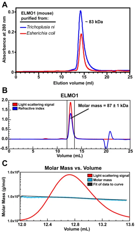
ELMO1 purified from bacteria and from insect cells was subjected to size exclusion chromatography on a Superdex 200 10/300 GL (GE Healthcare) analytical column. The molecular weight was estimated by comparing the peak elution volume to the peak elution volumes of a set of globular proteins of known molecular weight applied to the same column under similar conditions.
Light scattering measurements were made with a Wyatt DAWN EOS light scattering instrument (Wyatt Optilab refractometer, and Wyatt dynamic light scattering module) interfaced to an AKTA FPLC (GE Healthcare). 100 µl of the Dock2/ELMO1 complexes at 2 mg/ml were loaded onto a Superose 6 10/300 GL column (GE
Healthcare) pre-equilibrated in 20 mM HEPES pH 7.5, 150 mM NaCl, 2 mM DTT and 0.02% NaN3 at 25°C. For ELMO1 samples, 100 µl at 4.5 mg/ml was loaded onto a Superdex 200 10/300 GL column (GE Healthcare) pre-equilibrated with the same buffer as above. Data collection and analysis was performed with ASTRA software version 4.90.08 (Wyatt technologies). The refractive increment (dn/dc) was set at 0.185 for each protein in the molecular mass calculations, based on the premise that dn/dc is constant for unmodified proteins [111]. Experiments were repeated 3-5 times.
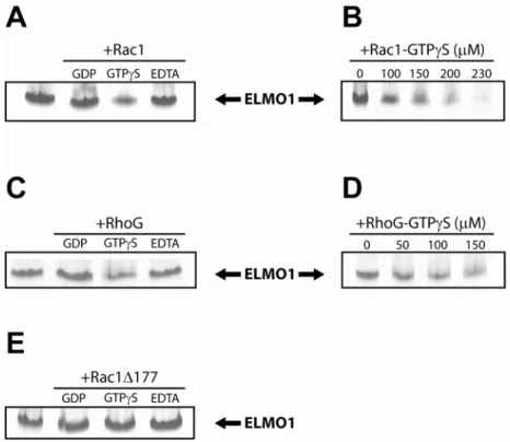
Native PAGE assays
GTPases were pre-loaded with the indicated nucleotide (or lack thereof) by incubating with EDTA at 2 times the concentration of MgCl2 in the protein buffer (usually 2 mM EDTA) and 10-fold molar excess of desired nucleotide for 30 minutes at room temperature. Loading reactions were then stopped by addition of 2.5-fold excess of MgCl2 over the concentration of EDTA (usually 5 mM MgCl2), except in the case of the nucleotide-depleted forms, where buffer containing 2 mM EDTA and no MgCl2 was used.
GTPases were incubated at indicated concentrations with ELMO1 for 15 minutes at room temperature, then were put on ice and loaded onto a 12.5% or 20% PhastGel (GE Healthcare) in combination with Native gel buffer strips (GE Healthcare), using a 6-well comb which loads 4µl of sample per well. Gels were run with the manufacturer’s suggested protocols for 12.5% or 20% native gels. Proteins were visualized by Coomassie brilliant blue staining.
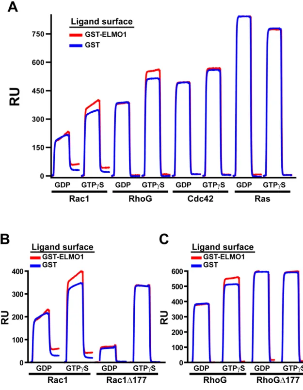
Surface plasmon resonance assays
Utilizing a BIACORE 3000 surface plasmon resonance (SPR) instrument (GE Healthcare), anti-GST antibody was coupled to a CM5 chip per the manufacturer’s instructions (GE Healthcare). Equal response units of indicated, GST-fusion ligand proteins were loaded onto the chip by brief injections of protein over the antibody surface. Interactions of Rho GTPases at a concentration of 10 µM were analyzed using the KINJECT command. Graphs of the response units are plotted with curves for the ligand protein of interest (ELMO1 or ELMO2) overlaid on the corresponding curve for the GST-only ligand surface to identify interactions.
Exchange assays – loading
Exchange assays monitoring loading of GTPase with fluorescent nucleotide was carried out in 20 mM HEPES, 200 mM NaCl, 10 mM MgCl2, 1 mM DTT, 100 nM BODIPY-FL-GDP and 10% glycerol at 10°C. GTPases were added to 2 µM and 100 nM Dock2/ELMO1 complex or 20 mM EDTA were added as exchange factors. Loading of BODIPY-FL-GDP onto the GTPase was monitored by an increase in fluorescence by a Perkin-Elmer LS-55 fluorimeter with excitation and emission wavelengths of 503 and 511, respectively. Addition of EDTA was used to confirm the GTPase was active. Rac and RhoG purifications are described above. All other GTPases were purified as described [112].
Rac-binding immunoprecipitation
Purified Dock2/ELMO1, Flag-Rac1, HA-Rac1 and Rac1Δ177 were added as indicated to appropriate reactions. 450 pmoles Dock2/ELMO1 was used per reaction, and 450 pmoles of the tagged Rac1 construct was used per reaction. 900 pmoles of Rac1Δ177 was used per reaction. Proteins were diluted to equivalent concentrations in 20 mM HEPES pH 7.5, 150 mM NaCl, 5 mM EDTA, and allowed to incubate with each other, as indicated, for 15-30 minutes at 25°C. Anti-HA antibody-coupled beads (Roche) or Anti-Flag M2 antibody-coupled beads (Sigma) were washed with wash buffer (20 mM HEPES pH 7.5, 150 mM NaCl, 5 mM EDTA, 0.25% NP-40, 100 µg/ml BSA) and 25 µl of appropriate beads were used per reaction. Wash buffer was added to reactions to bring the volume up to 500 ml and the tubes were incubated at 4°C on a rotator for 90 minutes. Beads were collected by centrifugation at 6000 x g for 30 seconds, and supernatant was removed by aspiration. Beads were washed 3 times with 1 ml wash buffer and protein
was eluted by boiling in 30 µl SDS-PAGE gel-loading dye. Samples were loaded onto an SDS-PAGE gel, and bound proteins were visualized by staining with Coomassie brilliant blue.
Results
Dock1, Dock2 and ELMO1 domain architecture
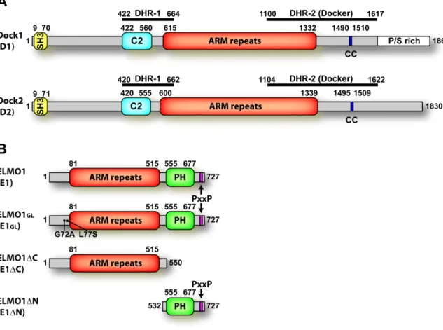
To determine if there are additional predicted domains within the sequences of Dock-family members and ELMO proteins, and to help define the domain boundaries of published, predicted domains, we used a combination of multiple sequence alignments, secondary structure prediction programs, tertiary structure prediction programs and threading algorithms. Others have reported that ELMO1 contains a PH domain, a leucine-zipper motif, a PxxP motif, and Armadillo repeats. We found, through our analysis, that the PH domain boundaries predicted by SMART are reasonable, if a bit extended, and thus, we labeled the PH domain as consisting of residues 555-677 (Fig. 3B). BLAST analysis of the PH domain of ELMO1 finds the PH domain of PLC-δ to have the highest homology with that region. We did not find convincing evidence, however, for the predicted leucine zipper motif, and believe it to be unlikely to exist within the predicted PH domain of ELMO1, which is where a potential leucine zipper was identified [40]. The PxxP motif at the C-terminus is conserved amongst the different ELMO proteins (1, 2 and 3) and across species and is located within residues 707-717 of mouse ELMO1 (Fig. 3B). Reportedly, ELMO1 contains Armadillo (ARM) repeats, which are regions of about 35-50 amino acids that form three alpha helices per repeat. Each repeat stacks with others, usually forming a superhelix of helices [113,114]. We
also found evidence for the ARM repeat region in the ELMO proteins; however, we would define the boundaries of the ARM repeat region differently than published. The N-terminal region of mouse ELMO1 was repeatedly aligned with SCOP family a.118.1 (ARM repeats [115]) when using a meta-server which compiles data from a variety of prediction and threading programs (http://www.bioinfo.pl/meta). Upon examination of the models and alignments built by the threading algorithm, 3D-PSSM, and several secondary structure prediction programs, we determined that the most likely boundaries for the ARM repeat region span residues 81-515 (Fig. 3B).
We performed a similar domain analysis for Dock1 and Dock2. Previously-predicted domains for Dock proteins were largely based on homology with other family members, since only a few domains had been predicted with a high degree of confidence. The SH3 domain of Dock1 and Dock2 is readily identified by programs such as SMART, and the boundaries are labeled accordingly. The DHR-1 and DHR-2 regions have not been identified as structural domains, but as regions of homology as their names suggest (Dock-homology-region-1 or 2). The limits of the DHR-1 region are labeled based on the literature (Dock1 422-664) [26,38], and the limits of the DHR-2 region are also based on the literature, but with refinements from our own multiple sequence alignments and secondary structure predictions (Dock2 1100-1617) [25,26,28]. The proline and serine-rich region at the C-terminus of Dock1 is shown as identified from the sequence; Dock2 does not have a noticeable proline/serine rich region (Fig. 3A) [33,35,53,116].
To delineate other domains within the Dock protein sequences, we used portions of the sequences outside of defined domains to aid the prediction programs by
eliminating easily-defined domains. Within these sequences, domains were often