DIVERSITY WITHIN THE MASTER REGULATORY p53 TRANSCRIPTIONAL NETWORK: IMPACT OF SEQUENCE, BINDING MOTIFS AND MUTATIONS
Jennifer J. Jordan
A dissertation submitted to the faculty of the University of North Carolina at Chapel Hill in partial fulfillment of the requirements for the degree of Doctor of Philosophy in the
Curriculum in Genetics and Molecular Biology.
Chapel Hill 2008
Approved by:
Michael A. Resnick, PhD William K. Kaufmann, PhD Norman E. Sharpless, MD Brian Strahl, PhD
Abstract Jennifer J. Jordan
Diversity within the master regulatory p53 transcriptional network: impact of sequence, binding motifs and mutations
(Under the direction of Michael A. Resnick, PhD)
ACKNOWLEDGEMENTS
As the p53 universe is forever expanding, so are the number of individuals who I am truly thankful to for their support throughout graduate school and in the development of my scientific career. First, I would like to thank the members of my dissertation committee William Kaufmann, Charles Perou, Norman Sharpless, and Brian Strahl. I appreciate their time and scientific guidance.
I would like to thank the current and past members of the Resnick lab for their support and making the lab an enjoyable and stimulating atmosphere in which to work. A special thanks to the p53 crew, Daniel Menendez and Alberto Inga. Both are enthusiastic scientists with whom it was a pleasure to work.
I would like to acknowledge the support and guidance of my mentor Michael Resnick who has my fullest respect as a scientist, but more importantly as a person. I will remember with fondness and gratitude the hours of quality blue chair sessions that were interrupted with chocolate breaks and Cajun popcorn. I am sincerely grateful for the opportunity I was provided within his lab.
A special thanks to my family, whose support and encouragement throughout this process was genuinely appreciated.
TABLE OF CONTENTS
LIST OF TABLES... ix
LIST OF FIGURES ... x
LIST OF ABBREVIATIONS ...xiii
Chapter I. INTRODUCTION ... 1
p53 from oncogene to tumor suppressor ... 2
Regulation and activation of p53 protein ... 4
p53 interaction with its consensus RE ... 7
p53 structure ... 9
References... 15
II. ISOGENOMIC IN VIVO DIPLOID YEAST SYSTEM TO ANALYZE TRANSACTIVATION CAPACITY OF THE MASTER REGULATOR P53 ... 23
Abstract... 24
Introduction ... 26
Results ... 36
Discussion... 41
References... 66
III. NONCANONICAL DNA MOTIFS AS TRANSACTIVATION TARGETS BY WILD TYPE AND MUTANT p53 ... 70
Introduction ... 72
Results ... 75
Discussion... 88
References... 119
IV. ALTERED FUNCTION p53 MISSENSE MUTATIONS ASSOCIATED WITH BREAST CANCER CAN HAVE SUBTLE EFFECTS ON TRANSACTIVATION ... 127
Abstract... 128
Introduction ... 129
Results ... 134
Discussion... 140
References... 167
V. Con-A: A SUPER-TRANSACTIVATING RESPONSE ELEMENT FOR p53 TRANSACTIVATION ... 173
Abstract... 174
Introduction ... 175
Results ... 176
Discussion... 184
References... 203
VI. DISCUSSION ... 204
Small changes in sequence can lead to dramatic difference in transactivation... 206
Noncanonical REs expand the p53 transactivational network ... 207
Functional mutations diversify the p53 transcriptional network ... 209
References... 212 VII. MATERIALS AND METHODS... 214
LIST OF TABLES
Table
2.1 ICORE insertion sites in p53 cDNA and window of modification... 55
2.2 Response element sequence, strength and rank of transactivation
capacity... 56
2.3 Toxic and super-transactivating mutations display altered function in the haploid and diploid rheostatable systems ... 62
3.1 Transactivation capacity of WT and mutant p53 towards canonical and
noncanonical REs ... 112 4.1 p53 missense mutations associated with breast cancers: functional
status, frequency, and features... 149
LIST OF SUPPLEMENTAL TABLES
Supplemental Table
S4.1 Response element sequence and biological function of associated
target gene... 160
S4.2 Functional status determined by in vivo transactivation assays vs.
structure based predictions ... 161
S4.3 Oligonucleotides with p53 mutation of interest for CORE
LIST OF FIGURES
Figure
1.1 The tumor suppressor p53 is a master regulatory gene... 12
1.2 Phenotypic diversity within the p53 transcriptional network ... 13 1.3 p53 binds cooperatively to response element sequences as a
dimer of dimers ... 14 2.1 Yeast as an in vivo test tube to determine mammalian p53
transactivation capacity... 48 2.2 Rheostatable promoter to address p53 transactivation potentials... 49
2.3 Qualitative plate color assay; Quantitative luciferase assay... 50 2.4 Isogenomic diploid yeast system to investigate transcriptional
capacity of p53 towards many REs at various p53 levels ... 52 2.5 Delitto perfetto site-directed in vivo mutagenesis system ... 53
2.6 Inducible expression of p53 under the rheostatable GAL1 promoter ... 57 2.7 Differential transactivation capacities of WT and mutant p53 towards
REs at variable expression observed with the phenotypic color assay ... 59 2.8 Quantitative assessment of p53-induced transactivation from the
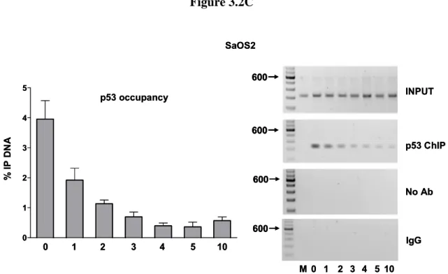
p21-5’ RE in vivo... 60 2.9 p53 protein stability following glucose pulse rate ... 61 2.10 Sequence specific differences in WT p53 transactivation capacity from REs does not correlate to the biological function of the target gene... 63 2.11 In vivo transactivation vs. in vitro binding ... 65 3.1 Weak REs can function synergistically when separated by a spacer... 100 3.2 Spacer decreases p53 transactivation, promoter occupancy and
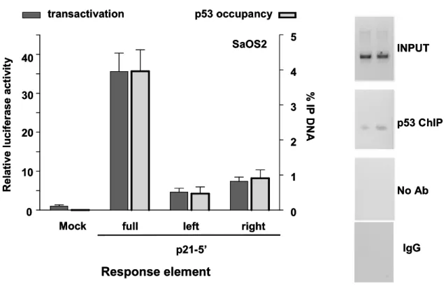
3.4 A ¾-site can function as a noncanonical RE in p53 transactivation ... 109
4.1 Functional fingerprints of mutant p53 reveal subtle transactivational Differences... 151
4.2 Assessment of WT and mutant p53 transactivation towards the p21-5’ RE using a luciferase assay in growing cells ... 152
4.3 Transactivation from the GADD45 RE distinguishes altered function mutants from WT p53 ... 153
4.4 Altered function mutations at the same codon in the tetramerization domain display different transactivation capacities ... 154
4.5 Change-in-spectrum p53 missense mutations can eliminate REs from the p53 transcriptional network ... 155
5.1 Three phases to transactivation by p53 ... 188
5.2 WT p53 transactivation is similar from the Con-A and p21-5’ REs at high levels of p53 expression... 190
5.3A WT p53 transactivation from Con-A vs p21-5’ REs at low levels of expression ... 191
5.3B Low level transactivation is p53-dependent ... 192
5.4A p53 tetramerization is required for the super-transactivation at low levels of p53 ... 193
5.4B Super-transactivation at basal levels of p53 requires a full-site RE ... 194
5.5A Super-transactivation at low levels of p53 requires a CATG core ... 195
5.5B Flanking sequences affect basal transactivation by WT p53 ... 196
5.6 Crystal I: a second super-trans RE from which p53 can transactivate at low levels ... 197
5.7 Small increases in spacer affects Con-A super-trans response at low levels of p53... 198
5.9B Compromised tetramerization diminishes basal response ... 201 5.9C Transactivation by the toxic mutant V122A ... 202
LIST OF SUPPLEMENTAL FIGURES
Supplemental Figure
S3.1 Increase in spacer decreases transactivation by p53 family members... 114
S3.2 Transactivation from noncanonical ¾-site REs in haploid yeast... 116 S3.3 Mutant p53 expression ... 118
S4.1 Functional fingerprint of 21 altered-function p53 missense
mutations. ... 156
S4.2 WT and mutant p53 expression... 158 S4.3 Transactivation from the p21-5’ RE by p53 missense mutations with
LIST OF ABBREVIATIONS
APAF-1 apoptosis protease activating factor 1 BAX BCL2-associated X protein
BRCA breast cancer susceptibility gene
ChIP chromatin immunoprecipitation
Con consensus
CORE COunterselectable REporter cassette CYC1 iso-1-cytochrome C
DBD DNA binding domain
ER estrogen receptor
FASAY functional analysis of separated alleles in yeast
FH familial history
GADD45 Growth Arrest and DNA Damage-inducible
GAPDH glyceraldehyde-3-phosphate dehydrogenase
HER2+ human epidermal growth factor-2 positive IARC International Agency for Research on Cancer LFL Li-Fraumeni-like syndrome
LFS Li-Fraumeni syndrome MDM2 mouse double minute
MMP2 Matrix metalloproteinase-2
nt nucleotides
P53AIP1 p53-regulated Apoptosis-Inducing Protein 1
P53R2 p53-inducible ribonucleotide reductase small subunit 2
PCNA proliferating cell nuclear antigen
PIDD p53 induced protein with death domain PUMA p53-upregulated modulator of apoptosis
PWM position-weight matrices
RE response element
RGC ribosomal gene cluster RNAP II RNA polymerase II
SAGA Spt/Ada/Gcn5/acetyltransferase
SEM standard error of measurement SNP single nucleotide polymorphism
TBP TATA-binding protein TP53 tumor suppressor p53
TIGAR TP53 induced glycolysis and apoptosis regulator
UAS upstream activating sequence VEGF vascular endothelial growth factor
CHAPTER 1
p53 from oncogene to tumor suppressor
While the discovery of TP53 in 1979 provoked an interest in a protein involved in the
role of cancer development, the natural function of p53 in tumorigenesis was thought to be
that of an oncogene since it was first identified complexed with the SV40 large T protein in
transformed cells [1-3]. The understanding that p53 was a tumor suppressor began to arise
years later when wild type (WT) p53 was found to suppress transformation of cells, whereas
mutant p53 was associated with cellular transformation [4, 5]. Furthermore, genetic
alterations in p53, which mostly occurred as point mutations were found frequently in human
cancers [6-9]. In agreement with Knudson’s two-hit hypothesis for tumor suppressors [10],
the p53 knockout mouse developed normally, but had an increased incidence of tumors
where WT, heterozygous and homozygous mice developed neoplasias at ~18, 9 and 6
months, respectively [11].
Several major findings contributed to the understanding that p53 executed its role as a
tumor suppressor through functioning as a transcription factor. The p53 protein was found to
be induced in response to DNA damage or cellular stress to stimulate either cell cycle arrest
or apoptosis [12-15]. Stimulation of these biological responses was dependent upon the
ability of p53 to bind DNA, where loss in DNA binding resulted in the loss of its ability to
suppress growth [16-22]. In addition, an intact transactivation domain and transactivation
activity were necessary for p53 to function as a tumor suppressor [20, 21, 23, 24].
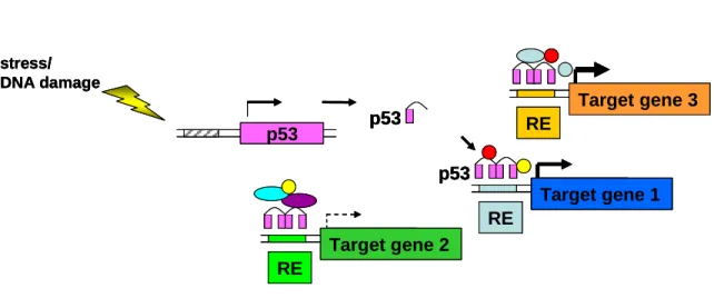
Currently, it is accepted that p53 plays a critical role as a master regulatory gene by
directly controlling the transcriptional regulation of greater than 100 target genes in a
sequence-specific manner in response to intra- and extra-cellular stresses (Figure 1.1) [25,
interconnected within the p53 transcriptional network. Aberrations within the p53
transcription network are a hallmark of cancers, where mutations in p53 itself are found in
greater than 50% of all cancers [27]. To date over 24,800 somatic p53 mutations and ~400
germline p53 mutations have been reported within the International Agency for Research on
Cancer’s (IARC) TP53 mutations database [27]. In terms of transactivation, p53 binds to a
consensus binding sequence [RRRCWWGYYY (n) RRRCWWGYYY] (where R=purine,
W= A or T, Y= pyrimidine and n is 0-13 bases) [28-30] to recruit the basal transcriptional
machinery and additional cofactors in order to regulate the expression of the associated target
gene [31]. However, p53 does not recognize and regulate each of its downstream targets in
an equivalent fashion (Figure 1.2)[32]. Differential induction is dependent upon the target
gene of interest, stress stimuli and cell type and is influenced by many factors including the
chromatin environment, post-translational modifications, availability of transcriptional
cofactors, and concentration or duration of activated p53 protein [26, 32, 33]. At the DNA
level, regulation of target genes is dependent on the sequence of response elements (RE),
number of REs in a promoter region, distance of the RE from the transcriptional start site and
intrinsic binding with a RE [26].
The Resnick lab recently established, using an isogenic, in vivo yeast-based model
system, that these latter influences can lead to greater than a 1000-fold variation in
p53-dependent transactivation from individual REs derived from human target genes [34].
Variation in gene regulation by p53 has led to the postulation of the master gene of diversity
concept [26] which can be applied to many sequence-specific transcription factors. Using a
piano analogy, the master regulatory gene, p53 is the hand that plays a chord on the keys of a
regulated and is dependent on both the genes regulated and strength of regulation.
Functional mutations in the hand can alter the “selection of keys” by altering binding,
spectrum of REs recognized, and/or the intensity of regulation [26].
Regulation and activation of p53 protein
The p53 protein is a 393 amino acid modular protein comprised of two N-terminal
transactivation domains (residues 1-73), a proline rich domain (PXXP, residues 63-97), a
core DNA binding domain (DBD) (residues 94 - 292), a tetramerization domain (residues
324-355) and a C-terminal regulatory domain (residues 360 – 393) [35, 36]. Within the cell,
p53 is constitutively expressed, but the protein is kept at low concentrations (estimated at
~1000 molecules/cell [37]) through several negative regulators, the most classic of which is
the auto-inhibitory feedback loop formed with one of its own target genes, MDM2 (mouse
double minute or the human homolog, HDM2). The MDM2 protein inhibits p53-dependent
transactivation by regulating p53 protein through two mechanisms. First, it can bind to the
amino terminal transactivation domain of p53 (residues 18-23) to inhibit interactions between
p53 and the basal transcriptional machinery or transcriptional cofactors [38-40]. Second, it
can function as a ring finger E3-ubiquitin ligase to ubiquitinate p53 on its C-terminal lysines
which targets p53 for proteasome degradation [41-44].
Recently, MDMX (HDMX/MDM4 in humans), a ring finger protein related to
MDM2 but lacking the E3 ligase activity, has been identified as an additional negative
regulator of p53. Similar to MDM2, MDMX represses p53-dependent transcription through
binding the N-terminus transactivation domain of p53. Under “non-stressed” conditions,
auto-ubiquitination thus increasing MDM2 dependent degradation of p53 [45-48]. Several
additional E3 ligases (i.e., COP1 and Pirh2) and de-ubiquitinating proteins (i.e., HAUSP)
have also been implicated as having a role in regulating p53 protein levels [49, 50].
Following DNA damage or cellular stress, a cascade of signaling pathways induce
post-translational modifications in p53 which have been postulated to contribute to the
stabilization of the protein. For example, phosphorylation of p53 at Ser15 by DNA-PK was
proposed to directly interfere with interactions between p53 and MDM2 [51, 52].
Phosphorylation of Ser15, Ser20 and Thr18 has also been proposed to decrease p53-MDM2
interactions indirectly by enhancing p53 interactions with co-activators such as the histone
acetyl transferases, p300 and CBP [53]. Additional post-translational modifications in the
C-terminal of the protein, such as Ser392 phosphorylation were found to contribute to shifting
the tetramer-monomer equilibrium towards the tetramer formation which is necessary for p53
transcriptional activity [37, 54].
However, several studies have shown that mutations which render a residue unable to
be modified, such as Ser20Ala do not necessarily prevent accumulation of the p53 protein
[55]. Current research has implied the rapid degradation of MDM2 following damage or
stress may play as imperative a role in p53 activation as modifications in p53 itself.
Following damage, phosphorylation of MDM2 at Ser395 by ATM was shown to abolish the
direct interactions with p53 [56-58]. Furthermore, phosphorylation of MDM2 has been
postulated to impede the MDMX-MDM2 interactions which results in the E3 ligase
specificity of MDM2 being drawn away from p53 and toward itself and MDMX [46].
These findings have sparked ongoing debates as to whether post-translational
promoter selection to specify or fine tune p53-dependent regulation and consequently a
particular biological response [58]. Currently, there are approximately 40 upstream effectors
identified which function to post-translationally modify p53 at over 30 residues (primarily in
the N-terminal transactivation domains and C-terminal) [59]. The concept that specific
post-translational modifications in p53 impact its ability to differentially regulate target genes has
been coined the “barcode hypothesis” [33]. For instance, one specific barcode which occurs
after p53 stabilization at high doses of DNA damage is the phosphorylation of Ser46. This
post-translational modification has been shown to specifically enhance p53 binding to and
activation of the apoptotic associated target, p53AIP1; mutagenesis of the residue mitigated
transactivation from p53AIP1, but not other REs including p21, MDM2, p53R2 and Noxa
[33, 60].
In addition to the post-translational modifications, co-factors, small molecules, and/or
other interacting binding proteins have been shown to influence the promoter selectivity of
p53 to elicit specific biological responses. ASPP1 and ASPP2 (apoptosis stimulating protein
of p53) as well as p53 family members p63 and p73 have been shown to enhance p53
binding toward some apoptotic REs, such as Bax [61, 62]. Whereas, HZF (hematopoietic
zinc-finger) binds the p53 DBD to augment p53 binding toward REs associated with cell
cycle arrest, such as p21 and 14-3-3σ [63]. Other proteins have been identified which
channel or recruit p53 to selected downstream targets. For example, BRCA1 has been shown
p53 interaction with its consensus RE
The consensus sequence which p53 binds consists of two decamer half-sites
[RRRCWWGYYY (0-13)n RRRCWWGYYY], where R = purine, Y = pyrimidine, W=A/T,
and which can be spaced by up to 13 nucleotides apart [28-30]. Unlike for other
transcription factor binding sites, such as NFκB, high evolutionary conservation between
species is not observed for the p53 response element sequence [65]. The p53 consensus is
highly degenerate where variation in sequence is tolerated at most positions with the
exception of the conserved C and G at positions 4 and 7, respectively [26]. Interestingly,
single nucleotide polymorphisms, or SNPs within the promoter RE sequences have been
found to include or exclude sequences from the p53 transcriptional network [66].
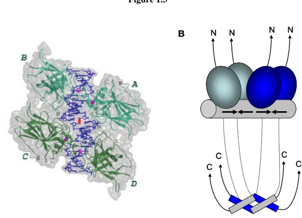
Each decamer is comprised of two inverted quarter sites arranged in a head-to-head
fashion. p53 cooperatively binds to the consensus site as a dimer of dimers, or tetramer
protein where the two core DBDs of a dimer bind to consecutive quarter-sites (Figure
1.3)[67-73]. Upon binding to the RE, p53 induces a conformational change in the DNA by
bending the RE. The degree of the angle is dependent upon the particular sequence of the
RE, where REs with a stronger affinity toward p53 are associated with larger bending angles
[71, 74]. Interestingly, various proteins such as HMG have been found to augment p53
binding to REs by inducing bends in the DNA [75].
While tetramerization is not a requirement for sequence-specific binding by the DBD
[76], it is required for efficient transactivation from the consensus sequence [77-80].
Arrangement of the quarter sites in a head-to-head orientation is essential for binding which
results in transactivation. p53 can bind to a RE sequence comprised of quarter sites arranged
in competitive binding assays, WT p53 was not able to transactivate from these sites [81].
These findings suggest in addition to sequence, the orientation of binding elements can also
influence p53 dependent transactivation.
Previous in vitro studies proposed modification of the C-terminal domain (i.e.,
phosphorylation of Ser392 [35]) was required to alter a “latent” p53 to an “active” p53 in
order to disrupt its negative effect on transactivation and stimulate p53 sequence-specific
binding [16, 22, 82]. However, recent structural studies revealed the conformation of p53
was similar in both the latent and active states [73, 83]. While the C-terminal of p53 was
previously believed to inhibit p53 binding, its non-sequence specific, structure-based DNA
interactions, which are highly dependent on the DNA topology, are now postulated to
facilitate p53 sliding on DNA as proposed by a linear diffusion based search for REs [76,
84-87].
Additionally, in vitro chromatin assembly and in vivo ChIP studies show p53 can bind
to promoters prior to its induction or modification [59, 73, 88, 89]. Such active binding has
favored the concept of a “selective context model” over a “selective binding model” [33]. In
the selective context model, expression of a downstream target is reliant not on p53 binding
per se, but on gene-specific requirements for transactivation. Thus, individual targets must
overcome specific hurdles in the transcription process or “filters” which may require more or
less transcriptional coregulators for activity [33]. Such filters may be reliant on whether or
not the promoter region is accessible to the general transcriptional machinery. In the case
where the promoter is not accessible, p53 binding can increase the promoter accessibility
through recruitment of chromatin remodeling factors, histone acetyltransferases and/or
pre-initiation complexes by interacting with mediator complexes or recruiting basal transcription
factors. In addition, p53 had been demonstrated to play a role in reinitiating paused
polymerases [35]. Thus, downstream target genes such as p21 which have a pre-initiation
complex, as well as a paused RNAP II at the promoter may require the recruitment of a
smaller amount of transcription cofactors to overcome any “filter” than a RE which is lacking
RNAP II. Importantly, the availability of the cofactors required to overcome such filters may
be cell-type or stimuli-specific.
p53 structure
Although structures for the individual domains of the protein have been solved
independently the exact structure of the p53 tetramer and how two dimer proteins interact in
terms of protein-protein and protein-DNA interactions remains controversial. The highly
disordered segments of the N- and C- termini which flank the structured core DNA binding
and tetramerization domains, as well as the intrinsic instability of the protein has impeded the
ability to form crystals of a p53 tetramer binding to a full-length p53 consensus sequence
[83].
The basic structure of the core DNA binding domain (DBD) was derived from a
crystal structure solved by Cho et al., which had three p53 core domain monomers bound to a
consensus half-site [90]. The p53 DBD was shown to consist of a β-sandwich which provides a scaffold for two large β-loops (L2 and L3) and a loop-sheet-helix motif which compose the DNA binding surface [90, 91]. The loop-sheet-helix motif binds to the major
groove of targeted DNA through several residues, whereas the L3 loop interacts with the
both the L2 and L3 loops are coordinated by a zinc ion (contacted by residues Cys176,
His179, Cys238 and Cys242) which is required for the stabilization of the protein, as well as,
the sequence-specific DNA binding [92].
Recently, Kitayner et al., solved the crystal structure of the WT p53 DBD bound to
three dodecamer sequences (consensus decamer half-sites separated by a 2 nt spacer)(Figure
1.3) [73]. While the overall structure was similar to that solved by Cho et al., intriguingly,
the DNA-protein interface was found to vary with the specific sequence encountered. For
example, residue K120 contacts the second purine of the response element sequence;
however the nature of the contact is dependent upon the specific base present [73, 93].
The DNA binding domain is linked to the tetramerization domain through ~35
flexible amino acids [68]. The tetramerization domain of p53 is composed of a β-strand
(amino acids 326-333) and an α-helix (amino acids 335-354) that form a “V-shape” structure
through a hinge region, or tight turn at residue 334. In the simplest terms, two monomers
form dimeric proteins through interactions between the α-helices and β-strands, where the
two β-strands from opposite monomers form an anti-parallel β-sheet. The dimers then
associate through hydrophobic interactions between the anti-parallel α-helices to form
tetramers [67, 68, 70, 94-97].
Regardless of the exact nature of the protein binding, tetrameric p53 has displayed a
great capacity to bind many RE variants to differentially regulate the activity of downstream
targets. However, all binding is not equivalent nor will all binding result in transactivation.
The overall goal of this study is to further contribute to the understanding of how p53
functions as a sequence-specific transcription factor by further defining the “rules of p53
to address what constitutes a functional response element and how organization of the
binding motif influences transactivation. Understanding the interaction of p53 with various
RE sequences under conditions which render all other factors (such as chromatin and
transcriptional cofactors) essentially neutral may elucidate how different p53-DNA
interactions alter the potential of p53-dependent transactivation and possibly dictate the
remaining factors required for efficient transactivation. Furthermore, analysis of the impact
of p53 mutations upon transactivation will help to elucidate the role of p53 as a master
Figure 1.1
TARGET GENE ONCOGENIC
STIMULI GENOTOXIC
FACTORS NON-GENOTOXICFACTORS
STABILIZED, ACTIVATED, LOCALIZED TO THE NUCLEUS POST-TRANSLATIONAL MODIFICATIONS Mdm2 mdmx Transcription factors Cell Responses Response Element
R=purine; Y=pyrimidine; W=A or T; N=spacer of 0-13 bp R R R C W WG Y Y YNR R R C W W G Y Y Y
p53 stability Apoptosis DNA repair Cell cycle arrest TARGET GENE ONCOGENIC STIMULI GENOTOXIC
FACTORS NON-GENOTOXICFACTORS
STABILIZED, ACTIVATED, LOCALIZED TO THE NUCLEUS POST-TRANSLATIONAL MODIFICATIONS Mdm2 mdmx Transcription factors Cell Responses Response Element
R=purine; Y=pyrimidine; W=A or T; N=spacer of 0-13 bp R R R C W WG Y Y YNR R R C W W G Y Y Y R=purine; Y=pyrimidine; W=A or T; N=spacer of 0-13 bp
R R R C W WG Y Y YNR R R C W W G Y Y Y
p53 stability Apoptosis DNA repair Cell cycle arrest
Figure 1.2
p53 p53
stress/ DNA damage
p53
ADE2
RE
Target gene 1
ADE2
RE
Target gene 2
ADE2
RE
Target gene 3
p53 p53
stress/ DNA damage
p53
ADE2
RE
Target gene 1
ADE2
RE
Target gene 2
ADE2
RE
Target gene 3
p53 p53
stress/ DNA damage
p53
ADE2
RE
Target gene 1ADE2 RE
Target gene 1
ADE2
RE
Target gene 2
ADE2
RE
Target gene 3
Figure 1.3
A B
C C
C C
N N N N
A B
C C
C C
N N N N
C C
C C
N N N N
References
1. Robins, H., et al., The first twenty-five years of p53 research., in 25 years of p53 research, P. Hainaut and K.G. Wiman, Editors. 2005, Springer: Dordrecht, The Netherlands. p. 1-25.
2. Linzer, D.I., W. Maltzman, and A.J. Levine, The SV40 A gene product is required for the production of a 54,000 MW cellular tumor antigen. Virology, 1979. 98(2): p. 308-18.
3. Lane, D.P. and L.V. Crawford, T antigen is bound to a host protein in SV40-transformed cells. Nature, 1979. 278(5701): p. 261-3.
4. Zambetti, G.P. and A.J. Levine, A comparison of the biological activities of wild-type and mutant p53. Faseb J, 1993. 7(10): p. 855-65.
5. Hinds, P., C. Finlay, and A.J. Levine, Mutation is required to activate the p53 gene for cooperation with the ras oncogene and transformation. J Virol, 1989. 63(2): p. 739-46.
6. Rotter, V., et al., Chromosomal assignment of the murine gene encoding the transformation-related protein p53. Mol Cell Biol, 1984. 4(2): p. 383-5.
7. Nigro, J.M., et al., Mutations in the p53 gene occur in diverse human tumour types. Nature, 1989. 342(6250): p. 705-8.
8. Benchimol, S., G. Matlashewski, and L. Crawford, The use of monoclonal antibodies for selection of a low-abundance mRNA: p53. Biochem Soc Trans, 1984. 12(4): p. 708-11.
9. Baker, S.J., et al., Chromosome 17 deletions and p53 gene mutations in colorectal carcinomas. Science, 1989. 244(4901): p. 217-21.
10. Knudson, A.G., Jr., Mutation and cancer: statistical study of retinoblastoma. Proc Natl Acad Sci U S A, 1971. 68(4): p. 820-3.
11. Donehower, L.A., et al., Mice deficient for p53 are developmentally normal but susceptible to spontaneous tumours. Nature, 1992. 356(6366): p. 215-21.
12. Maltzman, W. and L. Czyzyk, UV irradiation stimulates levels of p53 cellular tumor antigen in nontransformed mouse cells. Mol Cell Biol, 1984. 4(9): p. 1689-94.
14. Kastan, M.B., et al., Participation of p53 protein in the cellular response to DNA damage. Cancer Res, 1991. 51(23 Pt 1): p. 6304-11.
15. Hall, P.A., et al., High levels of p53 protein in UV-irradiated normal human skin. Oncogene, 1993. 8(1): p. 203-7.
16. Hupp, T.R., et al., Regulation of the specific DNA binding function of p53. Cell, 1992. 71(5): p. 875-86.
17. Farmer, G., et al., Wild-type p53 activates transcription in vitro. Nature, 1992. 358(6381): p. 83-6.
18. Kern, S.E., et al., Identification of p53 as a sequence-specific DNA-binding protein. Science, 1991. 252(5013): p. 1708-11.
19. Pietenpol, J.A., et al., Sequence-specific transcriptional activation is essential for growth suppression by p53. Proc Natl Acad Sci U S A, 1994. 91(6): p. 1998-2002.
20. Milner, J., E.A. Medcalf, and A.C. Cook, Tumor suppressor p53: analysis of wild-type and mutant p53 complexes. Mol Cell Biol, 1991. 11(1): p. 12-9.
21. Sturzbecher, H.W., et al., A C-terminal alpha-helix plus basic region motif is the major structural determinant of p53 tetramerization. Oncogene, 1992. 7(8): p. 1513-23.
22. Kim, E. and W. Deppert, The complex interactions of p53 with target DNA: we learn as we go. Biochem Cell Biol, 2003. 81(3): p. 141-50.
23. Chao, C., et al., p53 transcriptional activity is essential for p53-dependent apoptosis following DNA damage. Embo J, 2000. 19(18): p. 4967-75.
24. Jimenez, G.S., et al., A transactivation-deficient mouse model provides insights into Trp53 regulation and function. Nat Genet, 2000. 26(1): p. 37-43.
25. Ko, L.J. and C. Prives, p53: puzzle and paradigm. Genes Dev, 1996. 10(9): p. 1054-72.
26. Resnick, M.A. and A. Inga, Functional mutants of the sequence-specific transcription factor p53 and implications for master genes of diversity. Proc Natl Acad Sci U S A, 2003. 100(17): p. 9934-9.
28. el-Deiry, W.S., et al., Definition of a consensus binding site for p53. Nat Genet, 1992. 1(1): p. 45-9.
29. Funk, W.D., et al., A transcriptionally active DNA-binding site for human p53 protein complexes. Mol Cell Biol, 1992. 12(6): p. 2866-71.
30. Tokino, T., et al., p53 tagged sites from human genomic DNA. Hum Mol Genet, 1994. 3(9): p. 1537-42.
31. Liu, G. and X. Chen, Regulation of the p53 transcriptional activity. J Cell Biochem, 2006. 97(3): p. 448-58.
32. Menendez, D., et al., Changing the p53 master regulatory network: ELEMENTary, my dear Mr Watson. Oncogene, 2007. 26(15): p. 2191-201.
33. Espinosa, J.M., Mechanisms of regulatory diversity within the p53 transcriptional network. Oncogene, 2008.
34. Inga, A., et al., Differential transactivation by the p53 transcription factor is highly dependent on p53 level and promoter target sequence. Mol Cell Biol, 2002. 22(24): p. 8612-25.
35. Laptenko, O. and C. Prives, Transcriptional regulation by p53: one protein, many possibilities. Cell Death Differ, 2006. 13(6): p. 951-61.
36. McKinney, K. and C. Prives, Regulation of p53 DNA binding, in 25 years of p53 research, P. Hainaut and K.G. Wiman, Editors. 2005, Springer: Dordrecht, The Netherlands p. 27-49.
37. Sakaguchi, K., et al., Effect of phosphorylation on tetramerization of the tumor suppressor protein p53. J Protein Chem, 1997. 16(5): p. 553-6.
38. Kussie, P.H., et al., Structure of the MDM2 oncoprotein bound to the p53 tumor suppressor transactivation domain. Science, 1996. 274(5289): p. 948-53.
39. Haupt, Y., Y. Barak, and M. Oren, Cell type-specific inhibition of p53-mediated apoptosis by mdm2. Embo J, 1996. 15(7): p. 1596-606.
40. Wu, X., et al., The p53-mdm-2 autoregulatory feedback loop. Genes Dev, 1993. 7(7A): p. 1126-32.
41. Haupt, Y., et al., Mdm2 promotes the rapid degradation of p53. Nature, 1997. 387(6630): p. 296-9.
43. Grossman, S.R., et al., Polyubiquitination of p53 by a ubiquitin ligase activity of p300. Science, 2003. 300(5617): p. 342-4.
44. Nakamura, S., J.A. Roth, and T. Mukhopadhyay, Multiple lysine mutations in the C-terminal domain of p53 interfere with MDM2-dependent protein degradation and ubiquitination. Mol Cell Biol, 2000. 20(24): p. 9391-8.
45. Stad, R., et al., Hdmx stabilizes Mdm2 and p53. J Biol Chem, 2000. 275(36): p. 28039-44.
46. Wang, Y.V., et al., Quantitative analyses reveal the importance of regulated Hdmx degradation for p53 activation. Proc Natl Acad Sci U S A, 2007. 104(30): p. 12365-70.
47. Uldrijan, S., W.J. Pannekoek, and K.H. Vousden, An essential function of the extreme C-terminus of MDM2 can be provided by MDMX. Embo J, 2007. 26(1): p. 102-12.
48. Stommel, J.M. and G.M. Wahl, A new twist in the feedback loop: stress-activated MDM2 destabilization is required for p53 activation. Cell Cycle, 2005. 4(3): p. 411-7.
49. Brooks, C.L. and W. Gu, p53 ubiquitination: Mdm2 and beyond. Mol Cell, 2006. 21(3): p. 307-15.
50. Li, M., et al., A dynamic role of HAUSP in the p53-Mdm2 pathway. Mol Cell, 2004. 13(6): p. 879-86.
51. Shieh, S.Y., et al., DNA damage-induced phosphorylation of p53 alleviates inhibition by MDM2. Cell, 1997. 91(3): p. 325-34.
52. Appella, E. and C.W. Anderson, Post-translational modifications and activation of p53 by genotoxic stresses. Eur J Biochem, 2001. 268(10): p. 2764-72.
53. Dumaz, N. and D.W. Meek, Serine15 phosphorylation stimulates p53 transactivation but does not directly influence interaction with HDM2. Embo J, 1999. 18(24): p. 7002-10.
54. Sakaguchi, K., et al., Phosphorylation of serine 392 stabilizes the tetramer formation of tumor suppressor protein p53. Biochemistry, 1997. 36(33): p. 10117-24.
55. Wu, Z., et al., Mutation of mouse p53 Ser23 and the response to DNA damage. Mol Cell Biol, 2002. 22(8): p. 2441-9.
57. Tao, W. and A.J. Levine, P19(ARF) stabilizes p53 by blocking nucleo-cytoplasmic shuttling of Mdm2. Proc Natl Acad Sci U S A, 1999. 96(12): p. 6937-41.
58. McLure, K.G. and M.B. Kastan, 20 years of DNA damage signaling to p53, in 25 years of p53 research, P. Hainaut and K.G. Wiman, Editors. 2005, Springer: Dordrecht, The Netherlands. p. 53-71.
59. Szak, S.T., D. Mays, and J.A. Pietenpol, Kinetics of p53 binding to promoter sites in vivo. Mol Cell Biol, 2001. 21(10): p. 3375-86.
60. Oda, K., et al., p53AIP1, a potential mediator of p53-dependent apoptosis, and its regulation by Ser-46-phosphorylated p53. Cell, 2000. 102(6): p. 849-62.
61. Samuels-Lev, Y., et al., ASPP proteins specifically stimulate the apoptotic function of p53. Mol Cell, 2001. 8(4): p. 781-94.
62. Flores, E.R., et al., p63 and p73 are required for p53-dependent apoptosis in response to DNA damage. Nature, 2002. 416(6880): p. 560-4.
63. Das, S., et al., Hzf Determines cell survival upon genotoxic stress by modulating p53 transactivation. Cell, 2007. 130(4): p. 624-37.
64. MacLachlan, T.K., R. Takimoto, and W.S. El-Deiry, BRCA1 directs a selective p53-dependent transcriptional response towards growth arrest and DNA repair targets. Mol Cell Biol, 2002. 22(12): p. 4280-92.
65. Horvath, M.M., et al., Divergent evolution of human p53 binding sites: cell cycle versus apoptosis. PLoS Genet, 2007. 3(7): p. e127.
66. Tomso, D.J., et al., Functionally distinct polymorphic sequences in the human genome that are targets for p53 transactivation. Proc Natl Acad Sci U S A, 2005. 102(18): p. 6431-6.
67. Clore, G.M., et al., Refined solution structure of the oligomerization domain of the tumour suppressor p53. Nat Struct Biol, 1995. 2(4): p. 321-33.
68. Lee, W., et al., Solution structure of the tetrameric minimum transforming domain of p53. Nat Struct Biol, 1994. 1(12): p. 877-90.
69. McLure, K.G. and P.W. Lee, How p53 binds DNA as a tetramer. Embo J, 1998. 17(12): p. 3342-50.
71. Balagurumoorthy, P., et al., Four p53 DNA-binding domain peptides bind natural p53-response elements and bend the DNA. Proc Natl Acad Sci U S A, 1995. 92(19): p. 8591-5.
72. Wang, Y., et al., Interaction of p53 with its consensus DNA-binding site. Mol Cell Biol, 1995. 15(4): p. 2157-65.
73. Kitayner, M., et al., Structural basis of DNA recognition by p53 tetramers. Mol Cell, 2006. 22(6): p. 741-53.
74. Nagaich, A.K., E. Appella, and R.E. Harrington, DNA bending is essential for the site-specific recognition of DNA response elements by the DNA binding domain of the tumor suppressor protein p53. J Biol Chem, 1997. 272(23): p. 14842-9.
75. McKinney, K. and C. Prives, Efficient specific DNA binding by p53 requires both its central and C-terminal domains as revealed by studies with high-mobility group 1 protein. Mol Cell Biol, 2002. 22(19): p. 6797-808.
76. Wang, Y., et al., p53 domains: identification and characterization of two autonomous DNA-binding regions. Genes Dev, 1993. 7(12B): p. 2575-86.
77. Hainaut, P., A. Hall, and J. Milner, Analysis of p53 quaternary structure in relation to sequence-specific DNA binding. Oncogene, 1994. 9(1): p. 299-303.
78. Friedman, P.N., et al., The p53 protein is an unusually shaped tetramer that binds directly to DNA. Proc Natl Acad Sci U S A, 1993. 90(8): p. 3319-23.
79. Wang, P., et al., p53 domains: structure, oligomerization, and transformation. Mol Cell Biol, 1994. 14(8): p. 5182-91.
80. Stenger, J.E., et al., Formation of stable p53 homotetramers and multiples of tetramers. Mol Carcinog, 1992. 5(2): p. 102-6.
81. Thukral, S.K., et al., Discrimination of DNA binding sites by mutant p53 proteins. Mol Cell Biol, 1995. 15(9): p. 5196-202.
82. Anderson, M.E., et al., Reciprocal interference between the sequence-specific core and nonspecific C-terminal DNA binding domains of p53: implications for
regulation. Mol Cell Biol, 1997. 17(11): p. 6255-64.
83. Tidow, H., et al., Quaternary structures of tumor suppressor p53 and a specific p53 DNA complex. Proc Natl Acad Sci U S A, 2007. 104(30): p. 12324-9.
85. Kim, E. and W. Deppert, The versatile interactions of p53 with DNA: when flexibility serves specificity. Cell Death Differ, 2006. 13(6): p. 885-9.
86. Cain, C., et al., The N terminus of p53 regulates its dissociation from DNA. J Biol Chem, 2000. 275(51): p. 39944-53.
87. Kim, E., et al., Influence of promoter DNA topology on sequence-specific DNA binding and transactivation by tumor suppressor p53. Oncogene, 1999. 18(51): p. 7310-8.
88. Espinosa, J.M. and B.M. Emerson, Transcriptional regulation by p53 through intrinsic DNA/chromatin binding and site-directed cofactor recruitment. Mol Cell, 2001. 8(1): p. 57-69.
89. Kaeser, M.D. and R.D. Iggo, Chromatin immunoprecipitation analysis fails to support the latency model for regulation of p53 DNA binding activity in vivo. Proc Natl Acad Sci U S A, 2002. 99(1): p. 95-100.
90. Cho, Y., et al., Crystal structure of a p53 tumor suppressor-DNA complex: understanding tumorigenic mutations. Science, 1994. 265(5170): p. 346-55.
91. Joerger, A.C., et al., Structures of p53 cancer mutants and mechanism of rescue by second-site suppressor mutations. J Biol Chem, 2005. 280(16): p. 16030-7.
92. Pavletich, N.P., K.A. Chambers, and C.O. Pabo, The DNA-binding domain of p53 contains the four conserved regions and the major mutation hot spots. Genes Dev, 1993. 7(12B): p. 2556-64.
93. Veprintsev, D.B. and A.R. Fersht, Algorithm for prediction of tumour suppressor p53 affinity for binding sites in DNA. Nucleic Acids Res, 2008. 36(5): p. 1589-98.
94. Miller, M., et al., The oligomerization domain of p53: crystal structure of the trigonal form. FEBS Lett, 1996. 399(1-2): p. 166-70.
95. Chene, P., P. Mittl, and M. Grutter, In vitro structure-function analysis of the beta-strand 326-333 of human p53. J Mol Biol, 1997. 273(4): p. 873-81.
96. Mittl, P.R., P. Chene, and M.G. Grutter, Crystallization and structure solution of p53 (residues 326-356) by molecular replacement using an NMR model as template. Acta Crystallogr D Biol Crystallogr, 1998. 54(Pt 1): p. 86-9.
CHAPTER 2
ISOGENOMIC IN VIVO DIPLOID YEAST SYSTEM TO ANALYZE
Abstract
As a sequence-specific master regulatory gene, p53 controls the differential
expression of target genes within its extensive transcriptional network. In response to
cellular stress and DNA damage, p53 directly regulates genes involved in apoptosis, cell
cycle, angiogenesis and DNA repair from promoter response elements (REs). However,
the mechanisms of regulation from the various promoter REs targeted by p53 remain
unclear, particularly the relationship between p53 binding to target RE sequence and
transactivation. To specifically address the role of RE sequence variation in transactivation
by WT and mutant p53 proteins at various levels of p53 expression, we developed an in
vivo yeast-based reporter system. The chromosomal position for all of the human derived
REs was identical and the number of p53 molecules/cell could be varied over a
hundred-fold using an integrated GAL1::p53 construct that is sensitive to levels of galactose in the
medium. Evaluation of transactivation capacity was based on colony or enzyme based
color reporters. Both reporters exploit a “rheostatable” promoter system for p53 expression
and utilize the “delitto perfetto” in vivo mutagenesis approach for rapid inclusion of target
REs upstream of a reporter and the development of mutant p53s. This system expands a
plasmid-based haploid yeast system previously developed in the Resnnick lab to
systematically evaluate the contribution of RE sequence and p53 expression level towards
p53 differential transactivation.
Consistent with our previous findings, transactivation by WT p53 differs between
REs where small differences in the 20 to 30 bp target RE sequence can contribute
significantly to levels of transactivation. Regardless of “strength of binding” or biological
for all REs except from a novel sequence Con-A, described in chapter 5, which displays
high levels of transactivation at low levels of p53 expression. These results challenge
current notions that the ability of p53 to transactivate from various REs is simply due to
differences in RE binding affinities. Our results suggest that differences in binding affinity
for the various REs and on-rates may not be the principal driver of p53 transactivation
specificity. Therefore, the role of sequence in p53 transactivation must be addressed using
Introduction
Yeast as a model system to study p53
With its ease of genetic manipulation and cost effective measures as a research
tool, the budding yeast Saccharomyces cerevisiae has become a prominent model system to
study various human diseases [1]. Fundamental aspects of cancer including, but not limited
to DNA replication [2], cell cycle checkpoints [3], nuclear trafficking [4] and mechanisms
of drug resistance [5] have been addressed in this smaller eukaryotic in vivo test tube,
sometimes referred to as an “honorary mammal” due to its conservation of genes, signaling
and cellular pathways [6, 7].
Prior to the identification of the p53 consensus sequence, Schärer and Iggo
capitalized on the finding that a p53::GAL4 fusion protein could function as a DNA binding
transactivator in mammalian cells [8] to develop a transcription assay for human p53 in
yeast [9]. The assay utilized a gap repair technique which allowed a linear p53 cDNA
fragment (WT or mutant p53) to fill a gapped plasmid containing flanking regions of the
p53 gene by homologous recombination. This resulted in the generation of a p53
expression plasmid where p53 was under the control of the inducible GAL1 promoter
(repressed under glucose; activated in 2% galactose). Transactivation was assessed by β
-galactosidase activity on X-gal (5-bromo-4-chloro-3-indolyl-beta-D-galactopyranoside)
plates where β-gal activity was dependent on the ability of the heterologous p53, which is
not found in yeast, to drive the expression of a lacZ reporter. The lacZ reporter was under
the control of the minimal iso-1-cytochrome C (CYC1) promoter which had its upstream
activating sequence (UAS) replaced by a 33 base pair sequence that p53 was previously
stimulated production of the β-galactosidase enzyme which in turn hydrolyzed X-gal into
galactose and 4-chloro-3-brom-indigo.
Two important findings arose from this study. First, human p53 (and murine p53)
could utilize the conserved transcriptional machinery in yeast, where transactivation of a
reporter was found to be dependent on the orientation and number of copies of the binding
site used. Second, yeast could be used as a tool to assess the function of p53 and
discriminate between alleles that could support transactivation from those that were
deficient, or temperature-sensitive for transactivation. In addition, this study also
demonstrated immuno-reactivity methodologies, which screened for p53 mutations based
on the ability of the protein to bind conformation sensitive p53 antibodies, could not predict
allele functionality. Schärer and Iggo showed some p53 mutants that were
indistinguishable from WT p53 in terms of antibody binding were transcriptionally
inactive. In contrast, mutants, such as E285K which preferentially recognized epitopes
associated with altered protein conformation (i.e., PAb240) were partially active for
transactivation [9].
Analysis of tumor associated p53 mutations in yeast
With the discovery that p53 could utilize the conserved transcriptional machinery in
yeast, efforts were expanded in this model system to study additional aspects of p53’s role
as a transcription factor including the following: classification of cancer-associated alleles,
second-site suppressors, temperature sensitivity, dominant-negative and gain-of-function
activity, chemotherapeutic or small molecule reactivity, and structure-function analysis
mammalian systems, to determine p53 functional status from specific binding elements
through various reporters which were either integrated into the genome or plasmid-based.
The FASAY assay or functional analysis of separated alleles in yeast provides a
means to screen tumor samples for p53 mutations that alter the transcriptional activity in
comparison to WT p53 [15]. The first FASAY assay was originally developed as a colony
growth assay [15]. RT-PCR products converted from potential carrier mRNA were used to
fill a gapped plasmid resulting in placement of the p53 cDNA under the constitutively
expressed ADH1 promoter;the plasmid could be selected for with the LEU2 marker. [Note:
p53 expression levels were moderate from the ADH1 levels in comparison to GAL1 and
did not induce growth suppression as observed with expression from GAL1 [16, 17]]. Use
of a centromeric yeast plasmid ensured the colonies contained plasmids derived from one
replication event such that a single allele of the p53 gene was analyzed for functionality.
Transforming the p53 expression vector along with a reporter plasmid containing the HIS3
gene expressed under a p53-responsive promoter (the ribosomal gene cluster, RGC RE
upstream of the CYC1 minimal promoter) and selectable marker (TRP1) allowed for the
functionality of p53 alleles to be determined through histidine prototrophy. Thus, only p53
alleles which were capable of transactivating from the target binding element provided
proficient growth on His- plates, where inactive mutant alleles would not proliferate.
Flaman et al., further modified the yeast functional assay to screen for the presence
of somatic or germline p53 mutations in tumor samples, blood samples, and cell lines [18].
Similar to Ishioka et al. [15], the approach utilized unpurified RT-PCR products from
tumor or cell line p53 mRNA samples in a gap repair assay to construct a p53 expression
based on colony pigmentation. Placement of 3 copies of the RGC p53 RE upstream of the
CYC1 promoter resulted in transcription of ADE2 driven by the interaction of p53 with the
REs. Transcription of the ADE2 gene [encoding
phosphoribosylamino-imidazole-caroxylase [19]] resulted in large, white colonies. Whereas, mutations in p53 that
abrogated the protein’s ability to transactivate from the RGC REs rendered the ADE2 gene
inactive. This resulted in small, red colonies on plates with low, growth-limiting levels of
adenine [200 ug/mL] due to the disruption in adenine biosynthesis (Figure 2.3) [19].
Interestingly, the color assay could also identify partial function or temperature-sensitive
mutations which generated pink colonies, for example the mutants V272L and H214R.
Results from these yeast screens on a p53 mutant’s functional status were consistent
with those obtained from similar assays in mammalian cells [20]. Yet, the yeast system
provided the opportunity to screen a larger number of p53 alleles for function within a
simpler model. Several limitations in the system occur in that mutations in the p53
promoter are not detected nor are mutations which cause alternative splicing or those that
occur in the 3’ or 5’ untranslated regions [12, 13]. Furthermore, these screens relied on the
transcriptional activity from a single RE assuming functionality of p53 was an
all-or-nothing event where the ability of p53 to bind DNA in a sequence-specific fashion equated
to transactivation, a key issue addressed in the present study.
Differential transactivation from REs by WT and mutant p53
Several yeast studies expanded the FASAY assays to analyze transactivation from
multiple REs factoring in the possibility that a mutant p53’s transactivation potential may
mutations may retain some function as a transcription factor towards a subset of REs and/or
under particular conditions.
Di Como and Prives analyzed WT p53 and 20 p53 mutants, including hotspot and
non-hotspot residues, for transactivation activity towards 9 REs (p21, mdm2, GADD45,
Cyclin G, Bax, IGF-BP3 Box A and Box B, RGC and a consensus sequence SCS [which
we refer to as Con-A]) based on the HIS prototrophy growth assay [16]. Interestingly,
transactivation activity for WT p53 (expressed from the ADH1 promoter) was dependent
on both temperature and the RE, where p53 could transactivate from p21, SCS/Con-A,
MDM2, GADD45 and cyclin G to induce normal growth rates, from RGC and Bax to
induce reduced growth rates, but could not transactivate from the IGF-BP3. Assessment of
mutant transactivation revealed that some mutant p53s could transactivate target genes in a
manner that was dependent on the RE and temperature. Hotspot missense mutations lacked
transactivation capability, whereas among non-hotspot mutations there was loss-of-function
or retained function. Importantly, this study showed several cases where transactivation
was not equivalent to in vitro binding. For example, two mutations (V143A and
M160I/A161T) could transactivate from p21 in vivo, but in EMSA assays could not bind
the RE in the presence of the RE alone or in the presence of several p53 antibodies which
were thought to modify p53 to enhance its ability to bind target sequences. In addition,
C277Y was found to bind, but not transactivate from the SCS or RGC REs. These
observations were unexpected since the prevailing view was that strength of transactivation
would reflect the strength of p53 affinity in vitro to a specific DNA sequence.
The hypothesis that partial function mutations would be able to transactivate from
the R175P mutation could activate p21 to induce G1 cell cycle arrest, but not Bax
associated apoptosis in vivo mammalian cells [21, 22]. Flaman et al., explored this finding
by analyzing the ability of 51 p53 missense mutations to transactivate from p21, bax or 4
consecutive bax REs and stimulate the ADE2 reporter in yeast [23]. Thirty-nine of the
mutations were loss-of-function mutations that were unable to transactivate from any of the
REs examined (i.e., V272L, R249S and C277Y). However, similar to the phenotype
determined for R175P, 8/51 mutations were able to transactivate from the p21 RE, but not
Bax to produce white pigmented colonies with the ADE2 reporter (i.e., K120R, R181H and
R283H). In addition, 4 mutations were able to transactivate from p21 and multiple copies
of the Bax RE, but not a single copy of the Bax RE (i.e., R175C, R175L and R181L).
To determine if the loss of or change in transactivation activity was explained by
loss of binding to the REs, Flaman et al. performed bandshift analysis on WT p53 and
several of the contact (C277Y and R283H) and conformation (R181L and V272L) mutant
proteins [23]. Interestingly, R181L and R283H bound to the p21 probe to levels
comparable to WT p53, whereas V272L and C277Y displayed reduced levels of binding.
Examination of binding to the Bax probe revealed that all the mutations had reduced levels
of binding in comparison to WT p53 with the exception of C277Y which had lost its ability
to bind the Bax sequence in vitro. While the authors reasoned the loss of transactivation by
the mutations from the Bax RE, but not p21 RE was caused by a lower affinity for the
DNA sequence, the finding that binding was not completely lost, but merely abated by the
mutant proteins suggests that binding alone may not be sufficient for transactivation by
Campomenosi et al. continued to analyze mutations for selective or discriminant
transactivation capacities from the p21, Bax and PIG3 REs with the ADE2 assay [24]. A
screen of 77 mutations revealed 12 p53 missense mutations that could transactivate from
the p21 RE, but not Bax or PIG3. Four additional mutations were found to be able to
transactivate from the p21 RE comparable to WT p53, but transactivated from either the
Bax or PIG3 REs to reduced levels. Unexpectedly, this screen found mutations outside of
the DNA binding domain resulted in discrimination towards target REs (S99Y, S99F) or
cause loss-of-transactivation function (R337G and Q354E) suggesting residues in the
transactivation domain or tetramerization domain also influenced the p53 tertiary
configuration necessary for adequate interactions with DNA and/or other proteins. Finally,
four BRCA associated p53 missense mutations (T150I, G199R, R202S and S215C) were
found to be indistinguishable from WT p53 in their transactivation capacity.
Rheostatable promoter for controlled, inducible expression
While yeast systems could discriminate between alleles that were active or inactive
for function, the overexpression of p53 did not allow for a discrepancy between silent or
subtle, altered-function mutations as in the case of the BRCA-associated p53 mutations. In
such studies, p53 was often overexpressed from either constitutively active (i.e. ADH1), or
inducible promoters (i.e., GAL1) which may not always recapitulate physiologically
relevant conditions (Figure 2.1)[25]. The Resnick lab has contributed to the study of
sequence-specific transcription factors from specific target sequences by developing an
isogenic plasmid-based haploid yeast system which utilizes a rheostatable rather than
assessment of a sequence-specific transcription factor at various protein concentrations
from a single copy of a target RE upstream of a reporter (Figure 2.2) [26-28] providing the
opportunity to address the responsiveness of REs to wild type and mutant p53 proteins.
This can be viewed as an in vivo opportunity to better address biochemical properties of the
two factors.
In the case of the p53 tumor suppressor, the p53 coding sequence is placed under
the control of the GAL1 promoter, which provides for inducible control of p53 expression
through variation in the amount of galactose in the medium. Transcription of the p53
cDNA is dependent on activation of the GAL1 promoter by GAL4[29, 30]. Gal4 binds to
an upstream activation sequence (UAS) of GAL1. In the absence of galactose, Gal80
interacts with Gal4 inhibiting its ability to function as an activator although it may bind the
UAS. Addition of galactose leads to Gal3 inhibition of the Gal80/Gal4 interaction,
allowing Gal4 to stimulate the activation of its target genes through association with the
SAGA(Spt/Ada/Gcn5/acetyltransferase) complex and subsequent recruitment of the TBP
(TATA-binding protein) and RNA polymerase II. Glucose prevents GAL1 expression
through a catabolite repression mechanism [25]
Importantly, it should be noted that complete repression of the GAL1 promoter
through GAL4 is not entirely achievable. Evidence of “leakiness” from the GAL1 promoter
becomes apparent when attempted inducible promoter fusions between GAL1 and a
particular gene of interest is toxic to the yeast cells even under “repressed”, or
non-inducing conditions. For example, attempts to fuse GAL1 with the bacterial DNA
endonuclease genes, PvuII or EcoRV result in cell death even when glucose is
the inefficient repair of the blunt end, double strand breaks each enzyme creates. Lewis et
al., demonstrated the basal levels of expression can be attenuated without compromising
the ability to strongly induce the GAL1 promoter and gene of interest when particular
mutations are introduced within the UAS that Gal4 binds [31].
The rheostatable, plasmid-based haploid yeast system was utilized to systematically
evaluate the contribution of RE sequence and p53 expression level towards p53 differential
transactivation. In addition, it was employed to address the consequences of mutations and
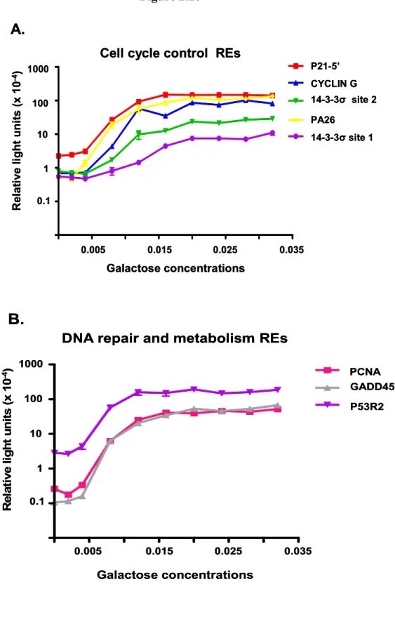
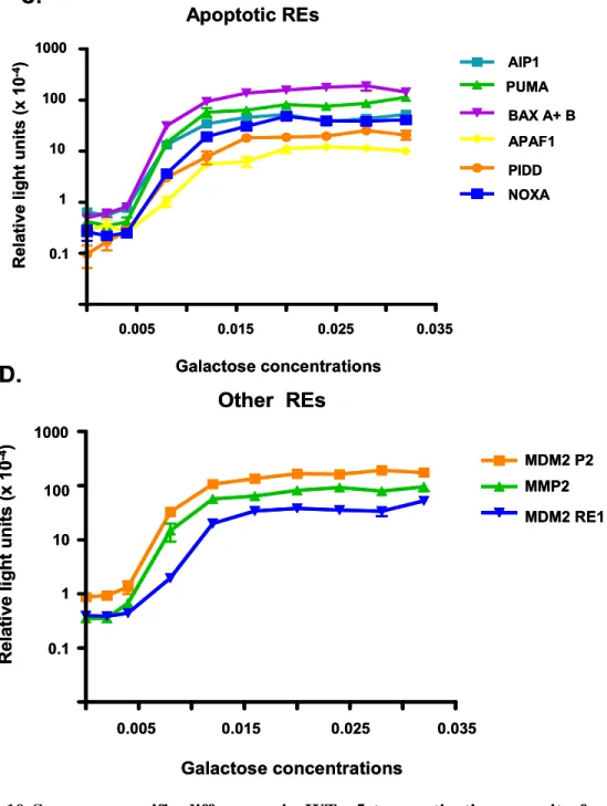
SNPs on the ability of p53 to function as a transcription factor [26, 27]. Twenty-six REs
(22 biological targets and 4 consensus sequences) were ranked for transactivation capacity
based on the ADE2 phenotypic color reporter assay (Figure 2.3). The assay was
subsequently modified to place ADE2 under the control of the minimal CYC1 promoter and
RE of choice at its natural loci on chromosome XV.
Transcriptional capacities of the REs were determined based on the minimal
relative amount of p53 required for a phenotypic change in pigmentation which ranged
from red to pink to white depending on the extent of transactivation. This assessment
revealed, under isogenic conditions that both the sequence and level of p53 protein
contributed to transactivation, where greater than a 1,000 fold difference at various protein
concentrations was observed between REs. The REs which were classified by
transactivation capacity as strong binders (i.e., p21-5’ and P53R2), modest inducers (i.e.,
NOXA and PA26) or poor transactivators (i.e., CFOS and PIG3) did not group according to
biological functions. Furthermore, a correlation between functional rank and statistical
prediction of binding energy of the REs was not observed. Importantly, the central core
transactivate from a RE. These observations suggested that an intrinsic property of the core
sequence “CATG” was required for strong levels of transactivation. (Binding studies
agreed with this finding suggesting the flexibility of the CATG sequence increases the
affinity of p53 towards an element [32].) Altering the CATG in the core to CTAG
dramatically reduced levels of transactivation by 20-fold. Finally, the rheostatable
promoter was used to unmask subtle transactivational differences between WT p53 and
several p53 missense mutations associated with breast cancer which had previously been
determined to be equivalent to WT in transactivation capacity under conditions of high
expression (including the T150I, G199R, R202S and S215C mutations discussed
above)[24]. Subsequent studies on p53 transactivation capacity utilizing the rheostatable
promoter revealed that variation in the p53 transcriptional network result from a matrix of
factors including p53 expression level, target binding sequence and mutations. These
findings culminated in the view that p53 is a “master regulatory gene of genetic diversity”
[27].
In order to further expand the understanding of p53’s role as a transcription factor
and to analyze how sequence and protein alterations contribute to the variability observed
within the p53 transcriptional network, we have modified the rheostatable system to
develop an integrated diploid yeast system. This system allows for a more sophisticated
and sensitive evaluation of the contribution of RE sequence, binding motifs and p53
expression on functionality and regulation within the p53 transcriptional network.
Furthermore, the system allows for a rapid assessment of the consequences of mutations
Results
Isogenomic system to address p53 transactivation from REs in diploid yeast
While many factors may determine the ability of p53 to differentially transactivate
individual genes in human cells including stress stimuli, post-translational modifications,
and transcriptional co-factors, the yeast system addresses the potential for wild type and
mutant p53 to bind and transactivate from various response elements derived from human
genes when placed in a constant chromatin environment. We have expanded the
rheostatable plasmid-based haploid yeast system to one based in diploid yeast to further
assess the transactivation capacities of p53 (WT or mutant) (Figure 2.4). This system
allows a single copy of a p53 variant to be rapidly assessed for transactivation capabilities
from many REs simply by taking advantage of the yeast mating types.
Two panels of modified S. cerevisiae strains were generated. The first was a set of
p53 host strains in which p53 (WT or mutant) is directed by a “rheostatable” GAL1
promoter that allows for controlled, over 200-fold, inducible expression of p53 in yeast
depending on the carbon source in the media (Figure 2.6). Importantly, similar to p53
protein accumulation in mammalian cells following stress [33], expression from the GAL1
promoter in yeast displays a graded transcriptional response such that there is a range of
activity from the promoter as opposed to a binary, or on/off response [34-36]. Biggar and
Crabtree [34] demonstrated through fluorescence-activated cell sorting (FACS)
experiments that expression of green fluorescent protein from a GAL1-GFP reporter within
a population of cells generated a single fluorescent peak where the intensity of the peak
was dependent upon the concentration of galactose supplemented in the media. Thus,
cells in the population respond (and within our system expressing an induced amount of
p53) rather than merely increasing the percentage of cells within the population expressing
the maximal level of protein.
The second set of strains of opposite mating type contained promoter REs upstream
of the minimal CYC1 promoter and either the ADE2 color reporter or the firefly luciferase
reporter [26]. To facilitate the construction of a large number of p53 mutants and REs at
chromosomally located target loci we employed the delitto perfetto system for in vivo
mutagenesis. Delitto perfetto utilizes oligonucleotides and targeted homologous
recombination to rapidly generate S. cerevisiae yeast strains with specific genetic
alterations [37-39].
Mating of the reporter and p53 host strains results in isogenic, diploid yeast that
enable the rapid assessment of the transactivation potential for WT or mutant p53 proteins
towards many individual REs in the p53 transcriptional network [27]. Importantly, all the
conditions in the cells are constant, i.e., isogenomic , where the only variables between
strains are the RE sequence, WT or mutant p53 and level of expression. The rheostatable
GAL1 promoter allows for controlled, over 100-fold, inducible expression of p53 in the
diploid yeast (Figure 2.6 A and B). A rough estimate of the number of p53 molecules in
each cell ranges from ~250 -500 at basal levels of expression to over 30,000 at 0.024%
galactose (Figure 2.6 C).
The ADE2 reporter provided a qualitative assay to determine transactivation
potential through the appearance of pigmentation in the stationary cells of colonies over