Homologous Recombination and Translesion
DNA Synthesis Play Critical Roles on
Tolerating DNA Damage Caused by Trace
Levels of Hexavalent Chromium
Xu Tian1☯, Keyur Patel1☯, John R. Ridpath1☯, Youjun Chen2, Yi-Hui Zhou3,4, Dayna Neo1, Jean Clement1, Minoru Takata5, Shunichi Takeda6, Julian Sale7, Fred A. Wright3,4,8*,
James A. Swenberg1,9, Jun Nakamura1*
1 Department of Environmental Sciences and Engineering, University of North Carolina at Chapel Hill, Chapel Hill, North Carolina, 2 Department of Neurology, UNC Neuroscience center, School of Medicine, University of North Carolina at Chapel Hill, Chapel Hill, North Carolina, 3 Bioinformatics Research Center, North Carolina State University, Raleigh, North Carolina, 4 Department of Biological Sciences, North Carolina State University, Raleigh, North Carolina, 5 Laboratory of DNA Damage Signaling, Department of Late Effects Studies, Radiation Biology Center, Kyoto University, Kyoto, Japan, 6 Department of Radiation Genetics Graduate School of Medicine, Kyoto University, Kyoto, Japan, 7 Medical Research Council Laboratory of Molecular Biology, Cambridge, United Kingdom, 8 Department of Statistics, North Carolina State University, Raleigh, North Carolina, 9 Curriculum in Toxicology, University of North Carolina at Chapel Hill, Chapel Hill, North Carolina
☯These authors contributed equally to this work.
*[email protected](JN);[email protected](FW)
Abstract
Contamination of potentially carcinogenic hexavalent chromium (Cr(VI)) in the drinking water is a major public health concern worldwide. However, little information is available regarding the biological effects of a nanomoler amount of Cr(VI). Here, we investigated the genotoxic effects of Cr(VI) at nanomoler levels and their repair pathways. We found that DNA damage response analyzed based on differential toxicity of isogenic cells deficient in various DNA repair proteins is observed after a three-day incubation with K2CrO4in
REV1-deficient DT40 cells at 19.2μg/L or higher as well as in TK6 cells deficient in polymerase delta subunit 3 (POLD3) at 9.8μg/L or higher. The genotoxicity of Cr(VI) decreased ~3000 times when the incubation time was reduced from three days to ten minutes. TK mutation rate also significantly decreased from 6 day to 1 day exposure to Cr(VI). The DNA damage response analysis suggest that DNA repair pathways, including the homologous recombina-tion and REV1- and POLD3-mediated error-prone translesion synthesis pathways, are criti-cal for the cells to tolerate to DNA damage caused by trace amount of Cr(VI).
Introduction
Chromium (Cr) is a naturally occurring element that exists in a variety of oxidation states between -2 to +6. Among these forms, Cr(III) is an essential trace element for normal
a11111
OPEN ACCESS
Citation: Tian X, Patel K, Ridpath JR, Chen Y, Zhou Y-H, Neo D, et al. (2016) Homologous
Recombination and Translesion DNA Synthesis Play Critical Roles on Tolerating DNA Damage Caused by Trace Levels of Hexavalent Chromium. PLoS ONE 11(12): e0167503. doi:10.1371/journal. pone.0167503
Editor: Kerstin Borgmann, Universitatsklinikum Hamburg-Eppendorf, GERMANY
Received: July 13, 2016
Accepted: November 15, 2016
Published: December 1, 2016
Copyright:©2016 Tian et al. This is an open access article distributed under the terms of the
Creative Commons Attribution License, which permits unrestricted use, distribution, and reproduction in any medium, provided the original author and source are credited.
Data Availability Statement: All relevant data are within the paper and its Supporting Information files.
carbohydrate, lipid and protein metabolism in humans [1]. On the other hand, Cr(VI) has been reported to cause cancer in laboratory animals and occupationally exposed workers [2] and thus draws great attention as a public health concern. Cr(VI) is widely used in various industrial applications including leather tanning, wood preservation, dye production, chrome plating, and alloy manufacturing. Industrial waste containing Cr(VI) may potentially result in the environmental pollution of soil, water and air. Incineration and gasoline usage can also lead to air and water pollution with Cr(VI) as a contaminant. In addition, Cr(VI) can be pro-duced naturally. For example, Cr(III) can be oxidized by Mn(III/IV) into Cr(VI) [2]. Cur-rently, there are hundreds of Superfund sites in the U.S. in which Cr is the major concern for contamination [3].
When taken up by sulfate channels, Cr(VI), which displays no direct DNA damage capabil-ity by itself, is reduced by ascorbate, glutathione, and cysteine, producing reactive intermedi-ates of Cr(V), Cr(IV) and reactive oxygen species (ROS) [4]. Cr(III), the final reduction product, can hardly penetrate the cytoplasmic membrane, thus leading to the massive accumu-lation of Cr inside of cells [2]. The reactive intermediates of Cr has been proposed to induce Cr-DNA adducts and protein-Cr-DNA crosslinks, thereby causing DNA strand breaks and double strand breaks and eventually introducing mutations and genome instability [2]. Besides Cr, ROS can also induce cytotoxicity and mutagenic effects in cells [4]. Due to its genotoxicity, inhaled Cr(VI) was classified as a known human carcinogen by the United States Environmen-tal Protection Agency (U.S. EPA) and the World Health Organization (WHO). With regard to carcinogenicity of Cr(VI) by ingestion, on the other hand, several of the epidemiological stud-ies present contradictory conclusions [5,6]. In the presence of organic molecules at acidic pH, Cr(VI) can be reduced to non-toxic Cr(III) very quickly, which reduces the concern of Cr(VI) as an ingested human carcinogen. A National Toxicology Program (NTP) conducted a two-year study showing that Cr(VI) is a rodent carcinogen when these rodents were administered extremely high doses (57,300μg/L or higher) of Cr(VI) in the drinking water [7]. The U.S. EPA has set the maximum contaminant levels (MCL) for total Cr in the human drinking water at 100μg/L. In California, MCL for Cr(VI) was set at 10μg/L [8], and Public health goals (PHGs) were established at 0.02μg/L [9]. Currently the U.S. EPA is re-evaluating these regula-tions [10]. In 2010, a report by the Environmental Working Group suggests that 89% of the water samples from U.S. cities are contaminated with Cr(VI) at levels ranging from 0.03 to 12.9μg/L [11]. The mechanism by which Cr(VI) causes genotoxicity is well characterized at concentrations equivalent or higher to current U.S. EPA regulatory levels. However, little information is available regarding the biological effects of Cr(VI) at doses lower than 100μg/L.
Because of the high level of homologous recombination and the relative ease of gene manip-ulation, DT40 cells, the chicken B-lymphocyte cells, have been widely used as a model system for higher vertebrate genetic functional studies [12,13]. With the fast replication speed and the strong phenotypic similarities with murine cells, it makes the DT40 isogenic cell line and its mutants deficient in various genes ideal for reverse genetic studies [14]. Recently, DT40 cells have also been successfully used to measure the genotoxicity of different chemicals [15–17].
Cells are constantly exposed to both endogenous and exogenous agents that may cause DNA damages, of which if not repaired might cause genome instability and induce mutations or cell death. Cells have developed multiple mechanisms to deal with different types of DNA damages[18]. Nucleotide excision repair [19] and Fanconi anemia genes [20] were previously reported to be essential for the removal of Cr(VI)-induced DNA damage or activated in human cells exposed with Cr(VI). Inter-strand DNA cross-links (ICLs) was reported caused by Cr(VI) exposure in the present of glutathione, one report suggests FANCD2, ERCC1 or XPF doesn’t impact cell sensitivity to Cr(VI)[21]. Non-homologous end-joining (NHEJ) repair pathways[22] was reported to be involved into the removal of Cr(VI) induced DNA damaged
in Saccharomyces cerevisiae and HR repair pathways were reported to be involved into the removal of Cr(VI) induced DNA damage in mammalian cells[23]. The Werner Syndrome Protein was also reported to be involved into Cr(VI) induced DNA damage[24]. To further investigate the mechanism of Cr(VI) induced DNA damage and repair pathways, in this study we screened a battery of DT40 mutant cells using genotoxicity profiling and found the DNA repair genes and pathways to be critical for cell survival when these cells were exposed to Cr (VI) at nanomolar concentrations. We also confirmed our results using human cancer knock-down cells and further studied the time- and dose- dependent genotoxicity and mutagenicity of nanomolar concentrations of Cr(VI).
Materials and Methods
Chemicals and reagents
The following chemicals and reagents were used in the study: potassium chromate (K2CrO4,
Sigma), 2,3-bis[2-methoxy-4-nitro-5-sulfophenyl]-2H-tetrazolium-5-carboxanilide inner salt (XTT) (Sigma), polybrene, puromycin (Sigma), 1-methoxy-5-methylphenazinium methyl sulfate (Sigma), TRIzol1RNA Isolation Reagent (Invitrogen), RPMI 1640 culture medium (Invitrogen), chicken serum (Invitrogen), penicillin/streptomycin (Invitrogen), fetal bovine serum (FBS) (Atlanta Biologicals), iScript™cDNA Synthesis Kit (Bio-Rad Laboratories, Inc.), SsoAdvanced™Universal SYBR1Green Supermix (Bio-Rad), and TransIT1-293 transfection reagent (Mirus Bio LLC).
Cell culture
Chicken DT40 and mutant cells were maintained as described previously [17]. Briefly, DT40 cells were maintained in non-phenol red RPMI 1640 medium with 10% fetal bovine serum, 1% chicken serum and 1% penicillin/streptomycin. The cells were incubated at 39.5˚C and 5% CO2with 95% humidity. The source of all DT40 knockout mutant cells is provided in Table 1
inS1 File. Human lymphoblastoid (TK6) cells were kindly provided by Dr. Rebecca Fry (Uni-versity of North Carolina at Chapel Hill). HeLa and 293T cells were obtained from the Line-berger Comprehensive Cancer Center at the University of North Carolina at Chapel Hill. TK6 cells were maintained in RPMI 1640 medium with 10% fetal bovine serum and 1% penicillin/ streptomycin. HeLa and 293T cells were grown in DMEM medium supplemented with L-glu-tamine, 10% fetal bovine serum and 1% penicillin/streptomycin. All human cells were main-tained at 37˚C with 5% CO2.
Lentiviral-mediated RNA interference
Analysis of DT40 cell-based DNA damage response
The DT40 cell-based DNA damage response was analyzed as previously reported [17] with minor modifications. Briefly, DT40 cells were suspended in culture medium (~750 cells per 75μL per well), seeded into 96-well plates, exposed to K2CrO4, and allowed to divide for ~7
cell cycles. After cultivation, cell viability was determined by XTT-based cell viability assays.
TK mutation assay
The thymidine kinase locus gene (TK) mutation assay was performed to quantify mutagenicity (214074). BackgroundTKgene mutants were removed from TK6 cells by growing them for 2 days in media containing CHAT followed by incubation for 1 day in media containing CHT. Cells were then seeded at 2.7×10⁵cells/ml in 20 ml serum containing media and were exposed to Cr6 for different periods. Following K2CrO4incubation, cells were washed three times with
sterile PBS, re-suspended in full serum containing media and cultured for 2 days to allow the development of mutations. The cells were maintained at 1.5×10⁶cells/ml or lower to avoid overgrowth. The cells were harvested by centrifugation, re-suspended in fresh media. For the TK-deficient mutant selection, the cells were cloned into 96-well plates at 40,000 cells/well in growth medium (0.125 ml) in the presence of the selective agent trifluorothymidine (TFT, final concentration 4μg/ml) for the detection of mutation frequency (MF). Cells were also seeded at a density of 1.0 cells/well into 96-well plates in the absence of TFT to determine plat-ing efficiency (PE). The plates were incubated at 37˚C in an atmosphere of 5% CO2 for 14 days. The colony formation was then scored. The plates containing TFT were then re-fed with TFT and incubated for an additional 14 days to score for the appearance of slow-growing TK mutants. The total mutation frequencies were calculated according to the Poisson distribution.
qPCR analysis
Total RNA was isolated using TRIzol1RNA Isolation Reagents, and an equivalent amount of RNA from each sample was used as template to make cDNA using the iScript™cDNA Synthe-sis Kit. qPCR was performed according to the standard protocol using SsoAdvanced™ Univer-sal SYBR1Green Supermix. Primers used in this study were listed in Table 2 inS1 File. Data were analyzed byΔΔCt method for relative quantifications, actin was used as internal control.
Statistical analysis
Data are reported as the mean±standard deviation of at least triplicate samples. Analysis of covariance (ANCOVA) was used to test for mean intercept differences and differences in the slopes of the linear dose-response curves in cell viability analyses between wild-type and a series of mutant cells. For display purposes, survival data were log-transformed, providing approximately normal errors, to calculate the lethal concentration 50 (LC50) values for each
cell line using Graphpad Prism 5 (La Jolla, CA).P-values were two sided and adjusted for mul-tiple comparisons as noted.
To represent the point of departure values for concentration-response, Benchmark dose (BMD) values [25] were obtained from the EPA BMD software v 2.4. These values represent one-standard deviation departures from control values and corresponding lower 5% BMDL values, using log10(concentration+0.5) to avoid taking logarithms of zero control values. For
determined from the logistic model outside of the 90% confidence interval reported by the BMD software.
Comparisons of BMD values from independent concentration-response curves were per-formed using z-statistics computed as z¼ ðBMD1 BMD2Þ= ffiffiffiffiffiffiffiffiffiffiffiffiffiffiffiffiffiffiffiffiSE2
1þSE22
p
;with standard errors inferred from the BMD confidence interval. One-sample t-tests were performed for comparisons of LC50across the cell lines in comparison to DT40 wilt-type cells using ratio
val-ues and in comparison to a null ratio of 1.0.
Results
Chicken DT40 cells deficient in either homologous recombination or the
error-prone translesion synthesis pathway are hyper-sensitive to Cr(VI)
To first determine the cytotoxicity of Cr(VI), we exposed chicken DT40 cells, a frequently used cell line for understanding DNA repair gene function studies, with Cr(VI) salt K2CrO4at
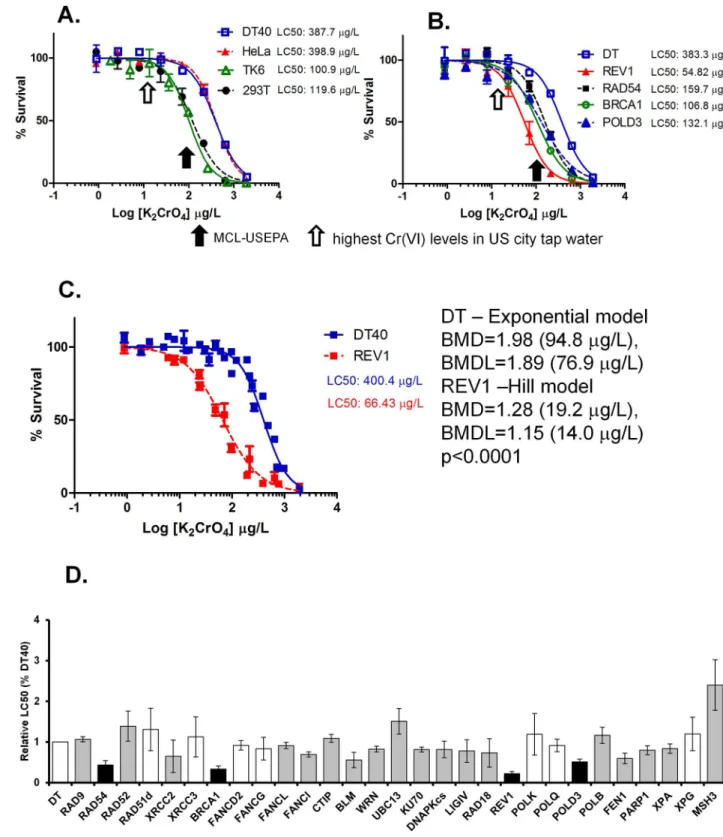
various concentrations ranging from 1 to 2000μg/L for approximately three consecutive days. We examined cell survival rate as a result of Cr(VI) treatment. The DNA damage response is a very broad term for the network of cellular pathways that sense, signal and repair DNA lesions. In the current study we use cell survival rate as an indication of toxicity and DNA damage response as shown previously [17]. At the dose of 100μg Cr/L (equivalent to 373μg K2CrO4/
L), which is the MCL for total Cr in drinking water as set by the U.S. EPA, DT40 cells showed substantial toxicity with ~50% survival rate compared with vehicle-treated cells. Breaking points (departure points from 100% survival rate) were detected at doses ranging between 20– 80μg K2CrO4/L (Fig 1A).
Cr(VI) exposure appears to lead to Cr-DNA adducts, protein-Cr-DNA crosslinks, or oxida-tive DNA damage resulting in genotoxicity [2]. Because DNA repair pathways are important for maintenance of genomic integrity, we hypothesized that cells deficient in a certain DNA repair pathway may show altered tolerance to K2CrO4treatment relative to wild type cells. To
test this hypothesis, a DNA damage response analysis for K2CrO4using isogenic DT40 mutant
cells was conducted in this study. DNA repair pathways screened in this study include base excision repair (BER), nucleotide excision repair (NER), mis-match repair (MMR), homolo-gous recombination (HR), nonhomolohomolo-gous end joining (NHEJ), translesion DNA synthesis (TLS), Fanconi anemia DNA repair pathway, cell-cycle checkpoint and RecQ helicases. The whole list and function of each gene is shown in Table 1 inS1 File.
Testing these mutant cells allowed us to identify the DNA repair pathways that are critical for cell survival in response to K2CrO4exposure. For a quantitative comparison of differential
toxicity between each mutants, we calculated the relative LC50(LC50of mutant cells divided by
the LC50of the parental DT40 cells). During this screening, we identified four genes, deletion
of any one of which led to hypersensitivity of the cells to K2CrO4treatment and significantly
reduced the relative LC50compared to parental DT40 cells (Fig 1D). These include two genes
involved in tanslesion DNA synthesis (TLS),REV1(relative LC50= 0.22,p<0.05) andPOLD3
(relative LC50= 0.51,p<0.05), and two genes involved in homologous recombination,BRCA1
(relative LC50= 0.33,p<0.05) andRAD54(relative LC50= 0.44p<0.05) (Fig 1B and 1D).
Among these four mutant cell lines,REV1-deficient DT40 cells are most sensitive to K2CrO4
treatment with an LC50at 66μg/L and a breaking point of cell survival at 19μg/L (= 5.2μg Cr/
L) compared with LC50at 400μg/L and breaking point at 95μg/L (= 25μg Cr/L) in wild type
hyper-Fig 1. K2CrO4reduces viability of human cells as well as DT40 cells deficient in certain DNA repair pathways at the maximum
contaminant levels (MCL) set by the U.S. EPA. Various human cells (A) and DT40 parental cells and their mutant cells (B) deficient in
REV1, BRCA1, or RAD54 were exposed to K2CrO4for ~3 days to determine their survival rates. Survival data were log-transformed giving
approximate normality using Prism 5. Each LC50value was then calculated. The black arrow indicates the maximum contaminant levels
(MCL) set by the U.S. EPA, while the open arrow shows the highest Cr(VI) levels detected in U.S. city water (12.9μg/L). (C) Point-of-departure analysis showed a significant difference (p<0.0001) in DT40 (log10(BMD) = 1.98 by the Hill model) vs. REV1-deficient cells (log10 (BMD) = 1.28 (19.2μg/L) by the Hill model). (D) DT40 cell-based DNA damage response analysis was performed for K2CrO4using a series
and hypo-snsitivity results, DT40 cell-based DNA damage response analysis demonstrate moderate sensitivity to K2CrO4in various mutants including cells deficient inXRCC2,
FANCL,FANCI,BLM,WRN,KU70,DNAPKcs,LIGIV,RAD18,FEN1,PARP1, andXPA.
Cr(VI) exposure leads to genotoxicity in human cells
While DT40 cells offer a relatively quick and facile method to analyze the DNA damage response to chemicals, one concern of this approach is that the effects observed in chicken cells may not replicate those in human cells. Therefore, we examined the genotoxicity of human cells induced by Cr(VI). As with DT40 cells, we continuously exposed several different lines of human cells including HeLa (cervical carcinoma epithelial cell line), 293T (kidney epi-thelial cell line) and TK6 (B-lymphoblast cell line) cells to Cr(VI) salt K2CrO4at various
con-centrations for three days and subsequently assessed cytotoxicity. Similar to DT40 cells, all of the human cell lines tested in this study showed a marked toxic response to Cr(VI) at 373μg K2CrO4/L (= 100μg Cr/L) with survival rate of 50%, 17%, and 10% for HeLa cells, 293T and
TK6 cells, respectively) (Fig 1A). We next investigated the DNA damage response of Cr(VI) exposure in human cells deficient in DNA damage repair genes. SinceBRCA1,RAD54,POLD3 andREV1are the genes most critical for chicken DT40 cell survival in the presence of Cr(VI), we investigated whether these genes are also critical for human cell survival. Knockdown of BRCA1,RAD54, andPOLD3in human cells was successfully established by lentiviral-mediated RNA interference (Fig 1 inS1 File). However, knockdown ofREV1led to severe cell death, and we were unable to carry out any further experiments usingREV1-deficient cells (data not shown). Similar to DT40 mutant cells, human cells deficient inPOLD3,RAD54orBRCA1 were also more sensitive to Cr(VI) than wild type cells infected with a non-target control virus, as indicated by reduced cell viability (Fig 2).BRCA1-deficient HeLa cells showed toxicity with an LC50of 52μg/L, andRAD54-deficient HeLa cells showed toxicity with an LC50of 79μg/L
compared with control HeLa cells with an LC50of 358μg/L (Fig 2A).POLD3-deficient TK6
cells showed toxicity with an LC50of 39μg/L and a breaking point at 9.8μg/L (= 2.6μg Cr/L)
compared with control TK6 cells with an LC50of 104μg/L and a breaking point at 34μg/L
(= 9.0μg Cr/L) (Fig 2B and 2C). Based on the results from the DT40 mutant cells and human mutant cells, we concluded that trace amount of Cr(VI) have the potential to cause a DNA damage response and genotoxicity during a three day continuous exposure, and homologous recombination (BRCA1andRAD54) and error-prone translesion synthesis (REV1and POLD3) are critical for cells to tolerate nanomolar levels of Cr(VI)-induced genotoxicity.
Cr(VI) exposure-induced genotoxicity is both time and dose dependent
It has been reported that efficient uptake of Cr(VI) into cells results in a massive accumulation of intracellular Cr during continuous 24-hour exposure [2,26,27]. Therefore, we hypothesized that trace amounts (i.e. nanomolar levels) of Cr(VI) exposure leads to a DNA damage response only by long-term, and probably not by short-term, incubation of K2CrO4. To address this
possibility, we incubated the most sensitive mutant cells observed in our study,REV1-deficient DT40 cells, with K2CrO4for durations of 10 min to 8 hours followed by extensive washing of
the cells to minimize residual extracellular K2CrO4levels. The cells were further incubated for
3 days in fresh media before assessing cell survival rate.REV1-deficient DT40 cells showed a represent standard deviation from at least three independent experiments. Student’s t-test was used to test for significance in mean LC50
values between DT40 parental cells and each mutant. Columns shaded in gray indicate significant differences (p<0.05) between parental DT40 cells and each mutant. Columns shaded in black show the cell lines (REV1, RAD54, POLD3, and BRCA1 mutants) that are markedly sensitive to K2CrO4. (p<0.05 for all four cell lines after Bonferroni adjustment for 32 tests).
drastic time-dependent increase in cell toxicity caused by K2CrO4(Fig 3A). A ten-minute
K2CrO4exposure resulted in cell toxicity with an LC50of ~24 x 10 3
μg/L, which is 465 times higher (less toxic) than a three-day continuous K2CrO4exposure (Fig 3A). We further found a
highly significant linear relationship between log-transformed incubation time and LC50
Fig 2. K2CrO4reduces the viability of human cells deficient in certain DNA repair pathways at Cr(VI) levels detected in U.S. city
water. HeLa cells (A) and TK6 cells (B) transiently or stably knocked down with shRNA against RAD54, BRCA1, or POLD3 were exposed to K2CrO4for ~3 days to determine their survival rate. (C) Point of departure analysis of TK6 indicated significant differences (p<0.0001) for
TK6 control (log10(BMD) = 1.53, exponential model) vs. POLD3 shRNA knock down (log10(BMD) = 0.99 (9.8μg/L), polynomial model). The black arrow indicates the maximum contaminant levels (MCL) set by the U.S. EPA. The open arrow shows the highest Cr(VI) levels detected in US city water.
Fig 3. The genotoxicity of K2CrO4in human and DT40 cells is drastically decreased when K2CrO4
incubation time is reduced. (A) REV1 ko DT40 cells were incubated with K2CrO4for 10 min to 8 hours
followed by extensive washing. The cells were further cultivated for 3 days in fresh medium without addition of K2CrO4to determine cell survival. (B) The exposure times (hours) were multiplicatively inversed followed by
log transformation (x-axis). LC50data for each exposure time were log transformed (y-axis). Linear regression
analysis was performed to determine the relationship between exposure time and LC50values (p<0.0001).
values (Fig 3B). Wild type TK6 cells also showed a time dependent toxicity with ~3000-fold higher LC50value upon ten-minute K2CrO4exposure than a three-day incubation (Fig 3C). It
is worth noting that, under these experimental conditions, wild type andPOLD3-deficient TK6 cells showed significantly different LC50values (Fig 3C). Upon 10 minute exposure to
K2CrO4, the LC50value is ~403 x 103μg/L for wild type cells and ~94 x 103μg/L for
POLD3-deficient cells. Upon exposure of cells to K2CrO4for 3 days, the LC50value is 135μg/L for wild
type cells and 48μg/L forPOLD3-deficient cells. Since POLD3 is involved in DNA damage repair, our results suggest that K2CrO4treatment leads to a time-dependent increase in
induc-tion of DNA damage in human cells.
The mutagenicity of Cr(VI) in human TK6 cells is both time and dose
dependent
Finally, we examined the mutagenic activity of K2CrO4and asked whether this activity is time
and dose dependent. We exposed human TK6 cells with K2CrO4at 180,000μg/L for 10 min.
This concentration of K2CrO4is very close to Cr(VI) concentration which has been reported
to cause oral and intestine cancers by drinking water in rodents [7]. We found that this K2CrO4exposure led to about a two fold increase in theTKgene mutation frequency of TK6
cells compared to that of vehicle-treated cells (Fig 4B). A similar increase in TK gene mutation was detected when cells were treated with K2CrO4at a concentration between 100 and 200μg/
L for 24 hours (Fig 4C), which is more than three orders of magnitude lower in concentration compared to a 10 min exposure with 180,000μg/L K2CrO4(Fig 4B). We then examined
whether prolonged exposure to K2CrO4at levels as low as the highest level of contamination
found in U.S. city water caused mutations in TK6 cells [11]. Cell growth inhibition in wild-type TK6 cells started at 1, 3, and 5 days when the cells were continuously exposed to K2CrO4
at 100, 50, and 25μg/L, respectively (Fig 4A). TK mutation frequency was markedly increased as exposure time increased from 1 day to 6 days with different threshold levels for each expo-sure condition (Fig 4C). These results clearly show that the genotoxicity introduced by Cr(VI) is dependent on both the concentration of Cr(VI) and the total exposure time. Our data also indicate that short-term Cr(VI) exposure is markedly less genotoxic than chronic exposure at the same Cr(VI) concentration.
When comparing a 6-day K2CrO4exposure to a 14-day K2CrO4exposure, a drop in the
mutation rate observed in the 14-day rate for the lowest active produced an apparent differ-ence in point of departure BMD values (p<0.05), however, after eliminating this point there was no evidence of a difference in mutation rates between 6-day and 14-day exposures (Fig 4D). Combining the mutation frequency results from 6- and 14-day K2CrO4exposures, a
sig-nificant increase in mutation rate was observed at 12.5μg/L (= 6.7μg Cr(VI)/L) or higher as compared to controls in TK6 cells (Fig 4E). These results suggest that cells may be equipped with a mechanism to tolerate further increases in mutations caused by trace amounts (25μg/L or lower) of K2CrO4for exposure periods of longer than 6 days.
Discussion
In the current study, we investigated the genotoxicity of low dose Cr(VI) in cultured chicken DT40 cells and human cells. We found that the genotoxicity of cells exposed to nanomolar followed by extensive washing. The cells were further cultivated for 3 days in fresh medium without addition of K2CrO4to determine cell survival. The survival curves were compared between 10-min and 3-day exposure
groups (p<0.0001). The green arrow indicates the Cr(VI) concentration (172,000μg/L) that causes an increase in oral cancer, a finding that was previously shown in an NTP rodent study [7].
Fig 4. The mutagenicity of K2CrO4in human TK6 cells is markedly decreased by reducing K2CrO4
incubation time. (A) Cumulative cell growth rates of TK6 cells were monitored during continuous incubation with K2CrO4at different concentrations for up to 6 days. (B) Mutation rates of TK6 cells exposed to K2CrO4for 10 min.
levels of Cr(VI) is both time and dose dependent, with higher dose or longer exposure leading to severer genotoxicity. We also identified four key components of DNA repair proteins, REV1,POLD3,BRCA1andRAD54, that are critical for tolerating the genotoxicity induced by Cr(VI). In addition, we found that prolonged (1–2 weeks) exposure to trace amounts (~10μg/ L) of K2CrO4causes increased mutations in human lymphoblastoid cells. However, the
geno-toxicity of Cr(VI), including the DNA damage response and mutagenesis, decreased ~3000 times when the incubation time was shortened from three days to 10 minutes. Our dose- and time-dependent Cr(VI) genotoxicity results, combined with the quick transit time of Cr(VI) in the digestive tracts of animals after drinking Cr(VI)-contaminated water could explain carci-nogenicity of Cr(VI) in drinking water in rodents only at super high concentrations.
Nucleotide excision repair, but not Fanconi anemia pathway, plays
critical roles on the tolerance to DNA lesions caused by K
2CrO
4in DT40
cells
While Cr(VI) is not reactive to DNA under physiological conditions, the intracellular reduc-tion of Cr(VI) towards Cr(III), leading to cause DNA damage[28]. The DNA lesions produced through reduction of Cr(VI) include, single and double strand breaks[29], oxidative DNA damage[28,30,31], ternary complex of endogenous reducing agent-Cr-DNA phosphodiester backbone[28,32], Cr-DNA interstrand crosslinks [33] and DNA-Cr-protein crosslinks [34]. Among various DNA repair pathways for counteracting these DNA lesions, nucleotide exci-sion repair pathway has been reported to be important for the removal or tolerance of Cr(VI)-induced DNA damage [19,35–37]. In an agreement with these reports, K2CrO4caused higher
toxicity in DT40 cells deficient in XPA compared to parental DT40 cells.
Fanconi anemia cells are well-known to be hyper-sensitive to DNA interstrand and DNA-protein crosslinking agents such as cisplatin, diepoxybutadiene, and formaldehyde [38,39]. Previous report showed that FANCA-deficient cells are hyper-sensitive to Cr(VI) in cell sur-vival with S-phase-dependent DSB formation using none-isogenic cell lines and Cr(VI) also induced FANCD2 monoubiquitination [20,40]. However, other group’s report demonstrated conflicting results using isogenic cultured cells in the presence of physiological levels of ascor-bic acid, which is a critical reducing element for Cr(VI)-induced DNA crosslinking lesionsin vitro[21]. In the DT40 cell-based DNA damage response analysis, we found that the cells defi-cient inFANCD2andFANCGwere not hypersensitive to K2CrO4treatment with marginal
sensitivity in cells deficient inFANCIandFANCL. Our results suggest that DNA crosslinks are not major contributors to the DNA damage response caused by trace amounts of Cr(VI) in the DT40 cell system under the condition we utilized.
experiments, the TK6 cells were treated with K2CrO4for 24 hours followed by washing and 2-day phenotype
expression period before TFT treatment (log10(BMD) = 2.01, Hill model). For 6-day exposure experiments, we continuously treated the cells with K2CrO4for 6 days by adding fresh K2CrO4into the culture medium when the cells
were subcultured. After the 6-day treatment, the cells were treated with TFT (log10(BMD) = 1.48, Hill model). The number of mutants was counted at 2 and 4 weeks after the first TFT treatment. Error bars represent standard deviation around the mean obtained from at least three independent experiments. The 1-day and 6-day BMD values are significantly different (P = 0.005). (D) Dose-response curves of mutation rates of TK6 cells exposed to trace amounts of K2CrO4were compared between 1-, 6- or 14-day treatment groups. For 1-day and 6-day
exposures, the mutation assays were performed as described in Fig 4C. For 14-day exposure experiments, the assays were conducted as described in the 6-day experiment except that the duration of K2CrO4treatment was
prolonged. Error bars represent standard deviation around the mean obtained from at least three independent experiments. (E) Since 6- and 14-day dose-response curves overlapped with each other in mutation rates and there was no significant difference between the mutation rates above the second active concentration, the mutation rate data were combined and analyzed as pooled results. (log10(BMD) = 1.02 (10.5μg/L), Hill model).
HR and TLS are critical for tolerating Cr(VI) exposure
In our DNA damage response assay using a battery of DT40 cells deficient in DNA repair genes, we found thatREV1,BRCA1,RAD54, andPOLD3are essential in rescuing Cr(VI)-induced DNA damage. This finding suggests that homologous recombination and the error-prone translesion synthesis are critical for DT40 cells to tolerate DNA damage caused by trace levels of Cr(VI). We have confirmed these results using human knockdown cells. Previous reports also demonstrated similar results using Saccharomyces cerevisiae deficient in various DNA repair genes with higher sensitivity ofRad18 Rad27,Rad50,Rad51,Rad52,Rad54, Rad59,Rev3, andRev1mutants to either Na2CrO4or CrO3[41,42] compared to their
wild-typecells. Homologous recombination is one of two primary DSB repair pathways and utilizes a series proteins including ATM, RAD54, and BRCA1 for repairing DSBs during S and G2 phases [18]. The mechanism by which Cr(VI) causes replication-associated DNA DSBs [43] appears to be due to the collapse of replication fork as well as conversion of single strand break to DSBs.
We found DT40 cells deficient in REV1 is the most sensitive cell line to K2CrO4among
DT40 mutants utilized in this study. As described above, yeast study also showed higher toxic-ity in the yeasts deficient in RAD18, REV3, and REV1 [41,42]. In addition, our study demon-strated POLD3-deficient cells were hyper-sensitive to K2CrO4with moderate toxicity of
RAD18 mutant cells. POLD3 is one of the subunits of replicative DNA polymerase delta and POLD2-POLD3 complex is believed to be involved in TLS by switching binding between POL-D1and translesion polymerase REV3 when encountered with DNA replication barrier [44, 45]. Besides, in our preliminary data PCNA mono-ubiquitination deficient mutation cells shows similar Cr(VI) sensitivity with REV1 deficient cells and REV1 C-terminal is critical for REV1’s function to tolerate Cr(VI) toxicity but not REV1 catalytically domain. Taken together, we are proposing the following mechanism of bypassing K2CrO4-mediated DNA lesions by
TLS pathway: 1) ternary Cr-DNA lesions stall DNA replication and activate RAD18-mediated TLS pathway; 2) RAD6-RAD18 complex monoubiquitinates PCNA; 3) low-fidelity TLS poly-merase bypass DNA lesions in the presence of scaffold protein REV1, leading to mutations; and 4) REV3 further extends DNA synthesis with POLD2 and POLD3 complex. It is worth-while to note that previous paper reported polymerase zeta-dependent chromium-induced mutagenesis in yeasts [46]. As with REV3, in the absence of either REV1, or POLD3, we believe Cr(VI)-mediated mutagenesis may be significantly decreased.
Supporting Information
S1 File. This file contains all Supporting Tables (1–2) andFig 1. Table 1 in S1 File. DT40
mutant cells used in this study. Table 2 in S1 File. Oligonucleotides for shRNA construction and QRT-PCR. Fig 1 in S1 File. Confirmation of knockdown efficiency. BRCA1, RAD54 and POLD3 knockdown cells were prepared and knockdown efficiency were measured by using qPCR. Data are presented as mean±SD; n = 3.
(PDF)
Acknowledgments
The authors thank Dr. Amy E. Clipperton-Allen at The Scripps Research Institute and Dr. Brian Pachkowski for critical reading of our manuscript.
Author Contributions
Formal analysis: XT YZ FW JN.
Funding acquisition: J. Swenberg JN.
Investigation: XT KP JR JC DN JN.
Methodology: JN.
Project administration: JN.
Resources: J. Sale ST MT.
Supervision: JN.
Validation: XT JN.
Visualization: XT JN.
Writing – original draft: XT YC JN.
Writing – review & editing: XT YC JN.
References
1. Anderson RA. Essentiality of chromium in humans. The Science of the total environment. 1989; 86(1– 2):75–81. PMID:2602941
2. Zhitkovich A. Chromium in drinking water: sources, metabolism, and cancer risks. Chemical research in toxicology. 2011; 24(10):1617–29. doi:10.1021/tx200251tPMID:21766833
3. Agency for Toxic Substances and Disease Registry Toxicological Profile for Chromium. US Depart-ment of Health and Human Services, Washington, DC. 2000.
4. Liu KJ, Shi X. In vivo reduction of chromium (VI) and its related free radical generation. Mol Cell Bio-chem. 2001; 222(1–2):41–7. PMID:11678610
5. Beaumont JJ, Sedman RM, Reynolds SD, Sherman CD, Li LH, Howd RA, et al. Cancer mortality in a Chinese population exposed to hexavalent chromium in drinking water. Epidemiology. 2008; 19(1):12– 23. doi:10.1097/EDE.0b013e31815cea4cPMID:18091413
6. Kerger BD, Butler WJ, Paustenbach DJ, Zhang J, Li S. Cancer mortality in chinese populations sur-rounding an alloy plant with chromium smelting operations. J Toxicol Environ Health A. 2009; 72 (5):329–44. doi:10.1080/15287390802529898PMID:19184749
7. National Toxicology P. Toxicology and carcinogenesis studies of sodium dichromate dihydrate (Cas No. 7789-12-0) in F344/N rats and B6C3F1 mice (drinking water studies). National Toxicology Program technical report series. 2008;(546: ):1–192. PMID:18716633
8. California Regulations Related to Drinking Waterhttp://www.waterboards.ca.gov/drinking_water/certlic/ drinkingwater/documents/lawbook/dwregulations-2015-07-16.pdf. July 16, 2015.
9. OEHHA Adopts First-in-the-Nation Public Health Goal For Hexavalent Chromium in Drinking Water
http://www.oehha.ca.gov/public_info/press/072611PRChrom6.pdfOffice of Environmental Health Haz-ard Assessment Oakland, California 94612. July 27, 2011
10. USEPA. IRIS Toxicological Review of Hexavalent Chromium (2010 External Review Draft). U.S. Envi-ronmental Protection Agency, Washington, DC, EPA/635/R-10/004A,. 2010.
11. Sutton R. Chromium-6 in U.S. Tap Waterhttp://static.ewg.org/reports/2010/chrome6/chrome6_report_ 2.pdf2010:1–23.
12. Suzuki A, Fukagawa T. Cell biological analysis of DT40 knockout cell lines for cell-cycle genes. Curr Protoc Cell Biol. 2011; Chapter 8:Unit 8.7. doi:10.1002/0471143030.cb0807s50PMID:21400701
13. Ishiai M, Uchida E, Takata M. Establishment of the DNA repair-defective mutants in DT40 cells. Meth-ods Mol Biol. 2012; 920:39–49. doi:10.1007/978-1-61779-998-3_4PMID:22941595
14. Sonoda E, Morrison C, Yamashita YM, Takata M, Takeda S. Reverse genetic studies of homologous DNA recombination using the chicken B-lymphocyte line, DT40. Philos Trans R Soc Lond B Biol Sci. 2001; 356(1405):111–7. doi:10.1098/rstb.2000.0755PMID:11205323
16. Ji K, Seo J, Liu X, Lee J, Lee S, Lee W, et al. Genotoxicity and endocrine-disruption potentials of sedi-ment near an oil spill site: two years after the Hebei Spirit oil spill. Environ Sci Technol. 2011; 45 (17):7481–8. doi:10.1021/es200724xPMID:21786741
17. Ridpath JR, Takeda S, Swenberg JA, Nakamura J. Convenient, multi-well plate-based DNA damage response analysis using DT40 mutants is applicable to a high-throughput genotoxicity assay with char-acterization of modes of action. Environmental and molecular mutagenesis. 2011; 52(2):153–60. doi:
10.1002/em.20595PMID:20839229
18. Ciccia A, Elledge SJ. The DNA damage response: making it safe to play with knives. Molecular cell. 2010; 40(2):179–204. doi:10.1016/j.molcel.2010.09.019PMID:20965415
19. O’Brien TJ, Brooks BR, Patierno SR. Nucleotide excision repair functions in the removal of chromium-induced DNA damage in mammalian cells. Mol Cell Biochem. 2005; 279(1–2):85–95. doi:10.1007/ s11010-005-8225-0PMID:16283517
20. Vilcheck SK, Ceryak S, O’Brien TJ, Patierno SR. FANCD2 monoubiquitination and activation by hexa-valent chromium [Cr(VI)] exposure: activation is not required for repair of Cr(VI)-induced DSBs. Muta-tion research. 2006; 610(1–2):21–30. doi:10.1016/j.mrgentox.2006.06.009PMID:16893675
21. Morse JL, Luczak MW, Zhitkovich A. Chromium(VI) causes interstrand DNA cross-linking in vitro but shows no hypersensitivity in cross-link repair-deficient human cells. Chemical research in toxicology. 2013; 26(10):1591–8. doi:10.1021/tx400293sPMID:24059640
22. Santoyo G, Strathern JN. Non-homologous end joining is important for repair of Cr(VI)-induced DNA damage in Saccharomyces cerevisiae. Microbiol Res. 2008; 163(1):113–9. doi:10.1016/j.micres.2007. 09.001PMID:17923397
23. Bryant HE, Ying S, Helleday T. Homologous recombination is involved in repair of chromium-induced DNA damage in mammalian cells. Mutation research. 2006; 599(1–2):116–23. doi:10.1016/j.mrfmmm. 2006.02.001PMID:16564059
24. Liu FJ, Barchowsky A, Opresko PL. The Werner syndrome protein functions in repair of Cr(VI)-induced replication-associated DNA damage. Toxicological sciences: an official journal of the Society of Toxicol-ogy. 2009; 110(2):307–18.
25. U.S.EPA/100/R-12/001. Benchmark Dose Technical Guidance.http://www.epa.gov/raf/publications/ pdfs/benchmark_dose_guidance.pdf. 2012.
26. Reynolds M, Stoddard L, Bespalov I, Zhitkovich A. Ascorbate acts as a highly potent inducer of chro-mate mutagenesis and clastogenesis: linkage to DNA breaks in G2 phase by mismatch repair. Nucleic acids research. 2007; 35(2):465–76. doi:10.1093/nar/gkl1069PMID:17169990
27. Messer J, Reynolds M, Stoddard L, Zhitkovich A. Causes of DNA single-strand breaks during reduction of chromate by glutathione in vitro and in cells. Free radical biology & medicine. 2006; 40(11):1981–92. 28. Zhitkovich A. Importance of chromium-DNA adducts in mutagenicity and toxicity of chromium(VI).
Chemical research in toxicology. 2005; 18(1):3–11. doi:10.1021/tx049774+PMID:15651842
29. Xie H, Wise SS, Holmes AL, Xu B, Wakeman TP, Pelsue SC, et al. Carcinogenic lead chromate induces DNA double-strand breaks in human lung cells. Mutation research. 2005; 586(2):160–72. doi:10.1016/ j.mrgentox.2005.06.002PMID:16112599
30. Sugden KD, Wetterhahn KE. Direct and hydrogen peroxide-induced chromium(V) oxidation of deoxyri-bose in single-stranded and double-stranded calf thymus DNA. Chemical research in toxicology. 1997; 10(12):1397–406. doi:10.1021/tx970135rPMID:9437531
31. Sugden KD, Campo CK, Martin BD. Direct oxidation of guanine and 7,8-dihydro-8-oxoguanine in DNA by a high-valent chromium complex: a possible mechanism for chromate genotoxicity. Chemical research in toxicology. 2001; 14(9):1315–22. PMID:11559048
32. Zhitkovich A, Voitkun V, Costa M. Formation of the amino acid-DNA complexes by hexavalent and triva-lent chromium in vitro: importance of trivatriva-lent chromium and the phosphate group. Biochemistry. 1996; 35(22):7275–82. doi:10.1021/bi960147wPMID:8679557
33. Bridgewater LC, Manning FC, Patierno SR. Base-specific arrest of in vitro DNA replication by carcino-genic chromium: relationship to DNA interstrand crosslinking. Carcinogenesis. 1994; 15(11):2421–7. PMID:7955085
34. Voitkun V, Zhitkovich A, Costa M. Complexing of amino acids to DNA by chromate in intact cells. Envi-ron Health Perspect. 1994; 102 Suppl 3:251–5.
35. Arakawa H, Tang MS. Recognition and incision of Cr(III) ligand-conjugated DNA adducts by the nucleo-tide excision repair proteins UvrABC: importance of the Cr(III)-purine moiety in the enzymatic reaction. Chemical research in toxicology. 2008; 21(6):1284–9. doi:10.1021/tx800046yPMID:18452313
37. Zecevic A, Hagan E, Reynolds M, Poage G, Johnston T, Zhitkovich A. XPA impacts formation but not proteasome-sensitive repair of DNA-protein cross-links induced by chromate. Mutagenesis. 2010; 25 (4):381–8. doi:10.1093/mutage/geq017PMID:20410141
38. Walden H, Deans AJ. The Fanconi anemia DNA repair pathway: structural and functional insights into a complex disorder. Annu Rev Biophys. 2014; 43:257–78. doi:10.1146/annurev-biophys-051013-022737
PMID:24773018
39. Ridpath JR, Nakamura A, Tano K, Luke AM, Sonoda E, Arakawa H, et al. Cells deficient in the FANC/ BRCA pathway are hypersensitive to plasma levels of formaldehyde. Cancer research. 2007; 67 (23):11117–22. doi:10.1158/0008-5472.CAN-07-3028PMID:18056434
40. Vilcheck SK, O’Brien TJ, Pritchard DE, Ha L, Ceryak S, Fornsaglio JL, et al. Fanconi anemia comple-mentation group A cells are hypersensitive to chromium(VI)-induced toxicity. Environ Health Perspect. 2002; 110 Suppl 5:773–7.
41. O’Brien TJ, Fornsaglio JL, Ceryak S, Patierno SR. Effects of hexavalent chromium on the survival and cell cycle distribution of DNA repair-deficient S. cerevisiae. DNA repair. 2002; 1(8):617–27. PMID:
12509285
42. Johnson AJ, Veljanoski F, O’Doherty PJ, Zaman MS, Petersingham G, Bailey TD, et al. Revelation of molecular basis for chromium toxicity by phenotypes of Saccharomyces cerevisiae gene deletion mutants. Metallomics. 2016; 8(5):542–50. doi:10.1039/c6mt00039hPMID:27146641
43. Ha L, Ceryak S, Patierno SR. Generation of S phase-dependent DNA double-strand breaks by Cr(VI) exposure: involvement of ATM in Cr(VI) induction of gamma-H2AX. Carcinogenesis. 2004; 25 (11):2265–74. doi:10.1093/carcin/bgh242PMID:15284180
44. Makarova AV, Stodola JL, Burgers PM. A four-subunit DNA polymerase zeta complex containing Pol delta accessory subunits is essential for PCNA-mediated mutagenesis. Nucleic Acids Res. 2012; 40 (22):11618–26. doi:10.1093/nar/gks948PMID:23066099
45. Lee YS, Gregory MT, Yang W. Human Pol zeta purified with accessory subunits is active in translesion DNA synthesis and complements Pol eta in cisplatin bypass. Proc Natl Acad Sci U S A. 2014; 111 (8):2954–9. doi:10.1073/pnas.1324001111PMID:24449906