Western University Western University
Scholarship@Western
Scholarship@Western
Electronic Thesis and Dissertation Repository
6-16-2017 12:00 AM
Cognitive and Non-Cognitive Dysfunction in a Mouse Model of
Cognitive and Non-Cognitive Dysfunction in a Mouse Model of
Alzheimer's Disease
Alzheimer's Disease
Wai-Jane V. Lee
The University of Western Ontario
Supervisor Dr. Marco Prado
The University of Western Ontario Joint Supervisor Dr. Vania Prado
The University of Western Ontario Graduate Program in Neuroscience
A thesis submitted in partial fulfillment of the requirements for the degree in Master of Science © Wai-Jane V. Lee 2017
Follow this and additional works at: https://ir.lib.uwo.ca/etd
Part of the Cognitive Neuroscience Commons
Recommended Citation Recommended Citation
Lee, Wai-Jane V., "Cognitive and Non-Cognitive Dysfunction in a Mouse Model of Alzheimer's Disease" (2017). Electronic Thesis and Dissertation Repository. 4592.
https://ir.lib.uwo.ca/etd/4592
This Dissertation/Thesis is brought to you for free and open access by Scholarship@Western. It has been accepted for inclusion in Electronic Thesis and Dissertation Repository by an authorized administrator of
Abstract
Sensitive and translational tasks that efficiently and accurately assess cognitive function
during pre-clinical trials would be useful in developing novel treatments for Alzheimer's
disease (AD) patients. The Bussey-Saksida touchscreens employ various tasks similar
to those used in humans to effectively evaluate high-level cognitive and executive
functions in mice. This face validity provides the best chance of successful cognitive
translation across species.
In our study, donepezil had minor effects on the performance of 5xFAD mice in the
5-CSRTT, a touchscreen task evaluating attention. Additionally, 5xFAD mice do not
demonstrate impairments in the PVD task, which assesses visual discrimination/
cognitive flexibility. However, the parameters recorded by the touchscreen apparatus
found latency differences in task response and reward collection – which led us to
uncover severe gait impairments in old 5xFAD mice. In summary, our findings suggest
that the 5xFAD mouse model can be used as an animal model of non-cognitive function
Keywords
Alzheimer’s Disease
Amyloid-Beta (A) Plaques
Acetylcholine
Acetylcholinesterase Inhibitors
Attention
Cognitive Flexibility
Donepezil
Gait
Locomotion
Pairwise Visual Discrimination (PVD)
Pathology
Reversal Learning
Touchscreens
Visual Discrimination
Vigilance
5x Familial Alzheimer’s Disease (5xFAD) Mouse Model
Co-Authorship Statement
Wai-Jane Virginia Lee performed all the experiments and analyses in this thesis except:
Talal Massod collected data for the pairwise visual discrimination touchscreen task in
male 5xFAD mice at 4 months (5xFAD: n=7, WT: n=7) and 7 months (5xFAD: n=8, WT:
n=7) in Figures 4-7 and 9 under the supervision of Dr. Marco A.M. Prado and Dr. Vania
F. Prado. Pump implants, some locomotion and gait experiments for male 5xFAD mice
Acknowledgements
First and foremost, I would like to thank my supervisors, Dr. Marco Prado and Dr. Vania
Prado. I am grateful for your endless support, encouragement and the time you invested
in me the last two years. Thank you both for supporting my ideas while pushing me to
think and evaluate all my data critically. Words cannot describe how blessed I feel to
have had you both as my supervisors. Your passion for research has truly been an
inspiration.
I would like to thank my advisory committee: Dr. Brian Allman (Neuroscience Program
committee representative), Dr. Julio Martinez-Trujillo, and Dr. Stan Leung for their
guidance and support throughout my masters.
I would also like to acknowledge and Dr. Arthur Brown for helping me to refine my
scientific writing, presentation and communication skills, while providing support and
advice along the way. I admire the genuine concern you have for all your students. I
would also like to thank Dr. Robert Gros for his expertise and for teaching me how to
perform osmotic pump implant surgeries on mice.
I would like to extend my gratitude all the members of the Prado Lab. Thank you all for
your patience in answering my questions and helping me to troubleshoot my protocols.
You all played an instrumental role in what I learned here. In particular, I would like to
thank Flavio Beraldo. Thank you for taking me in as a summer student when I was in
the second year of my undergrad – and for giving me the opportunity to follow your
every step. That was the first time I was exposed to research. I am also grateful that you
decided to pursue research if it weren’t for you. I would also like to thank Ornela Kljakic
and Rachel Lackie. You have both made the last two years (both in and outside the lab)
that much more enjoyable. Also, many thanks to Jacky Lou and Hanein Madlol, my
undergraduate volunteers for their assistance. Lastly, special thanks to Chris Fodor, Jue
Fan for sharing her genotyping expertise and Matthew Cowan for showing me how to
perform all the behavioural tasks, helping me with all the mice, the surgeries and, of
course, for the multitude of insightful conversations we had.
Many thanks to my brother Vincent, my mother and my father for their unconditional
love and support. I couldn’t have done this without you by my side. Last but not least, I
would like to thank Joshua Lee for taking care of me and providing the moral support I
Table of Contents
Abstract………... i
Keywords……….………... ii
Co-authorship Statement……… iii
Acknowledgements………..……... iv
Table of Contents....……….……… vi
List of Tables...……….…...x
List of Figures.……….…………. xi
List of Appendices.……….………. xiv
List of Abbreviations……….….. xv
1 Introduction 1.1 Alzheimer’s Disease... 1
1.1.1 The neuropathology of AD... 2
1.1.2 Genetic risk factors and Familial Alzheimer’s Disease... 4
1.1.3 The amyloid cascade hypothesis of AD... 5
1.1.4 The cholinergic hypothesis of AD... 6
1.1.5 Treating AD... ... 7
1.1.5.1 Donepezil ... ... 8
1.1.6 Visuospatial function and attention in AD... 9
1.1.7 Gait impairments in patients with AD... 10
1.1.7.1 The association between gait and attention.... 11
1.1.8 Mouse models of AD... 11
1.1.8.1 The 5xFAD mouse model of AD...... 12
1.1.8.1.1 Retinal degeneration in the 5xFAD mouse model... 13
1.2 The Bussey-Saksida Touchscreen System...... 15
1.2.1 The Pairwise Visual Discrimination Task... 16
1.2.2 The 5-Choice Serial-Reaction Time Task... 18
1.2.3 Using touchscreen tasks to identify cognitive deficits in mouse models of AD...19
2 Materials and Methods
2.1 5x Familial Alzheimer’s Disease Mouse Model... 22
2.2 Housing and Diet... 22
2.3 Genotyping Pdebrd1... 23
2.4 Administration of Donepezil... 24
2.4.1 Subcutaneous Osmotic Pumps... 24
2.4.2 Intraperitoneal Injection ... 26
2.5 Touchscreens... 27
2.5.1 Touchscreen Pre-training ... 27
2.5.2 Pairwise Visual Discrimination... 31
2.5.3 5-Choice Serial Reaction Time Task... 34
2.6 Open-Field Locomotion... 37
2.7 Gait Analysis... 38
2.8 Forelimb Grip Strength... 39
2.9 Transcardial Mouse Perfusion, Tissue Preservation and Slicing... 39
2.10 Thioflavin-S Stain... 40
2.11 Amyloid-Beta Immunohistochemistry... 40
2.12 To-Pro-3 Iodide Nuclear Stain... 41
2.13 Microscopy and Quantification... 41
2.14 Statistical Analysis... 41
3 Results 3.1 Visual Discrimination and Reversal Learning in 5xFAD Mice... 42
3.2 Genotyping Pdebrd1... 50
3.3 The Effects of Donepezil on Attention in 5xFAD mice... 53
3.3.1 Infusion of donepezil affects vigilance in the 5-CSRTT... 53
3.3.2 Performance of 5xFAD mice at the 0.6 stimulus length in the 5-CSRTT following donepezil injection... 59
3.4 Spontaneous Locomotor Activity in Male 5xFAD Mice... 62
3.4.1 Effect of donepezil on spontaneous locomotor activity in 5xFAD mice... 64
3.4.2 Locomotor activity in old 5xFAD mice... 66
3.4.3 Locomotor activity in 15-month old 5xFAD mice... 68
3.5 Gait Analyses in 5xFAD Mice... 69
3.5.1 Gait is not impaired in 10-month old 5xFAD mice... 70
3.5.2 Gait is impaired in 14-month 5xFAD mice... 74
3.5.3 The gait of 5xFAD mice at 14-months of age is mildly improved by intraperitoneal injection of donepezil... 79
3.5.4 Gait remains altered in 15-month 5xFAD mice... 83
3.6 Neuromuscular Strength is not Affected in 5xFAD Mice at 14-months of Age ... 88
3.7 Mild Food-Restriction Does Not Affect Amyloid Pathology in Male and Female 5xFAD Mice... 89
4 Discussion 4.1 5xFAD Mice Do Not Show Retinal Degeneration Due to the Pdebrd1 Allele ... 93
4.2 Visual Discrimination and Cognitive Flexibility is not Impaired in 5xFAD mice... 93
4.3 The Sustained Attention of 5xFAD Mice is Affected by the Injection of Donepezil... 95
4.4.1 Donepezil reduces anxiety in 5xFAD mice... 99
4.4.2 The effect of food-restriction on locomotion and gait... 100
4.5 Gait Impairments in 5xFAD Mice... 101
4.6 Food-restriction Had No Effect On Amyloid Pathology in 5xFAD Mice... 105
4.7 Conclusions, Significance and Future Directions... 106
5 References... 109
6 Appendices... 122
List of Tables
Table 1. Order of probe trial sessions at 0.6s, 0.8s, 1.0s and 1.5s for
individual subgroups in the 5-CSRTT... 37
Table 2. Static gait parameters in 10-month-old 5xFAD mice... 72
Table 3. Dynamic gait parameters in 10-month-old 5xFAD mice... 72
Table 4. Static gait parameters in 14-month-old 5xFAD mice... 76
Table 5. Dynamic gait parameters in 14-month-old 5xFAD mice... 77
Table 6. Static gait parameters in 14-month 5xFAD mice following donepezil treatment... 81
Table 7. Dynamic gait parameters in 14-month 5xFAD mice following donepezil treatment... 81
Table 8. Static gait parameters in 15-month 5xFAD mice... 86
List of Figures
Figure 1. Summary of touchscreen pre-training, PVD and 5-CSRTT schedules... 30
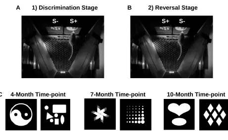
Figure 2. S+ and S- stimulus presentation and stimuli used in pairwise visual discrimination... 33
Figure 3. Illustration of the possible responses to the stimulus in 5-CSRTT... 36
Figure 4. 5-CSRTT experiment timeline... 36
Figure 5. Learning phase of the PVD Task... 46
Figure 6. Reversal learning phase of the PVD task at the 4-month time-point... 47
Figure 7. Reversal learning phase of the PVD task at the 7-month time-point... 48
Figure 8. Reversal learning phase of the PVD task at the 10-month time-point... 49
Figure 9. Representative Pdebrd1 genotyping results... 50
Figure 10. Reversal learning phase of the PVD task at the 10-month time-point separated by Pdebrd1 genotype... 52
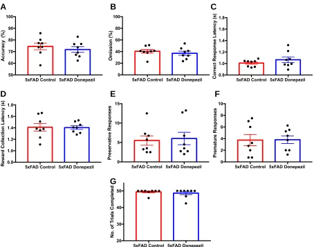
Figure 11. Number of trials completed by 5xFAD mice in the 5-CSRTT... 56
Figure 12. Performance and response measures of 5xFAD mice during the 5-CSRTT probe trial... 58
Figure 14. The vigilance of 5xFAD mice during the 0.6s stimulus length of the
5-CSRTT... 60
Figure 15. Performance and response measures of 5xFAD mice in the 0.6s
stimulus length of the 5-CSRTT... 61
Figure 16. Spontaneous locomotor activity of 10-month old 5xFAD mice... 63
Figure 17. Spontaneous locomotor activity of 5xFAD mice treated with
donepezil... 65
Figure 18. Spontaneous locomotor activity of 14-month-old 5xFAD mice... 67
Figure 19. Spontaneous locomotor activity of 15-month-old 5xFAD mice... 69
Figure 20. Step pattern and limb support measures in 10-month old 5xFAD
mice... 73
Figure 21. Base of support and weights in 10-month-old 5xFAD mice... 74
Figure 22. Step pattern and limb support measures in 14-month-old 5xFAD
mice... 78
Figure 23. Base of support and weights in 14-month-old 5xFAD mice... 75
Figure 24. Step pattern and limb support measures in 14-month 5xFAD mice
following donepezil treatment... 82
Figure 25. Base of support and weights in 14-month 5xFAD mice following
Figure 26. Step pattern and limb support measures in 15-month old male 5xFAD
mice... 87
Figure 27. Base of support and weights in 15-month 5xFAD mice... 88
Figure 28. Forelimb grip force in 14-month-old 5xFAD mice... 88
Figure 29. The effect of mild caloric restriction on 6E10 amyloid pathology in male
5xFAD mice at 6 months of age... 90
Figure 27. The effect of mild caloric restriction on 6E10 amyloid pathology in male
5xFAD mice at 6 months of age... 91
Figure 28. The effect of mild caloric restriction on Thioflavin-S amyloid pathology in
List of Appendices
Appendix 1. SOP Food restriction for adult mice (12 weeks or older)... 122
Appendix 2. SOP 2-Choice Pairwise Visual Discrimination Task Standard Operating
Procedure... 126
Appendix 3. SOP 5-Choice Serial Reaction Time Task Standard Operating
List of Abbreviations
Abbreviation Full Name
A Amyloid Beta
AD Alzheimer's Disease
ANOVA Analysis of Variance
ApoE Apolipoprotein E
APP Amyloid Precursor Protein
BACE -APP cleaving enzyme
CANTAB Cambridge Neurospsychological Test
Automated Battery
ChAT Choline Acetyltransferase transporter
CTF C-Terminal Fragment
DMSO Dimethyl Sulfoxide
NMDA N-methyl-D-Aspartate
PDEBrd1 Phosphodiesterase Subunit Beta -
Retinal Degeneration 1
PVD Pairwise Visual Discrimination
ROUT Regression Outlier Removal
VAChT Vesicular Acetylcholine Transporter
WT Wild-Type
5xFAD 5x Familial Alzheimer’s Disease
1 Introduction
1.1 Alzheimer’s Disease
Alzheimer’s disease (AD) is a disabling and fatal chronic disorder characterised by
progressive cognitive impairment – often beginning with memory loss and
neuropsychiatric symptoms such as depression, apathy and aggression (Li et al.,
2014b). The disease may progress gradually for several years before other cognitive
domains, such as language, executive function, visuospatial function, and attention are
affected (Perry and Hodges, 1999). Although the rate of decline can be extremely
variable, AD is usually fatal within 7-10 years of diagnosis (Dudgeon, 2010). The most
common cause of death among AD patients is pneumonia, which is hastened by the
marked inability of the patient to cough and move about normally (Dudgeon, 2010).
AD currently affects over 35.6 million individuals worldwide, accounting for
approximately half of the new cases of dementia diagnosed annually (Brookmeyer et
al., 2007; Prince et al., 2013). This number is predicted to double within the next 20
years (Prince et al., 2013). The majority of new cases of AD are sporadic, and tend to
occur in older age groups (Dudgeon, 2010). However, 5-7% of cases are early onset,
and can result from mutations in one of the many genes involved in amyloid processing
(Dudgeon, 2010). These cases of AD are referred to as familial Alzheimer’s disease.
The clinical diagnosis of AD is currently based on clinical history, neurological
examination and neurological tests. A criterion often used to diagnose AD (as stated by
requires these patients to demonstrate a loss of two or more of the following: memory,
language, calculation, orientation or judgment in the absence of other probable
diseases (Kawas, 2003). A more recent criterion suggested by McKhann et al. requires
patients that meet the criteria for dementia to also demonstrate a deterioration in
cognition along with one of the following: amnesia, impairments in language,
visuospatial function or executive function (McKhann et al., 2011). Again, this probable
diagnosis must be made in the absence of evidence pointing to other disease
(McKhann et al., 2011). These cognitive domains can be tested using the Mini-Mental
State Examination (MMSE), the Abbreviated Mental Test Score (AMTS) and objective
computerized Cambridge Neuropsychological Test Automated Battery (CANTAB).
Although expert clinicians correctly diagnose AD 70-90% of the time (Beach et al.,
2012; Kaye, 1998), a definitive diagnosis of AD requires a post-mortem confirmation,
with the presence of two histopathological features: amyloid plaques and neurofibrillary
tangles (Braak and Braak, 1991).
1.1.1 The neuropathology of AD
Alzheimer’s disease is characterized by the presence of amyloid plaques and
neurofibrillary tangles in the brain (Braak and Braak, 1991). Although these pathological
features are often seen in cognitively normal age-matched controls, the density and
distribution of these features differ (Bekris et al., 2010).
Amyloid plaques are composed of small A peptides that are processed from the
amyloid precursor protein (APP). APP is a type-I transmembrane protein that is
of physiological functions, including neurite outgrowth, synaptogenesis, protein
trafficking along the axon, transmembrane signal transduction, cell adhesion and
calcium metabolism (Zhang et al., 2011; Zheng and Koo, 2006). However, APP can
also have neurotoxic properties and other aversive effects (Zhang et al., 2011).
APP is processed by a series of sequential proteases into various fragments. A is
produced following the cleavage of APP by -secretase (also known as -APP cleaving
enzyme or BACE), forming soluble APP and a C-terminal fragment (CTF). Soluble
APP is thought to be involved in neuronal pruning and axonal cell death (Nikolaev et
al., 2009). Cleavage of APP by -secretase prevents A generation, as its cleavage site
is within the A domain – between lys16 and leu17 (Zhang et al., 2011). This forms
soluble APP and an C-terminal fragment (CTF). Soluble APP plays an important
role in neuronal plasticity/survival and in central nervous system development, where it
regulates neural stem cell proliferation (Zhang et al., 2011). The CTF and CTF are
further processed by -secretase to p83 and A40/42, respectively. P83 has no known
function and is rapidly degraded (Zhang et al., 2011). The CTF can be cleaved into
A40 or A42, where the number indicates the number of amino acids in the fragment.
At low levels, A has positive, modulatory roles on neurotransmission and memory,
while excessive levels of A leads to synaptic dysfunction and synapse loss (Zhang et
al., 2011). A42 is more hydrophobic and thus, more prone to fibril formation and
aggregation than A40 (Zhang et al., 2011). Under normal conditions, only 10% of the
A produced is A42 (Burdick et al., 1992). Alterations in this ratio has been suggested
Neurofibrillary tangles are composed of hyperphosphorylated filaments of the
microtubule-associated phosphoprotein tau. Tau projects from the surface of
microtubules, allowing them to interact with other cytoskeletal elements, cytoplasmic
organelles and proteins (Buée et al., 2000). Tau is regulated via phosphorylation, which
modulates the affinity between tau and the microtubules to allow for microtubule
assembly, affecting axonal morphology, growth and polarity (Buée et al., 2000). In AD,
the hyperphosphorylation of tau leads to the formation of fibrils which can then
aggregate within cells to form insoluble paired helical filaments, leading to
neurodegeneration (Buée et al., 2000).
1.1.2 Genetic risk factors and Familial Alzheimer’s Disease
A fraction of familial AD cases can be traced to mutations in proteins that affect amyloid
processing. Mutations in the gene encoding APP on chromosome 21 leads to the
abnormal formation of APP, affecting how it is processed (Goate et al., 1991; Mullan,
1992). To date, over 32 APP missense mutations have been identified, and the majority
of these mutations are located at the secretase cleavage sites (Goate et al., 1991;
Mullan, 1992). Examples include the Swedish (K670N, and M671L) and London (V717I)
mutations, which lead to an increase in the production of A and the development of AD
(Goate et al., 1991; Mullan, 1992). These mutations have been introduced in mice and
used to generate animal models of AD.
Mutations on chromosome 14 and chromosome 1 result in the partial loss of function of
presenilin 1 and 2, respectively (Bekris et al., 2010). Presenilins are major components
APP (De Strooper et al., 1998). Thus, mutations in these proteins affect APP
processing. Defects in presenilin 1 lead to the most severe forms of AD – with complete
penetrance, leading to early onset AD (Bekris et al., 2010). Meanwhile, mutations in
presenilin 2 are rarer, and are of lower penetrance than mutations in presenilin 1 (Bekris
et al., 2010).
The human apolipoprotein E (ApoE) gene on chromosome 19 exists as one of three
isoforms: 2, 3 and 4 (Wu and Zhao, 2016). In the brain, lipidated ApoE is responsible
for binding and removing aggregated A in an isoform dependent manner (Wu and
Zhao, 2016). The ApoE 2 polymorphism is very rare, and is considered to protect
against AD, as it enhances the ability of ApoE to clear A through a variety of
mechanisms (Conejero-Goldberg et al., 2014; Wu and Zhao, 2016). ApoE 3 has
recently been found to have neuroprotective effects as well (de-Almada et al., 2011).
However, the ApoE 4 polymorphism has been associated with both familial and
sporadic cases of late-onset AD (Bekris et al., 2010). ApoE 4 lipoproteins bind A with
a lower affinity than the other polymorphisms, thus possibly impairing A clearance (Liu
et al., 2013a).
1.1.3 The amyloid cascade hypothesis of AD
The amyloid cascade hypothesis of AD suggests that the accumulation and deposition
of A in the brain is a crucial step that ultimately leads to the development of AD
(Karran et al., 2011). This had been observed many years prior to other AD-related
the production and clearance of A peptides. This hypothesis is supported by genetic
mutations in the presenilins and APP leading to early-onset or AD in humans (Bekris et
al., 2010). Meanwhile, the ApoE 4 polymorphism, which affects the clearance of A,
can predispose an individual to AD (Liu et al., 2013a). In addition, soluble oligomers of
A42 taken from AD patients can cause neurodegeneration, inhibit long-term
potentiation and enhance long-term synaptic depression in the hippocampus of healthy
rats (Selkoe and Hardy, 2016). These oligomers are also capable of inducing tau
hyperphosphorylation and enhancing the toxicity of tau (Selkoe and Hardy, 2016).
However, treatments aimed at A in humans have failed – and in some cases,
treatment accelerated deterioration in cognition and reduced quality of life compared to
placebo controls (Karran et al., 2011). Furthermore, the amyloid cascade hypothesis
fails to consider the effect of tau on the development of the disease. Tau pathology itself
can cause neuronal loss as well (Karran et al., 2011). The temporal and mechanistic
relationships between A and tau pathology remain to be resolved (Karran et al., 2011).
1.1.4 The cholinergic hypothesis of AD
The cholinergic hypothesis of AD suggests that the disease is a result of the
degeneration of cholinergic neurons in the basal forebrain and the associated loss of
cholinergic transmission (Bartus et al., 1982). This degeneration is accompanied by a
decrease in choline acetyltransferase (ChAT; responsible for synthesizing acetylcholine)
and a reduction of acetylcholine release and reuptake in cortex and hippocampus
(Francis et al., 1999). Similar observations have also been made in mouse models of
cholinergic transmission has been implicated in a variety of cognitive functions, and that
anti-cholinergic drugs have amnestic effects and can reproduce memory deficits in
non-demented elderly patients add merit to this theory (Contestabile, 2011; Francis et al.,
1999). In addition, cholinergic mimetics proved to be particularly useful in treating the
symptoms and cognitive decline associated with AD in humans and mouse models
(Contestabile, 2011; Dong et al., 2009; Romberg et al., 2011). Thus, a majority of the
drugs used to treat patients with AD target the cholinergic system.
1.1.5 Treating AD
There is currently no cure for AD, and all pharmacological interventions are palliative.
Two classes of drugs are currently available for the treatment of patients with AD in
Canada: three cholinesterase inhibitors (donepezil, rivastigmine and galantamine) and
an NMDA (N-methyl-D-aspartate) receptor blocker (memantine). Cholinesterase
inhibitors block acetylcholinesterase, preventing the degradation of acetylcholine in the
synaptic cleft and enhancing cholinergic transmission (Birks, 2006). These drugs are
usually used to treat patients with mild to moderate AD, while memantine is often
prescribed to patients with severe AD or to patients that cannot tolerate the side effects
of cholinesterase inhibitors (Bishara et al., 2015). Memantine blocks excess NMDA
receptor activity – which is thought to result in neuronal injury and death – without
disturbing its normal neuroprotective attributes (Bishara et al., 2015). The
pharmacological treatments for AD are not always well tolerated, as they commonly
lead to side effects including: dizziness, nausea, vomiting, diarrhea and anorexia
(Bishara et al., 2015). These drugs also interact with a variety of other pharmaceuticals,
patients with other comorbidities even more difficult (Bishara et al., 2015). Thus, there is
a great need for the development of new pharmacological agents to treat patients with
AD. If interventions could delay the onset and progression of the disease for only 1
year, there would be nearly 9.2 million fewer cases of the disease in 2050 (Brookmeyer
et al., 2007)
1.1.5.1 Donepezil
Donepezil (Aricept) is a selective and reversible acetylcholinesterase inhibitor that binds
to the active site of acetylcholinesterase with high affinity – thus avoiding unintended
interactions with butyrylcholinesterase and other receptors (Kryger et al., 1999).
Donepezil preserves the levels of acetylcholine at the synaptic cleft, which has been
shown to protect against ischemic damage, glutamate excitotoxicity and A toxicity,
while also attenuating hippocampal and cortical neurodegeneration (Akasofu et al.,
2008; Cutuli et al., 2013).
Donepezil is usually prescribed to patients with mild to moderate AD (Bishara et al.,
2015). However, recent studies suggest that donepezil can also be used for moderate
to severe cases of AD in combination with memantine (Bishara et al., 2015; Howard et
al., 2012). Donepezil must be administered to a patient daily, and administration should
not be interrupted, as its effects are quickly lost and may not be fully regained when
1.1.6 Visuospatial function and attention in AD
Visuospatial function and attention are among the first cognitive domains to be affected
early in AD, and continue to decline as the disease progresses (Albert, 1996; Pal et al.,
2016; Perry and Hodges, 1999; Quental et al., 2013). Although there are clear
neuropathological correlations, the direct neurobiological correlates of these
impairments have yet to be determined (Li et al., 2014a; Perry and Hodges, 1999).
Visuospatial function involves the identification of a stimulus and its location, and can be
assessed in humans using the Visual Object and Space Perception (VOSP) battery,
which effectively evaluates visuospatial function while minimizing interference from
other cognitive domains (Quental et al., 2013). Studies have shown that patients with
AD score poorly on the VOSP battery when compared to controls, and that their scores
continue to drop as the disease progresses (Pal et al., 2016; Quental et al., 2013).
Visuospatial ability has also been found to be an important contributor to functional
status in AD patients (Fukui and Lee, 2009), and requires connectivity between the
prefrontal cortex and the striatum to remain intact (Brigman et al., 2013). Visual
discrimination has been shown to depend on the perirhinal cortex in rodent models –
the volume of which is significantly reduced in AD as well (Bussey et al., 2003;
Juottonen et al., 1998).
Attention has been suggested to be the first non-memory domain to be affected after
the initial amnesic stage of AD (Perry and Hodges, 1999). Cholinergic activity plays an
important role in attention (Sarter et al., 2005). The cholinergic neurons that project to
demanding tasks (Arnold et al., 2002), while lesions to this system impair attentional
function (Muir et al., 1996). These neurons also degenerate in patients with AD,
resulting in the loss of cholinergic transmission (Bartus et al., 1982).
Attention consists of three subtypes: selective, sustained and divided, and these are
differentially affected in AD. Selective attention refers to the ability to filter out random
stimuli, while sustained attention or vigilance refers to the ability to focus attention on a
task for unbroken periods of time (Perry and Hodges, 1999). Divided attention is where
an individual has to either focus their attention on a stimulus or multiple stimuli, or on
two separate tasks. Divided attention and some aspects of selective attention are
particularly vulnerable, while sustained attention usually remains intact for the early
stages of the disease (Perry and Hodges, 1999). The various domains of attention can
be tested by various tasks, including tasks in the CANTAB (Perry and Hodges, 1999).
1.1.7 Gait impairments in patients with AD
Patients with AD also experience gait disturbances, with cautious gait dominating in
patients with mild AD, and frontal gait disorders dominating in those with severe AD
(O’keeffe et al., 1996; Sala et al., 2004). These extensive gait impairments contribute to
the increase in fall risk and immobility in AD patients compared to age matched controls
(Amboni et al., 2013; Muir et al., 2012; Nutt, 2013). Immobility is associated with
changes in social behaviour, personality and deteriorations in mental health, along with
Cautious gait is a slow gait, with shortened steps and en bloc turns – which are defined
as turns where the individual keeps their head and trunk rigid, taking multiple steps
rather than twisting the body and pivoting the toes to turn (Nutt, 2013). These
individuals may also widen their base to increase stability (Nutt, 2013). Cautious gait is
an appropriate adaptation to real or perceived imbalance and is also commonly
observed in patients with Parkinson’s Disease (Nutt, 2013; O’keeffe et al., 1996).
Frontal gait disorders, or gait apraxia includes disturbances in trunk movements,
standing and walking that are not caused by any orthopedic abnormalities, muscle
wasting, arteriosclerosis in the lower limbs, neuromuscular deficits, ataxia, dystonias,
dyskinesias, psychiatric disease, side effects of drugs or cautious gait (Sala et al.,
2004). Several forms of gait apraxia have been reported in patients with AD, including
small steps, freezing of gait and disequilibrium (Nutt, 2013).
1.1.7.1 The association between gait and attention
Gait is increasingly considered to be more than just an automated motor activity.
Rather, gait requires a combination of executive function, attention and a judgement of
internal and external cues, which requires intact visuospatial function as well (Amboni et
al., 2013). There is a direct relationship between cognitive impairment and gait
abnormalities. When individuals are subject to a dual task paradigm, gait is
detrimentally affected. In this paradigm, individuals are asked to walk while performing a
concurrent cognitive or motor task, thus increasing competition for attentional resources
and forcing the brain to prioritize between the tasks (Amboni et al., 2013). In healthy
effects being observed in older adults (Lindenberger et al., 2000). However, in patients
with AD, the effect of dual task paradigms are much more pronounced, and an increase
in the complexity of the second task and/or the severity of cognitive impairment further
worsens gait measures (Amboni et al., 2013; Muir et al., 2012).
1.1.8 Mouse models of AD
Our knowledge of the autosomal dominant mutations leading to early onset AD have
allowed for the development of many animal models of the disease. These models have
been invaluable tools for identifying and characterizing molecular, cellular and
pathological changes that lead to the onset of AD (Newman et al., 2007). There are
over 20 strains of AD mouse models with mutations in APP, and many others with
mutations in tau and presenilins, causing these mice to develop the characteristic
plaques and tangles of AD (Newman et al., 2007). In addition, many of these mice
recapitulate various cellular and behavioural aspects of the disease.
1.1.8.1 The 5xFAD mouse model of AD
The 5xFAD mouse model of AD, developed by Oakley et al. (2006), co-expresses five
familial AD mutations: the Swedish (K670N/M671L), Florida (I716V) and London (V717l)
mutations of the amyloid precursor protein, and two mutations in presenilin 1 (M146L
and L286V). These mice are B6/SJL F1 hybrids and the genes were introduced by
site-directed mutagenesis and then subcloned into the mouse neuron-specific Thy1 (or
cluster of differentiation 90 – CD90) transgene cassette (Oakley et al., 2006). These
A42 in the brain and develop amyloid plaques and gliosis as early as 2 months of age
(Oakley et al., 2006), while the majority of AD mouse models take at least 6-12 months
to develop amyloid plaques (Spires and Hyman, 2005). The levels of A42 continue to
increase linearly and plaques begin to appear in the deep layers of the cortex and the
subiculum, moving throughout most of the cortex and hippocampus as the mice age
(Oakley et al., 2006). Plaques also eventually appear in the thalamus, brainstem and
olfactory bulb, although they are fewer in number (Oakley et al., 2006). Interestingly, the
cerebellum is often spared. These amyloid plaques are surrounded by activated
astrocytes and microglia (indicative of neuroinflammation), another hallmark of the AD
brain (Akiyama et al., 2000; Oakley et al., 2006). Gliosis increases with age in these
mice and closely follows the distribution of amyloid deposits (Oakley et al., 2006). This
leads to neurodegeneration and neuronal loss in specific regions of the brain – such as
cortical layer 1 and 5 – as the mouse ages, correlating with the deposition of amyloid
plaques and intraneuronal A (Oakley et al., 2006). Although 5xFAD mice recapitulate
amyloid pathology in AD relatively quickly, they do not develop neurofibrillary tangles or
appear to display hyperphosphorylated tau epitopes like some other APP transgenic
mice (Maarouf et al., 2013; Oakley et al., 2006).
Like AD patients, 5xFAD mice also demonstrate a significant reduction of ChAT, a
marker of cholinergic neurons (Devi and Ohno, 2010; Francis et al., 1999). Moreover,
the transplantation of ChAT+ basal forebrain cholinergic neurons derived from human
embryonic stem cells to the basal forebrain of 4-month-old 5xFAD mice was capable of
improving learning and memory in the Morris Water Maze (Yue et al., 2015). Also, Devi
the Y-maze with the partial knockdown of BACE1 (-APP cleaving enzyme 1) at 6
months but not at 15-18 months of age (Devi and Ohno, 2010).
With age, 5xFAD mice develop deficits in spatial memory on the Y-maze at
approximately 4-5 months, disturbances in learning in the Morris Water Maze beginning
at 9 months and severe motor impairments in the rota-rod at approximately 12 months
of age (Macdonald et al., 2013; Oakley et al., 2006; Schneider et al., 2014). In addition,
5xFAD mice also demonstrate changes in brain glucose metabolism, anxiety and
electroencephalogram (EEG) disturbances at 6 months of age (Macdonald et al., 2013;
Schneider et al., 2014). EEG recordings revealed that 5xFAD mice spend less time in
rapid-eye-movement sleep in relation to the total amount of sleep when compared to
wild-type controls (Schneider et al., 2014). In summary, 5xFAD mice develop amyloid
pathology, functional disturbances and behavioural deficits that make them a good
model for studying many aspects of AD.
1.1.8.1.1 Retinal degeneration in the 5xFAD mouse model
5xFAD mice have a mixed background: C57Bl6 and Swiss Jim Lambert (SJL). SJL mice
are homozygous for the recessive Pdebrd1 allele, which codes for the β-subunit of cGMP
phosphodiesterase on mouse chromosome 5 (Clapcote et al., 2005; Giménez and
Montoliu, 2001). Thus, F1 5xFAD mice should be heterozygous for the mutation. The
mutated allele is a nonsense mutation that decreases the transcription of the
phosphodiesterase, leading to retinal degeneration and blindness by wean age at
approximately 3 weeks and rendering mice homozygous for the Pdebrd1 allele
mutation is seen in FVB/NJ (Friend Virus B/ National Health Institute Jackson) mice as
well (Giménez and Montoliu, 2001).
1.2 The Bussey-Saksida Touchscreen System
High-level cognitive and executive functions in mouse models can be effectively
assessed using the Bussey-Saksida touchscreen system. This touchscreen system can
be used to study both impairments and enhancements in visual discrimination,
extinction, attention, impulsivity, compulsivity and a variety of other cognitive domains
(Bussey et al., 2012). The tasks can be identical to those used in humans – like those in
the CANTAB (Brigman et al., 2005; Bussey et al., 2001; Downes et al., 1989; Robbins
et al., 1994; Sahakian and Coull, 1993). This face validity provides the best chance of
successful cognitive translation across species. These tasks can be used to evaluate
various cognitive perturbations in mouse and rat models of disease, including those of
Alzheimer’s Disease, Parkinson’s disease, Schizophrenia, Attention Deficit Hyperactive
Disorder (ADHD), Obsessive Compulsive Disorder and addiction (Bussey et al., 2012).
This touchscreen system is automated, reducing variability and scope for error (Bussey
et al., 2001). The entire task – from stimulus presentation to reward provision – is
completely controlled by a computer program, minimizing experimenter interference,
inconsistencies and any other confounds. To switch between tasks, the experimenter
only needs to switch to the correct mask (which has task-specific windows allowing for
the mouse to see and respond to the task while reducing unintended responses) and
schedule. Automation also allows for the “in parallel” testing of many animals
Several parameters, such as accuracy (responding to the correct location), omissions
(failing to respond), perseverative responses (responding repeatedly to a previously
correct location; a measure of compulsivity), premature responses (responding before
the stimulus appears; also a measure of impulsivity), time to make a correct response
(correct response latency) and time to collect reward (reward collection latency) are
automatically recorded by the equipment during each task. The results can then be
exported to common data formats allowing for the creation of expandable databases to
facilitate increased data access and reproducibility.
Lastly, the tasks employed by these touchscreens are non-aversive and low stress,
providing a food reward for a correct response rather than punishing the animal for an
incorrect response (Bussey et al., 2012). The touchscreen chambers are also isolated
from the experimenter and its surroundings, minimizing distractions and other
stress-inducing stimuli as well.
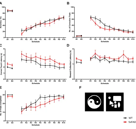
1.2.1 The Pairwise Visual Discrimination Task
Visual discrimination and cognitive flexibility can be assessed using the pairwise visual
discrimination (PVD) touchscreen task (Brigman et al., 2008; Bussey et al., 2008).
Impairments in both visual discrimination and cognitive flexibility have been reported in
the early stages of AD and have been suggested to serve as specific and accurate
prognostic markers of the disease (Albert, 1996; Pal et al., 2016; Quental et al., 2013).
In the PVD task, the mouse is presented with two stimuli and has to learn that one of
the stimuli (S+) leads to a reward while the other (S-) does not. The spatial location of
stimulus-reward association is reversed. The mouse then has to inhibit the response
acquired and associate the previously unrewarded stimulus with the reward (reversal
learning) and learn a new rule, providing an assessment of cognitive flexibility.
Performance of this task has been shown to be dependent on the prefrontal cortex in
rats and mice (Brigman and Rothblat, 2008; Bussey et al., 1997; Chudasama and
Robbins, 2003). Inactivation of the perirhinal cortex with either muscimol (a
gamma-Aminobutyric acid-A receptor blocker), AP5 ((2R)-amino-5-phosphonopentanoate, a
NMDA receptor antagonist) or scopolamine (a muscarinic receptor antagonist) impairs
performance in this task as well (Winters et al., 2010). Deficits in visual discrimination
learning and reversal in the PVD task have also been observed in NMDA receptor 2A
knockout mice (Brigman et al., 2008). AD patients also demonstrate a selective and
differential reduction of NMDA receptor levels in the brain (including receptor 2A), and
the levels of these receptors correlate with cognitive performance (Maragos et al., 1987;
Sze et al., 2001). In addition, the reduction of forebrain cholinergic tone in VAChT
Six3-Cre-flox/flox (vesicular acetylcholine transporter (VAChT) deficient) mice impairs cognitive
flexibility in the PVD task (Kolisnyk et al., 2013a). The expression of VAChT is
significantly reduced in AD patients, correlating with the severity of dementia (Efange et
al., 1997; Gilmor et al., 1999). Visual discrimination acquisition is impaired in 11-month
old rTg4510 mice as well (Harper et al., 2013). These mice express a human tau
mutation (Harper et al., 2013).
The PVD task can be administered multiple times, each with a different set of S+ and S-
stimuli. This allows for the longitudinal testing of animal models, allowing determination
1.2.2 The 5-Choice Serial Reaction Time Task
The 5-choice serial reaction time task (5-CSRTT), is mainly used to study attention,
which is impaired in AD patients (Bussey et al., 2012; Carli et al., 1983; Leonard, 1959;
Perry and Hodges, 1999). The task also gives information about impulsivity (premature
responses) and compulsivity (preservative responses). In the 5-CSRTT, mice are
required to scan a horizontal array of five screens for the presence of a brief,
randomized light stimulus and respond appropriately with a nose poke. The shorter the
duration of the stimulus, the higher the attentional demand required to successfully
perform the task (Bari et al., 2008).
Performance in the 5-CSRTT is impaired in mouse models of cholinergic dysfunction,
including M1 muscarinic receptor deficient mice (Bartko, 2011), ChAT-ChR2-EYFP mice
who express channel rhodopsin 2 (ChR2) under control of the choline acetyltransferase
(ChAT) (Kolisnyk et al., 2013b) and VAChTSix3-Cre-flox/flox mice (Kolisnyk et al., 2013a).
The accuracy of mice in the 5-CSRTT task is also significantly reduced by damage to
the prefrontal cortex and/or striatum (Muir et al., 2012; Passetti et al., 2003; Rogers et
al., 2001). However, performance in the task can be improved with drug treatment. For
example, the attentional impairments observed in triple transgenic (3xTG) mouse model
of AD, which is likely a result of pathological changes in the prefrontal cortex, was
reduced following treatment with the cholinesterase inhibitor donepezil (Romberg et al.,
2011). Galantamine has also been shown to improve performance of WT mice in this
task (Kolisnyk et al., 2013a). Other mouse models of AD demonstrate deficits in this
months of age (Romberg et al., 2013). These mice have the Swedish K670N/M671L
and the Indiana V717F APP mutations, leading to plaque formation by 3 months of age
(Chishti et al., 2001). Deficits in the 5-CSRT task have also been observed in male
5xFAD mice at 10 months of age (Masood, 2015). Masood (2015) observed a
significant decrease in percentage accuracy and the number of preservative responses,
along with delays in reward collection and task response.
1.2.3
Using touchscreen tasks to identify cognitive deficits in
mouse models of AD
Given the high prevalence and poor prognosis of AD, the development of novel
treatments for patients with AD is imperative. The drugs currently given to these
patients do not cure or prevent the progression of the disease, interact with drugs
commonly used to treat other comorbidities and are often extremely difficult to tolerate
(Bishara et al., 2015). Up to 85% of patients experience the adverse side effects of
these treatments and some even opt to discontinue treatment for this reason (Burns et
al., 2007). Unfortunately, the drug development process is tedious and costly; clinical
trials take many years and require several million dollars to complete. Between 2002
and 2012, 24 phase 1, 206 phase 2 trials and 83 phase 3 clinical trials were attempted,
with an overall success rate of 0.4% (Cummings et al., 2014). Most of these registered
trials aimed to improve cognition in AD patients, but failed to do so (Cummings et al.,
2014). Thus, the ability to predict whether a novel treatment can improve cognitive
function in humans during pre-clinical development would be greatly beneficial. This
cognitive function. Therefore, touchscreens have the potential to become a powerful
new platform for the pre-clinical evaluation of new pharmacological treatments directed
at cognitive function in AD patients.
Attention, visual discrimination and cognitive flexibility are commonly affected in patients
with AD (Albert, 1996; Pal et al., 2016; Perry and Hodges, 1999; Quental et al., 2013),
and all these parameters can be assessed in mice using touchscreen tasks.
Impairments in visual discrimination and attention have been observed in various
mouse models of cholinergic and glutamatergic dysfunction (Bartko, 2011; Brigman et
al., 2008; Kolisnyk et al., 2013a; Romberg et al., 2013; Winters et al., 2010), and a few
mouse models of AD (Harper et al., 2013; Romberg et al., 2011, 2013). However, little
remains known about the onset and presence of deficits in executive function on the
different mouse models of AD.
Romberg and colleagues were able to rescue attentional deficits in 3xTG mice following
treatment with donepezil (Romberg et al., 2011). Since our lab has also observed
attentional deficits in male and female 5xFAD mice at 10 months of age (Masood,
2015), the question remains whether donepezil can rescue the attentional impairments
1.3 Rationale and Hypothesis
We hypothesize that male 5xFAD mice develop reproducible age-dependent
deficits in the PVD and that deficits in the 5-CSRTT that can be ameliorated by
treatment with donepezil.
The overall objective of this thesis is to perform a longitudinal evaluation of visual
discrimination and cognitive flexibility in male 5xFAD mice, as the presence of cognitive
dysfunction is an important proof of the face validity of the 5xFAD mouse model. We
also want to determine if specific cognitive deficits can be rescued by donepezil as a
proof of principle. To address these objectives, the specific aims of this thesis are:
1) To perform a longitudinal evaluation of visual discrimination and reversal learning
in male 5xFAD mice at 4, 7, and 10 months of age.
2) To assess the effects of donepezil on cognition in male 5xFAD mice at 10 and 13
months of age using the 5-CSRTT.
3) To genotype the 5xFAD mice that have been subject to touchscreen evaluation
for the recessive PDEBrd1 allele to ensure that task performance is not affected.
4) To determine whether the mild food-restriction used to motivate mice to perform
touchscreen tasks affected amyloid pathology in male and female 5xFAD mice.
2 Materials and Methods
2.1 5x Familial Alzheimer’s Disease Mouse Model
The 5xFAD mice (B6SJL-Tg(APPSwFlLon,PSEN1*M146L*L286V)6799Vas/Mmjax, Jax
stock #006554) and age-matched wild-type controls (B6SJLF1/J, Jax stock #100012)
used in the PVD experiments were purchased from the Jackson Laboratory (Bar
Harbor, Maine) and delivered to the university. The 5xFAD mice used in the 5-CSRTT
experiments were bred at Western University. All mice used for the experiments were
tattooed in their tails at least a week prior to testing for identification purposes. Male
5xFAD mice and their age-matched wild-type controls (wild-type littermates were only
used for the 5-CSRTT) were used for all behavioral tasks, whereas male and female
5xFAD mice and their age-matched wild-type controls were used for pathological
analyses. All procedures were performed in accordance with the Canadian Council of
Animal Care guidelines at the University of Western Ontario with an approved animal
protocol (2008-127).
2.2 Housing and Diet
Mice that underwent behavioural tasks were singly housed without environmental
enrichment in a temperature and pressure controlled room with a 12-hour light/dark
cycle. Lights would turn on at 7:00am and shut off at 7:00pm daily. All behavioural tests
were conducted during the light phase of this cycle. Cages were changed biweekly.
percent of their original baseline adult weight (according to the Adult Mouse Food
Restriction Standard Operating Procedure – Appendix 1) to ensure that they would be
motivated to complete the task. The mice were put on food restriction prior to the start of
behavioural testing and the body weights were gradually lowered to 85% of their original
weight. All mice were weighed daily. Water was provided ad libitum.
For the pathology experiments, male and female 5xFAD mice were either
food-restricted to 85% percent of their original baseline adult weight (according to the Adult
Mouse Food Restriction Standard Operating Procedure – Appendix 1) or provided food
ad libitum for 2-3 months – until they were euthanized at 6 months of age. Water was
provided ad libitum in both cases. All mice were fed with Teklad Laboratory Animal
Chow (Envigo).
2.3 Genotyping Pdeb
rd1DNA was extracted from mouse ear tissue and amplified using the REDExtract-N-Amp
Tissue PCR Kit Protocol (Sigma-Aldrich, Oakville, Ontario). Polymerase chain reaction
(PCR) was done using the Bio-Rad T100 Thermal Cycler (Bio-Rad Laboratories,
Hercules, California) with a 500bp x 40 cycle schedule (94C x 3 minutes followed by 40
x [94C x 30 seconds] + 60C x 30 seconds + 72C x 30 seconds then 72C x 2
minutes). The tubes were held at 10C until use. The following reagents were used for
each sample: 5l of 2x premix, 0.5l of retinal degeneration (RD) 3 oligonucleotide
primer (concentration: 0.5 M; 28-mer, 5’-TGACAATTACTCCTTTTCCCTCAGTCTG-3’,
(concentration: 0.02 M; 28-mer, 5’-GTAAACAGCAAGAGGCTTTATTGGGAAC-3’,
accession number L02109, nucleotides 644 to 617) and 2.9l of RD6 oligonucleotide
primer (concentration: 14.5 M; 28-mer, 5’-TACCCACCCTTCCTAATTTTTCTCAGC-3’,
accession number L02110, nucleotides 2539 to 2512). RD3 and RD4 amplifies a 0.55kb
PCR product from the Pdebrd1 mutant allele, while RD3 and RD6 amplifies a 0.40kb
PCR product from the WT allele (Giménez and Montoliu, 2001). The PCR products are
then run on an agarose gel along with a 100bp ladder (Gene DireX, Frogga Bio,
Toronto, Ontario) and imaged with FluorChem Q (Alpha Innotec Corp., San Leandro,
California).
The positive control for Pdebrd1 was ear tissue from a Friend Virus B NIH Jackson
mouse (FVB/NJ; Jax stock #001800), an inbred strain of mouse known to be
homozygous for the Pdebrd1 mutation. This mouse was purchased from the Jackson
Laboratory (Bar Harbor, Maine). The control for the WT allele of Pdeb for this gel was
ear tissue obtained from a B6SJLF1/J mouse.
2.4 Administration of Donepezil
Donepezil hydrochloride (C6821-50mg) monohydrate was obtained from Sigma-Aldrich
(Oakville, Ontario) and rehydrated using saline and 10% dimethyl sulfoxide (DMSO) to a
dose of 2.5 mg/kg (for the osmotic pumps) or 1.0mg/kg for the intraperitoneal injections.
2.4.1 Subcutaneous Osmotic Pumps
The implantation of subcutaneous osmotic mini-pumps allows for the continuous,
weeks). Relative to bolus dosing (injection), the infusion of a modulating agent has the
potential to widen the therapeutic index of the agent, increase its efficacy and/or reduce
any side effects (Fara and Urquhart, 1984). This is especially true for substances with
short-half lives. The infusion of an agent also reduces the possibility of injuring or
subjecting laboratory animals to the additional stress of daily injections, possibly
confounding the results of subsequent behavioral analyses. Thus, osmotic mini-pumps
can be an effective alternative to daily injections when executing a moderately long-term
study.
Osmotic mini-pumps purchased from Alzet (Durect Cooperation, Cupertino, California)
are composed of 3 concentric layers: a semipermeable, rate controlling membrane, an
osmotic layer that contains a high concentration of sodium chloride and an impermeable
but flexible drug reservoir (http://www.alzet.com/products/guide_to_use/implantation_
and_explantation.html). Water enters the pump across the semipermeable membrane
due to the high concentration of sodium chloride in the osmotic layer. The entry of water
causes the osmotic chamber to expand, compressing the flexible reservoir and
delivering the drug solution through the flow moderator
(http://www.alzet.com/products/guide_to_use/implantation_and_explantation.html).
These pumps have been used in the evaluation of a variety of different modulating
agents in laboratory animals, including the effect donepezil in AD mouse models (Dam
et al., 2008; Spilman et al., 2014).
Twenty male 5xFAD mice were implanted with subcutaneous osmotic pumps (model
1004, Alzet, Durect Cooperation, Cupertino, California) designed to deliver the drug at a
pumps loaded with saline + 10% DMSO or donepezil. Donepezil was diluted and
prepared based on calculations done using the drug concentration calculator on the
Alzet website (http://www.alzet.com/products/guide_to_use/formulating.html). This was
based on the pump model, flow rate, weight of the mouse and the desired dose. The
pumps were loaded with a 1mL syringe (the appropriate needle was provided with the
pumps), capped with a pin, and then soaked in saline until they were implanted within
one hour. Mice were first anesthetized with 4% isoflurane at 1L/minute until
unconscious. Maintenance was done with 1.5% isoflurane. The dorsal side of the
mouse was sprayed with ethanol and then small incision was made on the right side
(near the shoulder of the mouse). The pump was inserted and the incision was sealed
with a wound clip. Mice were allowed to recover in their home cage under a heat lamp.
The mice were given two days to recover from pump insertion before beginning a
5-CSRTT probe trial. Any mice that pulled out their own pump were euthanized. One
mouse was dropped from the experiment for this reason. All surviving mice had their
pumps removed after 28 days.
2.4.2 Intraperitoneal Injection
At approximately 14 months of age, 5xFAD mice were randomly chosen to receive
either donepezil or saline + 10% DMSO for 5 consecutive days. After a two-day
washout, the cohort that received saline received donepezil and vice-versa. Donepezil
was administered via intraperitoneal injection at the lower right or left quadrant of the
abdomen to avoid damage to the urinary bladder and other abdominal organs. Mice
behavioural tests.
2.5 Touchscreens
The pairwise visual discrimination (PVD) and 5-choice serial reaction time tasks
(5-CSRTT) were conducted using the automated Bussey-Saksida Touchscreen System for
mice (Model 81426, Campden Instruments, Lafayette, Indiana), which consists of a
testing chamber housed within a ventilated sound and light-attenuating box. The
chamber has a house light, a tone generator, and stimuli are displayed on a LCD
monitor that is equipped with infrared sensors. The mouse views the stimuli through a
black mask that contains windows through which each stimulus can be seen. This also
prevents unintended responses by other body parts of the mouse. The mask used
depends on task being administered. The entire apparatus is controlled by the Abet II
Touch Software Version 2.20 (Lafayette Instrument Company, Lafayette, Indiana). The
schedules were designed and the data was collected using this software as well. Each
mouse was only run on one schedule at approximately the same time each day.
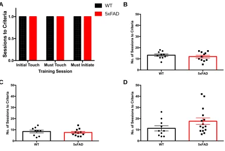
2.5.1 Touchscreen Pre-training
Prior to the commencement of either the PVD or the 5-CSRT task, mice are subject to a
basic training schedule (Figure 1A). Mice are habituated to the touchscreen chambers
for the first four days. Habituation 1 (day 1) lasts 10 minutes – all lights are tuned off
and no stimulus or reward is presented. Habituation 2a (day 2 and 3) lasts 20 minutes.
The food tray light will be on at the beginning of each trial and the mouse can complete
an unlimited number of trials within the 20-minute session. When the mouse enters the
Quebec) is dispensed. When the mouse leaves the food tray, the tray light turns off. The
tray light will turn back on after 10 seconds and another trial begins. Habituation 2b (day
4) is the exact same as habituation 2a, but it lasts 40 minutes. The mouse must be
removed from the cabinet once a habituation schedule is complete.
Following habituation, mice are subject to initial touch, where a white square (for
5-CSRTT) or image (for PVD; can be any image not designated for use in
discrimination/reversal) is displayed in one window pseudo-randomly (such that the
stimulus is not shown in the same position more than three times in a row) while the
other window(s) are left blank. The stimulus disappears after 30 seconds and reward is
delivered. If the mouse touches the screen where the white square is displayed while it
is still being displayed (within 30 seconds), a tone will be played and 3x the reward will
be delivered immediately (this is accompanied by a tone and the illumination of the tray
light). The mouse then has to collect the reward, turning the tray light off, and initiating
the inter-trial interval (20 seconds for PD, 5 seconds for 5-CSRTT) – after which another
trial begins. Collection of the reward starts the next inter-trial interval. The mouse must
complete 30 of these trials within 60 minutes and this usually only takes one session.
Otherwise, this schedule is repeated until criterion is achieved.
Initial touch is followed by must touch. In PVD, the stimulus is an image selected
pseudo-randomly (no image shown more than three times in a row) from a list of images
that do not include any of the images that will be used in the discrimination and reversal
trials. In 5-CSRTT, the stimulus remains as a white square. The position of the stimulus
is also chosen pseudo-randomly. The mouse must touch the stimulus to receive a
response if the mouse touches the blank screen(s). Collection of the reward starts the
next trial after the inter-trial period (20 seconds for PD, 5 seconds for 5-CSRTT). The
mouse must complete 30 of these trials within 60 minutes to reach criterion. If the
mouse does not reach criterion after 7 sessions for PVD or 5 sessions for the 5-CSRTT,
the mouse is retrained on initial touch until it reaches criterion. If the mouse does not
reach criterion after 7 sessions (PVD) or 5 sessions (5-CSRTT) of the second attempt of
must touch, it is removed from the study.
Must touch is followed by must initiate. For each trial, a free delivery of food is made,
and the tray light is illuminated. The mouse must nose poke and exit the reward tray
before a stimulus (same stimuli with the same criterion as those used in must touch;
task dependent) appears. The other window(s) remain blank and there is no response if
the mouse touches it. The mouse must touch the stimulus to receive a reward. This is
accompanied by a tone and the illumination of the tray light. After the mouse collects the
reward, the inter-trial interval (20 seconds for PD, 5 seconds for 5-CSRTT) begins. The
mouse must nose-poke and exit the reward tray before the next stimulus is displayed.
Thirty of these trials must be completed within 60 minutes to reach criterion. If the
mouse does not reach criterion after 5 sessions, the mouse is retrained on must touch
until it reaches criterion. For the second attempt, if the mouse does not reach the
criterion for must initiate within 5 sessions, it is removed from the study.
The last pre-training phase is punish incorrect, which is similar to must initiate – except
when the mouse touches a blank window, the house light will be turned on for 5
seconds and no reward will be given. After the light turns off, the mouse will not be able
complete ≥ 24/30 trials correctly within 60 minutes two days in a row. If the mouse
cannot reach criterion after 30 days, it is removed from the study.
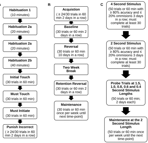
Figure 1. Summary of touchscreen pre-training, PVD and 5-CSRTT schedules. A) Flow chart of the touchscreen pre-training schedules, their duration and criterion. B) Flow chart of pairwise visual discrimination acquisition, baseline, reversal and retention reversal (7 and 10-month time-point only) which is repeated with a different pair of images at each time point. C) Flow chart of the 5-CSRTT training and probe trials at 1.5, 1.0, 0.8, 0.6 second stimulus lengths.
Habituation 1 (10 minutes) Habituation 2a (20 minutes) Habituation 2a (20 minutes) Habituation 2b (40 minutes) Initial Touch (30 trials in 60 min)
Must Touch (30 trials in 60 min)
Must Initiate (30 trials in 60 min)
Punish Incorrect ( ≥ 24/30 trials in 60 min 2 days in a row) A
Acquistion ( ≥ 24/30 trials in 60 min 2 days in a row)
Baseline (30 trials or 60 min 2
days in a row)
Reversal (30 trials or 60 min
10 days in a row)
Two Week Break
Retention Reversal (30 trials or 60 min 2
days in a row)
Maintenance (30 trials or 60 min once per week until
next time-point)
B 4 Second Stimulus
(50 trials or 60 min with ≥ 80% acuracy and ≤ 20% ommisions 3 days
in a row; must complete at least 30
trials)
2 Second Stimulus (50 trials or 60 min with
≥ 80% acuracy and ≤ 20% ommisions 3 days
in a row; must complete at least 30
trials)
Probe Trials at 1.5, 1.0, 0.8, 0.6 and 0.4 Second Stimulus
Lengths (50 trials or 60 min;
2 days each)
Maintenance at the 2 Second Stimulus
Length
(50 trials or 60 min once per week until the next