Sutapa Ghosh*, Shashi kumar Yadav
Nanomaterials Laboratory,Inorganic and Physical Chemistry Division, Indian Institute of Chemical Technology (IICT), Hyderabad, India.
*Corresponding author’s E-mail:[email protected]
Accepted on: 07-01-2014; Finalized on: 28-02-2014. ABSTRACT
New interpenetrating polymeric network (IPN) hydrogels based on Chitosan (C), poly (N-vinyl pyrrolidone) (PVP), Acrylic acid (AAc) and Acryl amide (A), cross linked with Glutaraldehyde (G) and N, N-methylene bis acryl amide (MBA), were prepared and investigated for better composition to achieve gastrointestinal controlled drug release by using a model drug Ampicillin trihydrate. Three different ratios of monomers 1:1, 1:3 and 3:1 (A:AAc) were used to optimize the better composition of hydrogels by using three different concentrations of Glutaraldehyde (0.5, 1.0 and 2.0 w/w) to control the overall porosity of the hydrogels. Fourier transform- infrared spectroscopy (FTIR), Thermo gravimetrical analysis (TGA) and Scanning electron microscopy (SEM) were performed for IPN hydrogels. Swelling studies and In-vitro release studies were carried out in pH 1.1 and pH 7.4 at 37°C to investigate possible site-specific drug delivery. It was concluded that F4 and F5 IPN hydrogels were the better compositions for controlled drug release in stomach due to their low swelling tendency and lower Ampicillin release at pH1.1 than at pH 7.4. Hence, this study may serve as a potential device for the delivery of drugs in which the primary target is the stomach or the upper small intestine.
Keywords: Acrylic acid, Acryl amide, Ampicillin trihydrate, Chitosan, Controlled drug release, FTIR swelling studies, Hydrogels.
INTRODUCTION
ecent advances in controlled release technology have made it possible to release drugs at a constant rate for long periods of time ranging from days to years.1 The failure of conventional treatments could be due to poor permeability of the antibiotics across the mucus layer or from sub therapeutic antibiotic concentrations at the site of infection after administration from conventional tablets or capsules. pH-sensitive swelling hydrogels seem to be useful for localized antibiotic delivery in the acidic environment of the gastric fluid. One of the most important advantages of these hydrogels is that formulations remain on the targeted site more time than conventional ones.2 Hydrogels are one of the promising and versatile materials with enormous possibilities and potential. In particular, controlled release systems are capable of delivering drug at constant rate over an extended period of time.3-5 Homopolymer alone cannot meet such divergent demand in terms of both properties and performance. Therefore, a composite or interpenetrating polymer network (IPN) of two or three different polymers would be a better approach.6
Today many natural polymers such as chitosan, chitin, gelatin, etc. and synthetic polymers such as poly (acryl amide), poly (acrylic acid), poly (N-isopropylacrylamide), and poly (Nvinylpyrrolidone) have been used extensively for preparing IPN. Chitosan (C) is a deacetylated derivative of chitin, a naturally occurring polymer. It is widely studied in pharmaceutical and biomedical fields because of biodegradability, biocompatibility and interesting structural properties (presence of amino and hydroxyl groups). Chitosan hydrogels can be used as
10
Poly (N-vinyl pyrrolidone) (PVP) is a polymeric compound that is widely used for textiles, cosmetics and toiletries, pharmaceuticals and medical applications.11,12 Poly acrylic acid (AAc) is known to be a good mucoadhesive and may increase the transit time of formulation. The polymers, composed of acrylic acid, have the ability to absorb a large amount of water and are used in many applications including ion exchange resins, personal hygiene products and membranes for hemodialysis, ultrafiltration, and controlled drug release. Polyacrylamide hydrogels and acryl amide-based matrices have been used in in-vitro and in-vivo systems and biocompatibility studies.
In the present work, an attempt has been made to evaluate the effect of composition of poly (acrylic acid & acryl amide) chitosan based hydrogel on pH sensitive drug release. Two different cross linkers Glutaraldehyde (G) and N, N’-methylene bis acrylamide (MBA) were used to control the network properties. IPN loaded with a model drug ampicillin trihydrate were used for drug release studies.
MATERIALS AND METHODS
Chemicals
Chitosan (C) (Mw = 600 000), poly (N-vinyl pyrrolidone) (PVP) (Mw = 40 000) was obtained from Calbiochem (Darmstadt, Germany), Acrylic acid (AAc), Acryl amide (A), N, N-Methelene bisacrylamide (MBA), Glutaraldehyde (G) and Ammonium persulfate (APS) as redox initiator were obtained from Merck (Darmstadt, Germany). Hydrochloric acid, potassium chloride, disodium hydrogen
The Effect of Composition of Poly (Acryl Amide & Acrylic Acid) – Chitosan Based
Hydrogel on pH Sensitive Drug Release: In-Vitro
R
purchased from Merck (Darmstadt, Germany).The model drug Ampicillin trihydrate was also purchased from Fluka (Steinheim, Switzerland). All chemicals were of analytical grade and were used as received. Double distilled water was used for all the experiments.
Preparation of IPN Hydrogels
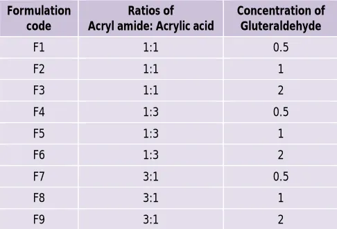
PVP and cross linkers were added to a solution of chitosan (C) (10mL, 1% w/v in 0.8% acetic acid) under continuous mixing then acrylic acid (AAc) or/and acryl amide (A) were added. Further ammonium persulfate solution (25 µL, 5%) was added to this mixture and continues the mixing for one hour. The mixture was placed in eppendroff of 1.5 ml volumes. A gel formed after 5 hr of reaction time at ambient temperature. After 24 hr, the IPN were obtained and washed with de-ionized water. These IPNs were dried in air and stored for further use. Chitosan, PVP and AAc or/and A was used in 1:1:25 weight ratios. The ratios (w/w) between Glutaraldehyde and chitosan in the IPN were 0.5, 1 and 2. The weight ratio of MBA to AAc or/and A was 1:50. Three different ratios of monomers 1:1, 1:3 and 3:1 [Acryl amide: Acrylic acid (A:AAc)] were used to optimize the better composition of hydrogels by using three different concentrations of Glutaraldehyde (0.5, 1.0 and 2.0 w/w) to control the overall porosity of the hydrogels shown in table 1 and remaining ingredients were kept constant in all the formulations.
Table 1: Composition of hydrogel formulations with different monomer and cross linker ratios
Formulation code
Ratios of Acryl amide: Acrylic acid
Concentration of Gluteraldehyde
F1 1:1 0.5
F2 1:1 1
F3 1:1 2
F4 1:3 0.5
F5 1:3 1
F6 1:3 2
F7 3:1 0.5
F8 3:1 1
F9 3:1 2
Characterization of Hydrogels
Fourier transforms infrared (FT-IR) analysis
FT-IR spectra of IPN hydrogels were recorded using a on a Nicolet Nexus 670 spectrometer equipped with a DTGS KBr detector in the range 4000 cm-1 to 500 cm-1 to determine their structure and intermolecular interactions. Thoroughly ground IPN samples were mixed with dried KBr, and discs were prepared by compression under vacuum. Spectra were recorded with a resolution of 1 cm−1.
Thermo gravimetric analysis (TGA)
Shimadzu 50 thermo gravimetric analyzer was used to determine the thermal stability of IPN. TGA was performed with 10 mg samples under nitrogen atmosphere with a nominal gas flow rate of 25 mL. Experiments were made at a heating rate of 10 °C min−1 until 600 °C.
Morphological examination
The morphology of porous structures was examined with a scanning electron microscope (SEM). IPN hydrogel composites were cut to expose their inner structure, coated with a thin layer of palladium gold alloy in Hummer I Sputter Coater Techniques, Alexandria, VA) and imaged in an SEM (JSM-840, JEOL USA, Peabody, MA). Evaluation of Hydrogels
Swelling Studies
To determine the pH-dependent swelling properties of air dried hydrogels, pre-weighed dry samples were immersed in buffers with pH 1.1 (0.1N HCl) and 7.4 (50mM sodium phosphate buffer) at 37oC. At predetermined time intervals, the swollen samples were removed from the solution, quickly wiped with filter paper to remove droplets on the surface and weighed.
In Vitro Release studies
Ampicillin trihydrate was selected as our model drug. Drug loading was carried out by adding 50mg Ampicillin
per gram of precursor IPN before
polymerization/crosslinking reactions. After polymerization, the hydrogels were optically transparent indicating complete solubility of the drug in the polymer matrix. The antibiotic containing IPN were then air dried. The in vitro release studies of the entrapped drug were carried out by placing the drug loaded IPN samples in simulated gastric fluid of pH 1.1 (0.1N HCl) at 37 °C. Sample of dissolution medium (5ml) were withdrawn at different time intervals (0.5, 1, 2, 4, 6hr), suitably diluted and assayed for Ampicillin by measuring at 256nm using a Shimadzu 160A UV-visible spectrophotometer (and a previously constructed calibration curve). The release media were replenished periodically with fresh solutions of pH 1.1 (5 mL). The percentage of released antibiotic was calculated from standard calibration curves and its corresponding release graph was constructed. The same release experiments were carried out in simulated intestinal fluid of pH 7.4 (50mM sodium phosphate buffer) at 37°C.
RESULTS AND DISCUSSION
Swelling studies
To investigate the time dependent swelling behavior of the IPN in buffer solutions with different pH values, dynamic swelling studies were performed.
Where M0 is the weight of dry gel at time 0 and Mt is the
weight of swollen gel at time t.
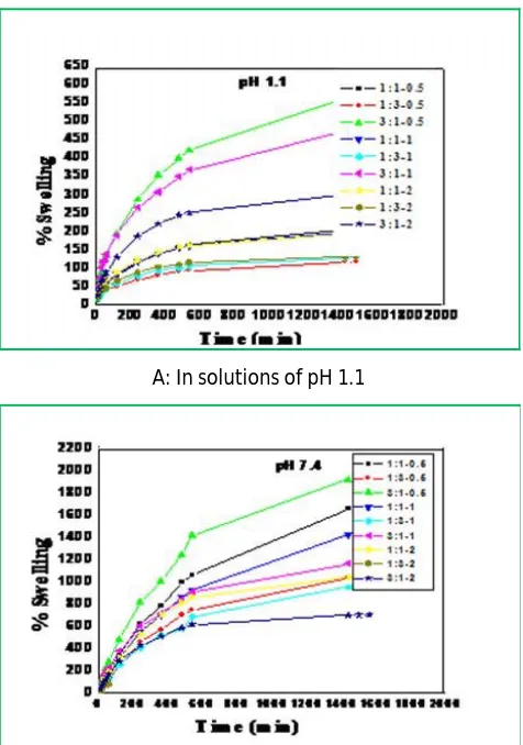
Swelling curves of IPN hydrogels with different crosslinker proportions in solutions of pH 1.1 and pH 7.4 at 37°C are shown in Figure 1 (A), (B) respectively. From these figures, it is observed that IPN formulations 1:3-0.5 (F4), 1:3-1(F5) are having lowest swelling degree compared to all other IPNs.
A: In solutions of pH 1.1
B: In solution of pH 7.4 at 37 °C.
Figure 1: Swelling curves of IPN hydrogels with different crosslinker proportions
Figure 2 shows that IPNs exhibit greater swelling at pH 7.4 when compared with that at pH 1.1. This can be explained by ionic interactions of the carboxyl groups of PAAc in intestinal pH conditions. Macromolecular chains in the IPN network swell extensively due to ionization of PAAc at pH 7.4 or higher values. The developed (ionized carboxylic acid groups) charges on the network lead to electrostatic repulsion among the charged –COO- groups, resulting in extensive swelling of the IPN. The pKa value for PAAc is 4.7513 above this value dissociation of the acid groups takes place in the structure, resulting in the expansion of the IPN network. This behavior can be exploited in swelling controlled releasing systems since C-P-AAc IPN
are polyelectrolyte capable of swelling at the pH of intestines.7,8
Figure 2: Comparison of swelling behavior of F4, F5 IPN hydrogels at pH 1.1 & pH 7.4
Hence, these IPNs can be used as the most suitable network structures for controlled drug delivery due to increased swelling with time. The percentage swelling S is given in Table 2 for IPN hydrogels.
Scanning Electron Microscopy study
Figure 3(a) shows the surface morphology of the swollen state IPN hydrogels with expansion of the polymer network. Figure 3b shows the surface morphology of the air-dried hydrogel membrane revealed a non porous nature.
Figure 3: SEM image of IPN hydrogel at (a) wet state (b) dry state
FT-IR analysis
between C=O groups of PVP and –OH groups of chitosan. The absorption bands at 1456 cm-1 can be assigned to the characteristic vibration of pyridine ring.14 From this spectral analysis, it can be concluded that a polymeric network is formed, due to crosslinks of chitosan with Glutaraldehyde and PAAc and PA with MBA, and also between chitosan molecules.
Figure 4: FTIR spectra of IPN hydrogel
Thermo gravimetric analysis
Thermograms of Fig. 5 shows dried samples of IPN hydrogels obtained under nitrogen atmosphere at a heating rate of 10°C min-1 from 0°C to 600°C. From this figure, we can say that initially up to 112°C can be attributed to the loss of bound water after that ranging from 197 to 368°C corresponds to chitosan partial degradation, finally ranging from 350 to 600°C represents the total degradation of chitosan. As can be seen, the thermal stability of IPN has increased dramatically with the increase in the Glutaraldehyde (G) concentration.
Figure 5: Effect of Glutaraldehyde (G) concentration on IPN hydrogel; A= G-0.5; B = G-1; C = G-2.
In-vitro Release studies
The release studies of IPN hydrogels were conducted by using different release media. The percent cumulative release (R) of ampicillin from IPN was calculated by:
R% = Mt/Mo*100
Where Mt and Mo are the amount of drug released at
time t and initial amount of drug present in the dry IPN, respectively. The values of R% are presented in Table 2.
Based on the swelling studies F4 and F5 IPNs were found to be as suitable compositions for drug loading and release studies because of their low swelling tendency compared with others. Figure 6 depicts the percent cumulative release of Ampicillin from IPN at pH 1.1 and pH 7.4, at 37°C and shows that drug release is pH dependent, the percentage cumulative release of Ampicillin from the IPN was higher in the basic medium than in the acidic medium. The diffusion of the drug molecules out of the IPN was enhanced because of swelling at higher pH. The extent of drug release increases as the hydrogel swelling increases due to the increase in pH, which leads to ionization of the carboxyl groups. It was observed that release results are very similar to swelling results. This parallel behavior is plausible since drug release from IPN into solutions is swelling-controlled. The release time for Ampicillin was found 6 hr at pH 1.1 and at pH 7.4.
Figure 6: In vitro Ampicillin release profiles from IPN F4 & F5 at pH 1.1 & 7.4.
Table 2: The values of Swelling equilibrium parameter (S%) & percent cumulative drug release (R%) of F4 & F5 IPNs at different pH values.
IPN Hydrogel S% R %
pH 1.1 pH 7.4 pH 1.1 pH 7.4
1:3-0.5 (F4) 118.32 1063.30 74.33 99.32
1:3-1 (F5) 131.93 966.42 81.96 97.23
CONCLUSION
network at pH 7.4. Consequently, F4 and F5 code IPN hydrogels developed in this study may serve as a potential device for the delivery of drugs in which the primary target is the stomach or the upper small intestine.
REFERENCES
1. Fischer W, Boertz A, Davis SS, Khosla R, Cawello W, Sandrock K, Cordes G, Investigation of the gastrointestinal transit and in vivo drug release of isosorbide-5-nitrate pellets, Pharm Res, 6, 1987, 480–485.
2. Torre PM, Enobakhare Y, Torrado G, Torrado S, Release of amoxicillin from polyionic complexes of chitosan and poly
(acrylic acid), Study of polymer/polymer
and polymer/drug interactions within the network structure, Biomaterials, 24, 2003, 1499-1506.
3. Jeong B, Choi YK, Bae YH, Zentner G, Kim SW, New biodegradable polymers for injectable drug delivery systems, J Control Release, 62, 1999, 109–114.
4. Kwok CS, Horbett TA, Ratner BD, Design of infection-resistant antibiotic-releasing polymers -II Controlled release of antibiotics through a plasma-deposited thin film barrier, J Control Release, 62, 1999, 301–311.
5. Peppas NA, Keys KB, Torres-Lugo M, Lowman AM, Poly (ethylene glycol)-containing hydrogels in drug delivery, J Control Release, 62, 1999, 81–87.
6. Burugapalli K, Bhatia D, Koul V, Choudhary V,
Interpenetrating polymer networks based on poly (acrylic
acid) and gelatin. I: Swelling and thermal behavior, J Appl Polym Sci, 82, 2001, 217–227.
7. Khalid MN, Agnely F, Yagoubi N, Grossiord JL, Couarraze G, Water state characterization, swelling behavior, thermal and mechanical properties of chitosan based networks, Eur J Pharm Sci, 15, 2002, 425–432.
8. Koseva N, Manolova N, Markova N, Radoucheva T, Rashkov I, Chitosan gel beads as drug carriers, Polym Bull, 43, 1999, 101–107.
9. Hejazi R, Amiji M, J. Chitosan-based gastrointestinal delivery systems, J Control Rel, 89, 2003, 151–165.
10. Oztop HN, Saraydin D, Cetinus S, PH–sensitive chitosan films for baker's yeast immobilization, Appl Biochem Biotech, 101, 2001, 239–249.
11. Ekici S, Isikver Y, Sahiner N, Saraydin D, Adsorption of some textile dyes onto crosslinked poly (N- vinylpyrrolidone), Adsorpt Sci Technol, 21, 2003, 651–659.
12. Peppas NA, Bures P, Leobandung W, Ichikawa H, Hydrogels in pharmaceutical Formulations. Eur J Pharm Biopharm, 50, 2000, 27-46.
13. Hu Y, Jiang X, Ding Y, Ge H, Yuan Y, Yang C, Synthesis and charecterisation of chitosan- poly (acrylic acid) nanoparticles, Biomaterials, 23, 2002, 3193-3198.
14. Wu KH, Wang YR, Hwu WH, FT-IR and TGA studies of polyvinylpyridine, Polym Degrad Stab, 79, 2003, 195-200.