Siva Kumar Ramamurthy*1, Chenchugari Sridhar2
1* Research scholar of Rayalaseema University Kurnool, Andhra Pradesh, India – 518007.
2 Professor & Director (Academics), Sri Padmavathi School of Pharmacy, Tiruchanoor, Andhra Pradesh, India – 517503. *Corresponding author’s E-mail:[email protected]
Received: 12-07-2019; Revised: 21-08-2019; Accepted: 02-09-2019.
ABSTRACT
The prime objective of this study is to biosynthesize Gold nanoparticles by using parthenium hysterophorous plant extract as a reducing agent and its characterization by spectroscopic techniques. A novel method was developed to prepare gold nanoparticles by using gold trichloride as a precursor and biosynthesis of gold nanoparticles was mediated by parthenium hysterophorous plant extract without the aid of external energy (high pressure and temperature). This new method involves simple techniques such as centrifugation, filtration and stirring. Gold nanoparticles formation was confirmed by analytical techniques such as UV-Visible spectroscopy, powder X-ray diffraction (XRD), Fourier Transforms Infrared Spectroscopy (FTIR) and by scanning electron microscopy (SEM) analysis. The XRD measurement showed that gold nanoparticles possess a typical face center cubic structure and the crystallite size of the synthesized gold nanoparticles was found to be 10.8 nm calculated by scherrer’s formula. The UV-Visible spectrum of gold nanoparticles shown well-defined excitation band at 534 nm which is the characteristic of gold nanoparticles surface plasmon resonance (SPR) and confirms the formation of gold nanoparticles with red color. The SEM images show agglomeration of gold nanoparticles clusters. The characteristic bands (3327.21, 1625.99 and 1377.17-cm) observed in the FTIR spectrum further confirmed synthesized nanoparticles are gold. An eco-friendly method was established to prepare gold nanoparticles with parthenium hysterophorous plant extract which is a novel approach without the aid of external energy. This method can be used in pharmaceutical industry for the synthesis of an antimicrobial agent / Therapeutic agents.
Keywords: Gold, nanoparticles, Parthenium hysterophorous, UV-Visible spectroscopy, SEM, XRD, FTIR.
INTRODUCTION
old has attracted much attention due to Gold nanoparticles (AuNPs) are considered nontoxic to human cells and presents higher stability when in contact with biological fluids.1 They also have remarkable potential application in the field of medicine like biological activities such as antimicrobial, antioxidant, etc.
Many studies also demonstrated the high antibacterial activity of gold nanoparticles connected to antibiotics such as ampicillin, vancomycin, cefaclor, and the antibacterial enzyme lysozyme against positive and Gram-negative bacteria.2
Over the years, there has been a marked improvement in the ability to synthesize gold nanoparticles. However, conventional methods normally involve the use of toxic compounds that are not appropriate for long-term environmental sustainability. There is a growing interest to prepare different type of nanoparticles by environmentally friendly methods that do not use toxic materials in the synthesis procedures.3–8 Synthesis of metal oxide nanoparticles using the medicinal plant extract is quite novel, which is effective at an affordable cost,9-13 without any external energy (high pressure, energy, temperature).
The medicinal plant parthenium hysterophorous (Feverfew) is traditionally used for vast pharmacological applications (such as treatment of fevers, migraine, headache, infertility, etc.). Among Greek and early
European herbalists, the parthenium herb has a long history of use in traditional and folk medicine. Parthenium
hysterophorous plant extract and gold both have
antimicrobial properties. Based on the above facts,
Parthenium hysterophorus plant was selected with gold
metal particle for green synthesis of gold nanoparticles. The objective of the study was to establish an easy method for biosynthesis of gold nanoparticles by using parthenium hysterophorus plant extract and its characterization by spectroscopic techniques.
Rationale for the study is its pharmaceutical use as antimicrobial agent. Easy method for synthesis without the aid of external energy such as high pressure and temperature. This study is a novel approach that describes the easy synthesis of gold nanoparticles without heat treatment, using parthenium hysterophorus plant extract. These gold nanoparticles were characterized by spectroscopic techniques for the confirmation of the formation of gold nanoparticles. This research study provides an established method for biosynthesis of gold nanoparticles which can be used as an antimicrobial agent in the pharmaceutical industry.
MATERIALS AND METHODS
Parthenium hysterophorus plant material
Flowers and leaves of parthenium hysterophorus plant were collected from the forest of Tirumala, Andhra Pradesh, India. Parthenium hysterophorus plant of family
Parthenium Mediated Synthesis of Gold Nanoparticles and its Characterization
G
Asteraceae was identified by the Department of Botany, Sri Venkateswara University, Tirupati with voucher number 1216. The plant was identified based on the leaves, lobed with fine soft hair, flowers on the top are small creamy colored with black colored seed. Based on the features of the plant it was confirmed as parthenium hysterophorus.
Preparation of parthenium hysterophorus plant extract
After the identification of the plant, the leaves and flowers were separated from the plant. The leaves and flowers were dried under dark and shady conditions, without exposing the material to sunlight. After drying, leaves and flowers were powdered in a mechanical grinder and the fine powder was collected by passing through sieve no 40. This powder is stored in a cool and dry place until its use. Plant powder was extracted in a number of solvents such as methanol, hexane, anhydrous sodium sulfate, acetone, chloroform, diethyl ether. Of all the solvents used, acetone is considered as the best solvent for the extraction of the compound from the leaves and flowers of parthenium
hysterophorus plant.
50 g of powdered parthenium hysterophorous plant material was weighed and carefully transferred into the round-bottomed flask of Soxhlet extractor. Then 250 ml of acetone was added, and the plant material was soaked in acetone for 24 h at room temperature. Then the acetone extract of the plant was filtered using Whatman no 1 filter paper. This supernatant is taken in a separate beaker.
Then the extract was evaporated under reduced pressure to obtain a residue. The residue was adsorbed on silica gel and subjected to column chromatography eluted with hexane and a mixture containing increasing amounts of ethyl acetate. The fraction eluted at 2% of ethyl acetate in hexane was collected separately concentrated and rechromatographed using silica gel column to obtain pure extract. This pure plant extract was used only for further analysis.
Biosynthesis of gold nanoparticles with parthenium
hysterophorous plant extract:
A quantity of 1g of parthenium hysterophorous plant extract was dissolved in 100 ml of de-ionized water and centrifuged for 15 min and filtered. Gold chloride trihydrate 0.75g (0,1 M) was used as the precursor for the preparation of gold nanoparticles. 40 ml of the extract of
parthenium hysterophorous was added dropwise in gold
precursor while stirring using a magnetic stirrer. In order to adjust the pH = 12 of the solution, sodium hydroxide (NaOH, 1 M) was added drop-wise while stirring. A red colored precipitate of gold was obtained, which is washed 2-3 times with de-ionized water, filtered and dried to obtain the gold nanoparticles.
RESULTS AND DISCUSSION
Characterization of gold nanoparticles:
a. Powder X-ray diffraction:
XRD was taken to examine the crystal structure and phase purity of synthesized gold nanoparticles using the extract
of parthenium hysterophorous plant without annealing.
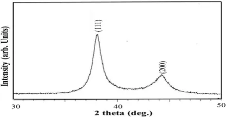
Fig. 1 shows the corresponding XRD pattern. The dominant peaks are corresponded to standard Bragg reflections (111) and (200) of face center cubic (FCC) lattice. The intense diffraction at 38.1 peak shows that the preferred growth orientation of gold was fixed in (111) direction. This XRD pattern is typical of pure gold nanocrystals [standard joint committee on powder diffraction standards (JCPDS) card no. 04-0784]. Further, the pattern shows a line broadening, which indicates the crystallite size reduced. The crystallite size of the material was calculated using the
Scherrer’s formula:
d = Kl/b cosq,
Where d is the crystallite size, K is the dimension less shape factor (0.94), l is the X-ray wavelength, b is the full width
half maxima (FWHM) and q is the Bragg’s angle. The
crystallite size of the synthesized gold nanoparticles was found to be 10.8 nm.
Figure 1: Room temperature powder XRD pattern of as-prepared gold nanoparticles using parthenium extract.
b. Scanning electron microscopy:
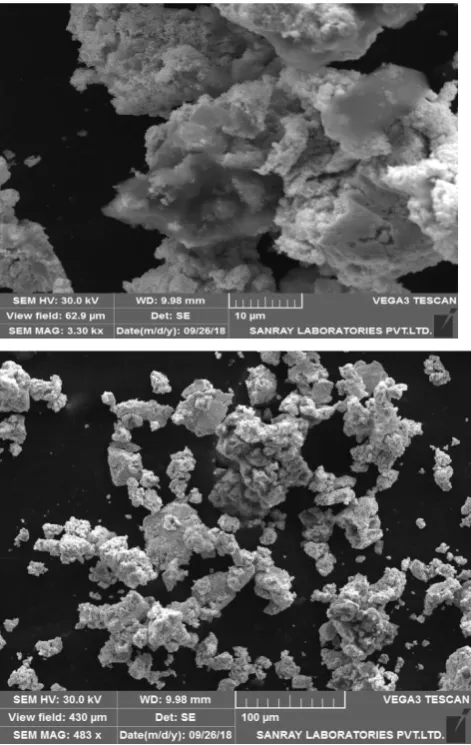
The SEM images show agglomeration of gold particles (fig. 2(a)). The magnified image is shown in fig. 2(b), it appears to be some of the particles are triangular and hexagonal in shape. Fig. 2 represents the morphology of the as-synthesized gold nanoparticles prepared by using
parthenium hysterophorous plant extract. Typical SEM
Figure 2: SEM images of as-prepared gold nanoparticles using parthenium extract at different magnitudes (a) 10 µm and (b) 100 µm.
c. UV–Visible Spectroscopy
UV–visible absorption spectrum as showed in fig. 3, is carried out to evaluate the potential optical properties of the as-prepared gold nanoparticles using parthenium
hysterophorous plant extract. For the UV–visible
absorption measurement, the as-prepared gold nanoparticles using parthenium hysterophorous plant extract sample is ultrasonically dispersed in absolute ethanol before the examination, using absolute ethanol as the reference. The spectrum was corrected for the solvent contribution.
The absorption spectrum of gold nanoparticles using
parthenium hysterophorous plant extract shows
well-defined excitation band at 534 nm which is in good agreement with the characteristic gold nanoparticles surface plasmon resonance (SPR). The SPR property of gold nanoparticles is evident with a characteristic absorption band in between 500 to 600 nm in the UV-Visible spectrum. This absorption band depends on the size and shape of the gold nanoparticles and the dielectric constant of the medium. Hence the present study results confirm the formation of gold nanoparticles.
Figure 3: Absorption of as-prepared gold nanoparticles using parthenium extract as a function of wavelength.
d. Fourier Transform Infrared Spectroscopy
Fourier Transforms Infrared Spectroscopy (FTIR) was used to identify the possible biomolecules responsible for the reduction of the gold ions and capping of the bio-reduced gold nanoparticles synthesized by parthenium plant extract.
The FT-IR spectrum below indicates characteristic three bands at about 3327.21, 1625.99 and 1377.17 cm-1. The band at 3327.21 cm-1 corresponds to N-H stretching or O-H stretching vibration. The peak 1377.17 cm-1 corresponds to C-N stretching of aromatic amine group. The band at 1625.99 cm-1 corresponds to C=O stretching vibrations due to the carbonyl group in the proteins of amide I group of proteins. The amide I band (between 1600 and 1700cm-1) is mainly associated with the C=O stretching vibration (70-85%) and is directly related to the backbone conformation. Gold nanoparticle might get bonded with protein through free amine group or carboxylate group. This infers that the synthesized gold nanoparticles are surrounded with protein and metabolite such as terpenoid that has functional groups of ketone, alcohol and carboxylic acids.
Figure 4: FT-IR spectrum of synthesized gold nanoparticles using parthenium extract.
DISCUSSION
In present time, nanoparticle synthesis approaches include three methods such as Physical methods, Chemical methods and Biological methods.14,15 Chemical methods
Wave number cm
-1
Trans
m
it
tance
include the reduction of chemicals,16 electrochemical procedures,17 and reduction of photochemicals.18
Physical and chemical methods are being used extensively for production of metal and metal oxide nanoparticles. However, this production requires the use of very reactive and toxic reducing agents such as sodium borohydride and hydrazine hydrate, which cause undesired detrimental impacts on the environment, plant and animal life it supports.
Nanoparticle synthesis by using plant extracts is eco-friendly, involves simple reactions or minimal conditions such as low temperature and extraction process.19 Hence Nanoparticle synthesis by using plant extracts is attracting much attention. Major benefits in the plant mediated synthesis of nanoparticles are plant secondary metabolites have pharmacological activity, opportunity for scale up, devoid of hazardous chemicals and their pharmaceutical applications.20 In addition, synthesis requirements like high temperature, pressure, energy are insignificant.
The green method of synthesizing metal nanoparticles involves platinum (Pt), gold (Au), silver (Ag), copper (Cu), and zinc (Zn), among whom Au exhibits a unique and tunable surface plasmon resonance (SPR).21 Gold nanoparticles possess different size, shape, and aggregation ability. Previous reports on gold nanoparticle synthesis comprise the use of Cassia auriculata,22 Medicago sativa,23 Aloe vera,24 Pelargonium graveolens,25
Tamarindo’s indica,26 Coriandrum sativum27 and Cymbopogon citratus.28 Gold nanoparticles show high biocompatibility and have a wide range of medical applications,29 drug delivery,30 gene delivery,31 and photo-thermal therapy.32 Hence Parthenium plant was selected to mediate the synthesis of gold nanoparticles.
XRD is used to examine the crystal structure and phase purity of synthesized gold nanoparticles. In the present study the XRD pattern of synthesized gold nanoparticles using the extract of parthenium hysterophorous plant showed standard Bragg reflections (111) and (200) of face center cubic (FCC) lattice of gold. These results are in good agreement with the previous reports on green synthesis of
gold nanoparticles that indicates similar Bragg’s reflection
for gold.33,34 This XRD pattern is typical of pure gold nanocrystals with JCPDS card no. 04-0784.
Gupta et al., reported that 35 four different intense peaks
at 2θ angle: 38.22, 44.42, 64.71, and 77.62 with Bragg
reflections corresponding to (111), (200), (220), and (311) in biomass- associated gold nanoparticles. Alternatively, only a single prominent peak was observed at 2θ angle:
38.22 with a Bragg reflection corresponding to (111) in extracellular gold nanoparticles. Our present findings are consistent with earlier studies that used biological methods to synthesize gold nanoparticles using plant extracts.36-38
The UV–visible spectroscopy is carried out to evaluate the
extract. The basic property of gold nanoparticle is that it has characteristic wine-red color in solution. In the present study during synthesis of parthenium plant mediated synthesis of gold nanoparticles, red color change was observed, which confirms the formation of gold nanoparticles. Wani et. al., 39 in his study stated that gold nanoparticles are known to exhibit at maximum in the range of 200 to 800 nm. This color formation belongs to the surface Plasmon vibration of the metal nanoparticles.
The red color is due to the SPR phenomenon of gold nanoparticles. SPR occurs when the incoming visible light strikes with the electron oscillating on the surface of the nanoparticle then at a certain wavelength, a resonance takes place where the frequency of the light matches with the frequency of the oscillation of the electron. 40,41 Hence SPR phenomenon is due to change in metal oxidation state where in Au+ was reduced to Au 0 and is mediated by the biomolecules present in Parthenium plant extract. The gold nanoparticles absorption band at 534 nm observed in the present study is characteristic SPR of gold nanoparticles and is in good agreement with previous reports. 42, 43, 44
The FT-IR spectrum in the present study indicates characteristic three bands at about 3327.21, 1625.99 and 1377.17-cm. The band at 3327.21 cm-1 corresponds to N-H stretching or O-H stretching vibration. The peak 1377.17 cm-1 corresponds to C-N stretching of aromatic amine
group. The band at 1625.99 cm-1 corresponds to C=O stretching vibrations due to the carbonyl group in the proteins of amide I group of proteins.
Similar observations were reported in the previous study of S. Vijayakumar et al., 21. The intense broad absorption peak at 3451 cm_1 represents the O-H stretching vibrations of phenols and carboxylic acids. The peak located at 1636 cm_1 was assigned to the C=O stretching in carboxyl or C=N
bending in the amide group. The band observed at 1387cm_1 was assigned to C-N stretching or the O-H bending and its shift to 1034cm_1 implicated the role of these groups in the interaction with chloroauric acid. Results in this study are consistent with the literature data.21, 45, 46
CONCLUSION
A novel eco-friendly method was established to synthesize Gold nanoparticles without the aid of external energy. Formation of these gold nanoparticles was confirmed by spectroscopic techniques. Gold nanoparticles is a proven antimicrobial agent, biocompatible and nontoxic to human cells. Hence the established method can be further scaled up for gold nanoparticle synthesis for pharmaceutical use.
REFERENCES
1. Zhang X, Gold nanoparticles: recent advances in the biomedical applications, Cell Biochem Biophys, 72, 2015, 771–775.
2. Payne JN, Waghwani HK, Connor MG, Hamilton W, Tockstein S, Moolani H, Chavda F, Badwaik V, Lawrenz MB, Dakshinamurthy R, Novel synthesis of kanamycin conjugated gold nanoparticles with potent antibacterial activity, Front Microbiol, 2016.
3. Poovathinthodiyil Raveendran, Jie Fu and Scott L Wallen, Completely “Green” Synthesis and Stabilization of Metal Nanoparticles, Journal of the American Chemical Society, 125, 2003, 13940-13941.
4. Narayanan Sreeja, Binulal Nelson Sathy, Ullas Mony, Manzoor Koyakutty, Shantikumar Vasudevan Nair and Deepthy Menon, “Biocompatible magnetite/gold nanohybrid contrast agents via green chemistry for MRI and CT bioimaging,”ACS applied materials & interfaces, 41, 2012, 251-260.
5. Anju Thangam, Sakthi Ramlakshmi, Pritam, Effect of ZnO nanoparticles against strains of Escherichia coli, Asian J Pharm Clin Res, 7, 2014, 202-206.
6. Subramani Srinivasan, Dhananjayan Indumathi, Mathiyazhagan Sujatha, Kathiroli Sujithra, Udaiyar Muruganathan, Novel Synthesis, characterization and antibacterial activity of silver nanoparticles using leaf extract
of Melothria maderaspatana (linn) cong, Int J Pharm Pharm
Sci, 8, 2016, 104-109.
7. Pramod kumar, Indrajit roy, Applications of gold nanoparticles in clinical medicine, Int J Pharm Pharm Sci, 8, 2016, 9-16.
8. Nachiyar V, Sunkar S, Prakash P, Biological synthesis of gold nanoparticles using endophytic fungi, Der Pharma Chemica, 7, 2015, 31-38.
9. Ramesh M, Anbuvannan M and Viruthagiri G, Green synthesis of ZnO nanoparticles using Solanum nigrum leaf extract and their antibacterial activity, Spectrochimica Acta Part A:Molecular and Biomolecular Spectroscopy, 136, 2015, 864-870.
10. Xiao L, Liu C, Chen X and Yang Z, Gold nanoparticles induce renal toxicity through reactive oxygen species, Food Chem Toxicol, 90, 2016, 76-83.
11. Rajeshkumar S, Anticancer activity of eco-friendly gold nanoparticles against lung and liver cancer cells, Journal of Genetic Engineering and Biotechnology, 14, 2016, 195–202. 12. C Nagajyothi, P & An, Tran Nguyen & Tvm, et al., Green route biosynthesis: Characterization and catalytic activity of ZnO nanoparticles, Materials Letters, 108, 2013, 160-163. 13. Mahendran Vanaja, Gnanadhas Gnanajobitha, Kanniah
Paulkumar, Shanmugam Rajeshkumar, Chelladurai Malarkodi and Gurusamy Annadurai, Phytosynthesis of silver nanoparticles by Cissus quadrangularis: Influence of physicochemical factors, Journal of Nanostructure in Chemistry, 3, 2013, 17-24.
14. Liu Z, Bucknall DG, Allen MG Inclined nanoimprinting lithography for 3D nanopatterning, Nanotechnology, 22, 2011, 225–302.
15. Mohanpuria P, Rana NK, Yadav SK, Biosynthesis of nanoparticles: technological concepts and future applications, J Nanoparticle Res. 10, 2008, 507–517. 16. Guzmán MG, Dille J, Godet S (2009) Synthesis of silver
nanoparticles by chemical reduction method and their antibacterial activity, Int J Chem Biomol,2, 2008.
17. Rodríguez-Sanchez ML, Blanco MC, López-Quintela MA, Electrochemical synthesis of silver nanoparticles, J Phys Chem B, 104, 2000, 9683–9688.
18. Sharma VK, Yngard RA, Lin Y, Silver nanoparticles green synthesis and their antimicrobial activities, Adv Colloid Interf Sci, 145, 2009, 83–96.
19. Goodsell DS, Bionanotechnology: lessons from nature. Wiley, Hoboken, 2004.
20. Abdel-Halim ES, El-Rafie MH, Al-Deyab SS (2011) Polyacrylamide/guar gum graft copolymer for preparation of silver nanoparticles, Carbohydr Polym, 85, 2011, 692 21. M.M.H. Khalil, E.H. Ismail, F. El-Magdoub, Biosynthesis of Au
nanoparticles using olive leaf extract, 1st nano updates, Arab. J. Chem, 5, 2012, 431-437.
22. V.G. Kumar, S.D. Gokavarapu, A. Rajeswari, T.S. Dhas, V. Karthicka, Z. Kapadia, et al., Facile green synthesis of gold nanoparticles using leaf extract of antidiabetic potent Cassia auriculata, Colloids Surf. B Biointerfaces, 87, 2011, 159-163. 23. J.L. Gardea Torresdey, K.J. Tiemann, G. Gamez, K. Dokkenn, I. Cano-Aguilera, L.R. Furen lid, et al., Reduction and accumulation of Gold (III) Medicago sativa alfalfa biomass: X-ray absorption spectroscopy, pH, temperature dependence, Environ. Sci. Technol, 4, 2000, 4392-4396. 24. S.P. Chandran, M. Chaudhary, R. Pasricha, A. Ahmad, M.
Sastry, Synthesis of gold nanotriangles and silver nanoparticles using Aloe vera plant extract, Biotechnol. Prog, 22, 2006, 577-583.
25. S.S. Shankar, A. Ahmad, R. Pasricha, M. Sastry, Bioreduction of Chloroaurate ions by geranium leaves and its endophytic fungus yields gold nanoparticles of different sizes, J. Mater. Chem, 13, 2003, 1822-1826.
26. B. Ankamwar, M. Chaudhary, M. Sastry, Gold nanotriangles biologically synthesized using tamarind leaf extract and potential application in vapor sensing, Metal-Organ. Nano-Metal Chem, 35, 2005, 19-26.
27. K.B. Narayanan, N. Sakthivel, Coriander leaf mediated biosynthesis of gold nanoparticles, Mater. Lett, 62, 2008, 4588-4590.
28. K. Murugan, C. Panneerselvam, A.T. Aziz, J. Subramaniam, P. Madhiyazhagan, J.S. Hwang, D. Lan Wang, D. Dinesh, U. Suresh, M. Roni, A. Higuchi, M. Nicoletti, M. Saleh Alsalhi, G. Benelli, Eco-friendly drugs from the marine environment: spongeweed-synthesized silver nanoparticles are highly effective on Plasmodium falciparum and its vector Anopheles stephensi, with little non-target effects on predatory copepods, Environ. Sci. Pollut. Res, 23, 2016, 6832-6839.
30. P. Ghosh, G. Han, M. De, C.K. Kim, V.M. Rotello, Gold nanoparticles in delivery applications, Adv. Drug Deliv. Rev, 60, 2008, 1307-1315.
31. D. Pissuwann, T. Niidome, M.B. Cortie, The forth coming applications of gold nanoparticles in drug and gene delivery systems, J. Control. Release, 149, 2011, 65-71.
32. P.K. Jain, I.H. El-Sayed, M.A. El-Sayed, Au nanoparticles target cancer, Nano Today 2, 2007, 18-29.
33. P. Nalawade, T. Mukherjee, S. Kapoor, Green synthesis of gold nanoparticles using glycerol as a reducing agent, Adv. Nanoparticles, 2, 2013, 78-86.
34. S. Vijayakumar et al, Therapeutic effects of gold nanoparticles synthesized using Musa paradisiaca peel extract against multiple antibiotic resistant Enterococcus faecalis biofilms and human lung cancer cells (A549), Microbial Pathogenesis, 102, 2017, 173-183.
35. Gupta S, Bector S, Biosynthesis of extracellular and intracellular AuNPs by Aspergillus fumigatus and A. flavus. Antonie Van Leeuwenhoek, 103, 2013, 1113–1123. 36. Gardea-Torresdey JL, Parsons JG, Gomez E, Peralta-Videa J,
Troiani HE, Santiago P, Jose Yacaman M: Formation and growth of Au nanoparticles inside live Alfalfa plants. Nano Lett, 2, 2002, 397–401.
37. Shankar SS, Rai A, Ankamwar B, Singh A, Ahmad A, Sastry M: Biological synthesis of triangular gold nanoprisms, Nat Mater, 3, 2004, 482–488.
38. Shankar SS, Ahmad A, Pasrichaa R, Sastry M: Bioreduction of chloroaurate ions by geranium leaves and its endophytic fungus yields gold nanoparticles of different shapes, J Mater Chem, 13, 2003, 1822–1826.
39. I.A. Wani, T. Ahmad, Size and shape dependant antifungal activity of gold nanoparticles, Colloids and Surfaces B: Biointerfaces, 101, 2013, 162– 170.
40. Daniel, M.C. and D. Astruc, Gold Nanoparticles: Assembly, Supramolecular Chemistry, Quantum-Size-Related Properties, and Applications Toward Biology, Catalysis, and Nanotechnology, Chemical Reviews, 104, 2004, 293-346. 41. Chen H, Wang Y, Wang Y, Dong S, Wang E, One-step
preparation and characterization of PDDA-protected gold nanoparticles, Polymer, 47, 2006, 763-766.
42. A.M. Fayaz, M. Girilal, R. Venkatesan, P.T. Kalaichelvan, Biosynthesis of anisotropic gold nanoparticles using Maduca longifolia extract and their potential in infrared absorption, Colloids Surf. B Biointerfaces, 88, 2011, 287 - 291.
43. K. Govindaraju, S.K. Basha, V.G. Kumar, G. Singaravelu, Silver, gold and bimetallic nanoparticles production using single cell protein (Spirulina platensis), J. Mater. Sci, 43, 2008, 5115 - 5122.
44. S.P. Dubey, M. Lahtinen, H. Sarkka, M. Sillanpaa, Bioprospective of Sorbusaucuparia leaf extract in development of silver and gold nanocolloids, Colloids Surf. B Biointerfaces, 80, 2010, 26 - 33.
45. Shankar SS, Rai A, Ankamwar B, Singh A, Ahmad A, Sastry M: Biological synthesis of triangular gold nanoprisms, Nat Mater, 3, 2004, 482–488.
46. Shankar SS, Ahmad A, Pasrichaa R, Sastry M: Bioreduction of chloroaurate ions by geranium leaves and its endophytic fungus yields gold nanoparticles of different shapes, J Mater Chem, 13, 2003, 1822–1826.